Article

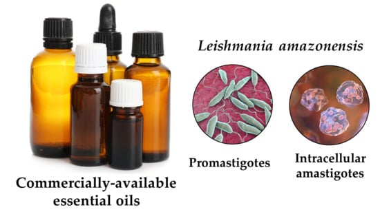
In-Vitro Evaluation of 52 Commercially-Available Essential Oils Against Leishmania amazonensis

1
Parasitology Department, Institute of Tropical Medicine “Pedro Kouri”, Havana 10400, Cuba
2
Aromatic Plant Research Center, 230 N 1200 E, Suite 100, Lehi, UT 84043, USA
3
Department of Chemistry, University of Alabama in Huntsville, Huntsville, AL 35899, USA
*
Author to whom correspondence should be addressed.
Molecules 2019, 24(7), 1248; https://doi.org/10.3390/molecules24071248
Submission received: 5 March 2019 / Revised: 25 March 2019 / Accepted: 29 March 2019 / Published: 30 March 2019
(This article belongs to the Special Issue Drug Discovery for Neglected Diseases)

Abstract

:
Leishmaniasis is a neglected tropical disease caused by members of the Leishmania genus of parasitic protozoa that cause different clinical manifestations of the disease. Current treatment options for the cutaneous disease are limited due to severe side effects, poor efficacy, limited availability or accessibility, and developing resistance. Essential oils may provide low cost and readily available treatment options for leishmaniasis. In-vitro screening of a collection of 52 commercially available essential oils has been carried out against promastigotes of Leishmania amazonensis. In addition, cytotoxicity has been determined for the essential oils against mouse peritoneal macrophages in order to determine selectivity. Promising essential oils were further screened against intracellular L. amazonensis amastigotes. Three essential oils showed notable antileishmanial activities: frankincense (Boswellia spp.), coriander (Coriandrum sativum L.), and wintergreen (Gualtheria fragrantissima Wall.) with IC50 values against the amastigotes of 22.1 ± 4.2, 19.1 ± 0.7, and 22.2 ± 3.5 μg/mL and a selectivity of 2, 7, and 6, respectively. These essential oils could be explored as topical treatment options for cutaneous leishmaniasis.

Graphical Abstract

1. Introduction

Leishmaniasis is a collection of parasitic diseases caused by several Leishmania species [1,2]. The disease is transmitted by several members of Phlebotominae sand flies. The genera Lutzomyia (New World) and Phlebotomus (Old World) are the only hematophagous vectors of the diseases. Leishmaniasis is considered by the World Health Organization to be a neglected tropical disease [3]. Depending on the Leishmania species, the disease can manifest itself in three main forms: cutaneous, mucocutaneous, and visceral. There are currently around 12–15 million people worldwide infected by Leishmania spp. with an estimated 350 million people at risk of acquiring leishmaniasis. Unfortunately, at the present time, there are no vaccines or prophylactic medicines available to prevent the disease. Current chemotherapeutic treatment options include sodium stibogluconate, meglumine antimoniate, conventional (deoxycholate) and liposomal amphotericin B, pentamidine isethionate, miltefosine and paromomycin. However, these agents suffer from severe side effects, poor efficacy, limited availability, high cost, or developing resistance [2,4]. Furthermore, treatment of leishmaniasis is often hampered by limited access to medicines and treatment options in developing countries where the disease is prevalent [5,6].
Essential oils are complex mixtures of volatile phytochemicals with numerous and varied biological properties [7,8]. Essential oils, particularly those that are readily available, may provide low cost treatment options for leishmaniasis. The antiparasitic activities of essential oils and their components have been demonstrated and previously reviewed [9,10]. Commercially available EOs have been screened for activity against a variety of human pathogens [11,12,13,14,15,16,17] as well as for cytotoxic activity [12,16]. In this work, we carried out in-vitro antileishmanial and cytotoxic screening of a selection of 52 commercially available essential oils against the promastigote form of Leishmania amazonensis and peritoneal macrophages from BALB/c, respectively. Promising antileishmanial essential oils were further screened against intracellular amastigotes.

2. Results and Discussion

A total of 52 commercially-available essential oils were screened for in-vitro antileishmanial activity against the promastigote form of L. amazonensis, as well as for cytotoxic activity against mouse peritoneal macrophages, the host cells for the amastigote form of the parasite. The antileishmanial and cytotoxic activities of the essential oils are summarized in Table 1. Of the essential oils tested, three showed notable antileishmanial activity and selectivity, frankincense oil (from the oleogum resin of Boswellia spp.), coriander oil (from the seeds of Coriandrum sativum L.), and wintergreen oil (from the leaves of Gualtheria fragrantissima Wall.). These three products were also active against the intracellular amastigote form of L. amazonensis (Table 2).
The cluster analysis revealed four clusters based on biological activities (Figure 1). Cluster 1 is made up of essential oils that showed good antileishmanial activity with little or no cytotoxicity. The very large cluster 2 can be described as essential oils that showed relatively weak antileishmanial activity and relatively weak cytotoxic activity, cluster 3 is composed of essential oils with both moderate antileishmanial activity but also with moderate cytotoxic activity, and cluster 4 is made up of essential oils that were very cytotoxic.
The active cluster (Cluster 1) can be subdivided into two sub-clusters made up of coriander (Coriandrum sativum) and frankincense (Boswellia spp.), Cluster 1a, and Cluster 1b, wintergreen (Gualtheria fragrantissima) and birch bark (Betula lenta L.). Frankincense is the oleogum resin from several Boswellia species. Commercial frankincense sources include B. sacra Flueck., B. carteri Birdw., B. frereana Birdw., B. papyrifera Hochst. and B. serrata Roxb. Commercial frankincense essential oil from doTERRA International is a proprietary blend of largely B. carteri, followed by B. sacra, B. papyrifera, and B. freriana. Analysis of the essential oil used in this investigation revealed that it is composed largely of monoterpenes, including α-pinene (41.2%), α-thujene (15.7%), limonene (9.4%), sabinene (4.5%), β-pinene (3.5%), and p-cymene (3.3%), as well as octyl acetate (5.4%) and β-caryophyllene (3.1%). Frankincense essential oil showed activity against promastigotes and amastigotes of L. amazonensis with IC50 values of <12.5 and 22.1 ± 4.2 μg/mL, respectively. Consistent with the antiparasitic activity of frankincense essential oil, Fujisaki and co-workers screened B. carteri essential oil against Plasmodium falciparum and the oil was shown to have an IC50 of 10 μg/mL [18]. α-Pinene itself has shown activity against both promastigotes (IC50 19.7 μg/mL) and amastigotes (IC50 16.1 μg/mL) of L. amazonensis [19].
Another essential oil rich in α-pinene was cypress (Cupressus sempervirens L.) with 49.7% α-pinene. Cypress essential oil gave an IC50 of 40.0 μg/mL against L. amazonensis promastigotes, but was too cytotoxic (CC50 27.2 μg/mL) against mouse peritoneal macrophages to be considered for further evaluation. Other major components in cypress essential oil were δ-3-carene (27.0%) and terpinolene (4.2%). Juniper (Juniperus communis L.) berry essential oil was also dominated by α-pinene (34.9%), with lesser quantities of myrcene (11.9%), sabinene (11.4%), β-pinene (7.9%), and terpinen-4-ol (4.5%). Juniper berry essential oil showed good activity against L. amazonensis promastigotes (IC50 27.4 μg/mL) and reduced cytotoxicity against mouse peritoneal macrophages (CC50 103.0 μg/mL). Thus, the presence of α-pinene seems to impart antileishmanial activity, but the activity is apparently enhanced or attenuated by other components in the essential oil.
Coriander is the fruit of C. sativum. Commercial coriander essential oil used in this study was obtained from dōTERRA International and the major components were linalool (73.5%), α-pinene (5.3%), and γ-terpinene (4.5%). A linalool-rich (73.2%) coriander essential oil was screened by Rondon and co-workers against Leishmania chagasi and was shown to be very effective against amastigotes (IC50 1.51 μg/mL), but much less active against the promastigotes (IC50 181 μg/mL) [20]. It is tempting to speculate that linalool might be the compound responsible for the antileishmanial activity. Indeed, Rosa and co-workers have shown linalool to be very active against both promastigotes (IC50 0.0043 μg/mL) and amastigotes (IC50 0.0155 μg/mL) of L. amazonensis [21]. However, the presence of linalool does not necessarily translate to a good antileishmanial profile. Pettitgrain (Citrus aurantium L.) leaf essential oil (25.4% linalool), lavender (Lavandula angustifolia Mill.) essential oil (34.4% linalool), and basil (Ocimum basilicum L.) essential oil (55.7% linalool), were only marginally active against L. amazonensis promastigotes (IC50 56.9, 70.7, and >200 μg/mL, respectively). There may be other components in these essential oils attenuating the antileishmanial activity of linalool.
Interestingly, wintergreen (Gualtheria fragrantissima) essential oil, which was dominated by methyl salicylate (99.7%), showed antileishmanial activity against both L. amazonensis promastigotes (IC50 20.7 μg/mL) and amastigotes (IC50 22.2 μg/mL). Betula lenta (birch bark) essential oil (99.9% methyl salicylate) was somewhat less active against L. amazonensis promastigotes (IC50 32.2 μg/mL). Considering the abundance of methyl salicylate in both wintergreen and birch essential oils, this compound is likely the active component.
Copaiba oils (not hydrodistilled essential oils) from several different species of Copaifera were screened by Santos and collaborators against L. amazonensis [22]. Antileishmanial activities against the promastigotes ranged from 5.0 to 22.0 μg/mL. Consistent with these results, copaiba oil in this study (50% β-caryophyllene) showed an IC50 of 17.2 μg/mL. The copaiba essential oil, however, was also too cytotoxic (CC50 23.3 μg/mL) to warrant further consideration. On the other hand, β-caryophyllene was found to be remarkably active against L. amazonensis amastigotes (IC50 1.3 μg/mL) but less toxic to murine macrophages (CC50 63.6 μg/mL) [23]. In contrast, antipromastigote activity of β-caryophyllene on L. amazonensis was reported to have an IC50 of 19 μg/mL [24]. β-Caryophyllene was also found to show excellent antileishmanial activity against L. infantum and L. major (IC50 1.06 and 1.33 μg/mL, respectively), with less cytotoxicity against Raw 264.7 mouse macrophage cells [25].
Rosemary (Rosmarinus officinalis L.) essential oil from Colombia (composition not reported) showed activity against L. braziliensis promastigotes with an IC50 of 17.4 μg/mL [26]. Similarly, rosemary essential oil from Tunisia (43.8% 1,8-cineole, 12.0% camphor, 11.5% α-pinene, 8.6% β-pinene, 4.8% camphene) was active against L. infantum (IC50 16.3 μg/mL) and L. major (IC50 20.9 μg/mL) promastigotes [25]. Conversely, a commercial rosemary essential oil obtained in Germany was inactive against L. major promastigotes (IC50 282 μg/mL) [11]. In this present study, commercial rosemary essential oil (45.9% 1,8-cineole, 12.0% α-pinene, 10.9% camphor, 6.3% β-pinene) was only marginally antileishmanial (IC50 89.7 μg/mL), but also marginally cytotoxic (CC50 83.4 μg/mL). Rosemary essential oils have shown great variation in composition with at least five different chemotypes, which likely result in different biological activities [27].
Mikus and co-workers have screened several essential oils and essential oil components for antileishmanial activity against L. major promastigotes [11]. These workers found commercial Melissa officinalis L. essential oil to show excellent activity against L. major promastigotes (IC50 7.0 μg/mL) with less toxicity against HL-60 (human leukemia) cells (CC50 25.5 μg/mL). Andrade and co-workers screened a commercial M. officinalis essential oil (37.2% neral, 52.0% geranial) and fount marginal activity against L. amazonensis promastigotes with an IC50 of 132 μg/mL [17]. In contrast, M. officinalis essential oil from Colombia was inactive against L. braziliensis promastigotes [26]. In this present work, M. officinalis essential oil showed antileishmanial activity (IC50 24.6 μg/mL) but also cytotoxicity (CC50 37.3 μg/mL) giving it an unfavorable selectivity index. The major components in M. officinalis essential oil were β-caryophyllene (13.4%), geranial (30.2%), and neral (23.1%). Citral, a mixture of geranial and neral, has shown antileishmanial activity against L. infantum, L. tropica, and L. major promastigotes with IC50 values of 42, 34, and 36 μg/mL, respectively [28]. However, citral has demonstrated in-vitro cytotoxic activity against several cell lines [29,30,31]. There are several chemotypes of M. officinalis based on essential oil composition, which likely account for the differences in biological activities. Nevertheless, the citral chemotype of M. officinalis has also shown in-vitro cytotoxicity [32].
Likewise, the unfavorable bioactivity profile of lemongrass (Cymbopogon flexuosus (Nees) Will. Watson) essential oil in this study is likely due to the abundance of geranial (49.9%) and neral (23.4%). However, Machado and co-workers found that citral-rich C. citratus as well as citral did not exhibit cytotoxic effects on either primary bovine aortic endothelial cells or RAW 264.7 macrophage cells [28]. Similarly, Santin and co-workers found both C. citratus essential oil and citral to be antileishmanial against promastigotes (IC50 1.7 and 8.0 μg/mL, respectively) and amastigotes (IC50 3.2 and 25.0 μg/mL, respectively) of L. amazonensis, but with lower cytotoxicity against J774G8 macrophage cells [33].
As has been appreciated, many pure components present in the tested EOs have exhibited some level of antileishmanial activity. In fact, it is known that the compounds present in the EOs can act synergistically in mixtures, increasing the intrinsic effects of the purified components. Therefore, we suggest the potential use of EOs as mixtures. In addition, we have also taken into account that the tested EOs are commercially available in their present forms, reducing the cost of a therapeutic alternative, which constitutes one of main drawbacks of current antileishmanial treatments. The notable antileishmanial effects and moderate cytotoxicities in the case of mixtures of compounds could be further explored in animal models of cutaneous leishmaniasis by intralesional or topical routes.

3. Materials and Methods

3.1. Essential Oils

The essential oils were obtained from commercial sources, dōTERRA International (Pleasant Grove, UT, USA), Ameo Essential Oils (Zija International, Lehi, UT, USA), Mountain Rose Herbs (Eugene, OR, USA), or Albert Vielle (Vallauris, France) and were previously analyzed by gas chromatography–mass spectrometry.

3.2. Parasites

Leishmania amazonensis parasites (Reference strain MHOM/77/BR/LTB0016) were kindly provided by the Department of Immunology, Oswaldo Cruz Foundation (FIOCRUZ), Brazil. The parasites were routinely isolated from mouse lesions (BALB/c mice) and maintained as promastigotes at 26 °C in Schneider’s medium (Sigma-Aldrich, St. Louis, MO, USA) containing 10% heat-inactivated fetal bovine serum (HFBS) (Sigma-Aldrich, St. Louis, MO, USA) and 100 U of penicillin and 100 μg streptomycin per mL as antibiotics. Parasite cultures were passaged every 3–4 days, but no longer used after the tenth passage after isolation from mice.

3.3. In-vitro Anti-promastigote Screening

Screening against L. amazonensis promastigotes was carried out as previously described [34,35,36]. Into each well in a 96-well plate, 50 μL medium (Schneider’s medium + HFBS + antibiotics) was added. Into the wells of column 2 and 7, 48 μL medium + 2 μL sample (dimethylsulfoxide solutions of essential oil, 20 mg/mL) were added. Five two-fold serial dilutions were carried out down each column to give final test concentrations of 12.5, 25, 50, 100 and 200 μg/mL. Exponentially growing parasites (4 × 10⁵) in medium were added to each well. Dimethylsulfoxide (DMSO) served as the negative control and pentamidine (Richet, Buenos Aires, Argentina) was used as a positive control. The plates were sealed with Parafilm® and incubated at 26 °C for 72 h. Afterward, 20 μL of a solution (5 mg/mL) of MTT (3-[4,5-dimethylthiazol-2-yl]-2,5-diphenyltetrazolium bromide) (Sigma-Aldrich, St. Louis, MO, USA) was added to each well. The plates were incubated for an additional 4 h, the supernatant was removed, and the formazan crystals were dissolved with 100 μL DMSO. The absorbance of each well was determined with a plate reader (Molecular Devices, San Jose, CA, USA) with a test wavelength of 560 nm and a reference wavelength of 630 nm from which median inhibitory concentrations (IC50) were calculated [37,38]. The IC50 values were determined from linear dose-response curves fit to the data using a linear equation model. Each test was carried out in triplicate, and the results expressed as means ± standard deviations (SD).

3.4. Mouse Peritoneal Macrophage Cytotoxicity Screening

The median cytotoxic concentrations (CC50) of the essential oils on mouse peritoneal macrophages (the host cells of Leishmania parasites) were determined as previously described [34,35,36]. Macrophages were collected from the peritoneal cavities of normal BALB/c mice and suspended in ice-cold RPMI 1640 medium (Sigma-Aldrich, St. Louis, MO, USA), supplemented with antibiotics. The cells were seeded in 96-well plates (3 × 10⁵ cells/well) and incubated at 37 °C for 2 h. Non-adherent cells were removed. Into the wells of column 2 and 7, 2 μL of essential oil solutions (as above) and 48 μL medium (supplemented with 10% HFBS and antibiotics) were also added. Two-fold serial dilutions down each lane were carried out to give final concentrations of 12.5–200 μg/mL. The macrophages were incubated for 72 h, after which the cytotoxicity was determined using the MTT assay (as above), using 15 μL/well. The CC50 was calculated for each compound in the same manner as the IC50 and selectivity indices (SI) for each essential oil were determined (CC50 for macrophages / IC50 for promastigotes).
Essential oils with an IC50 < 100 µg/mL and a SI ≥ 5 were selected for further evaluation against L. amazonensis amastigotes [34].

3.5. In-vitro Intracellular Anti-amastigote Screening

Median inhibitory concentrations (IC50) of the active essential oils on L. amazonensis amastigotes were carried out as previously described [34,35,36]. The peritoneal macrophages, obtained from BALB/c mice (as above), were seeded in 24-well culture plates at 106 cells/mL. The plates were incubated at 37 °C under a 5% CO2 atmosphere for 2 h. Non-adherent cells were removed, and stationary-phase L. amazonensis promastigotes were added (4:1 promastigote/macrophage ratio), and incubation continued for an additional 4 h. Free parasites were removed and 1000 μL RPMI completed medium was added to each well. Into the first well, 990 μL medium and 10 μL essential oil solution were added. Four two-fold dilutions were carried out to give final concentrations of 12.5, 25, 50 and 100 μg/mL. The plate was incubated at 37 °C under a 5% CO2 atmosphere for 48 h. The cell monolayers were fixed in absolute methanol, stained with Giemsa, and evaluated using a light microscope. The number of intracellular amastigotes was determined by counting 25 macrophages per sample. The results are expressed as percent reduction of infection rate (% infected macrophages × number amastigotes per infected macrophage) compared to that of the controls. The IC50 values were determined from the concentration-response linear curves. Each evaluation was carried out in triplicate and the results expressed as means ± SD.

3.6. Hierarchical Cluster Analysis

The chemical compositions of the commercial essential oils along with the antileishmanial and cytotoxic activities were used in an agglomerative hierarchical cluster (AHC) analysis. The essential oil compositions of the 52 commercially-available essential oils and the bioactivities were used to determine the associations between the essential oils and their activities using XLSTAT Premium, version 2018.5.53172 (Addinsoft, Paris, France). Dissimilarity was determined using Euclidean distance, and clustering was defined using Ward’s method.

4. Conclusions

This study has shown that the essential oils of frankincense, coriander, wintergreen, and birch have notable antiparasitic activities against Leishmania amazonensis with an acceptable SI with respect to cytotoxicity against mouse peritoneal macrophages. However, essential oils are complex mixtures of compounds, and the biological activities cannot necessarily be attributed to individual components; there are apparent synergistic and/or antagonistic interactions as well. Nevertheless, essential oils containing appreciable concentrations of α-pinene, linalool, or methyl salicylate should be investigated for antiparasitic activity.

Author Contributions

Conceptualization, W.N.S.; methodology, L.M.; formal analysis, L.M., P.S., and W.N.S.; investigation, L.M., I.H., P.S. and W.N.S.; resources, L.M.; data curation, W.N.S.; writing—original draft preparation, W.N.S.; writing—review and editing, L.M. and W.N.S.; project administration, W.N.S.

Funding

This research received no external funding.

Acknowledgments

W.N.S. is grateful to Zija International and dōTERRA International for generously donating essential oils. L.M. and W.N.S. carried out this work as part of the activities of the Research Network Natural Products against Neglected Diseases (ResNetNPND), (http://www.resnetnpnd.org/Start/). P.S. and W.N.S. contributed to this project as part of the activities of the Aromatic Plant Research Center (APRC, https://aromaticplant.org/).

Conflicts of Interest

The authors declare no conflict of interest.

References

  1. Torres-Guerrero, E.; Quintanilla-Cedillo, M.R.; Ruiz-Esmenjaud, J.; Arenas, R. Leishmaniasis: A review. F1000Research 2017, 6, 750. [Google Scholar] [CrossRef]
  2. Alvar, J.; Arana, B. Leishmaniasis, impact and therapeutic needs. In Drug Discovery for Leishmaniasis; Livas, L., Gil, C., Eds.; Royal Society of Chemistry: London, UK, 2018; pp. 3–23. [Google Scholar]
  3. World Health Organization. Neglected Tropical Diseases. Available online: https://www.who.int/neglected_diseases/diseases/en/ (accessed on 1 March 2019).
  4. Iqbal, H.; Ishfaq, M.; Wahab, A.; Abbas, M.N.; Ahmad, I.; Rehman, A.; Zakir, M. Therapeutic modalities to combat leishmaniasis, a review. Asian Pacific J. Trop. Dis. 2016, 6, 1–5. [Google Scholar] [CrossRef]
  5. Hailu, T.; Yimer, M.; Mulu, W.; Abera, B. Challenges in visceral leishmaniasis control and elimination in the developing countries: A review. J. Vector Borne Dis. 2016, 53, 193–198. [Google Scholar] [PubMed]
  6. Sunyoto, T.; Potet, J.; Boelaert, M. Why miltefosine—a life-saving drug for leishmaniasis—is unavailable to people who need it the most. BMJ Glob. Heal. 2018, 3, e000709. [Google Scholar] [CrossRef] [Green Version]
  7. Firenzuoli, F.; Jaitak, V.; Horvath, G.; Henri, I.; Bassolé, N.; Setzer, W.N.; Gori, L. Essential Oils: New Perspectives in Human Health and Wellness; Hindawi Publishing Corp.: London, UK, 2014. [Google Scholar]
  8. Başer, K.H.C.; Buchbauer, G. Handbook of Essential Oils: Science, Technology, and Applications; CRC Press: Boca Raton, FL, USA, 2010. [Google Scholar]
  9. Monzote, L.; Alarcón, O.; Setzer, W.N. Antiprotozoal activity of essential oils. Agric. Conspec. Sci. 2012, 77, 167–175. [Google Scholar]
  10. Bero, J.; Kpoviessi, S.; Quetin-Leclercq, J. Anti-parasitic activity of essential oils and their active constituents against Plasmodium, Trypanosoma and Leishmania. In Novel Plant Bioresources: Applications in Food, Medicine and Cosmetics; Gurub-Fakim, A., Ed.; John Wiley & Sons Ltd.: Oxford, UK, 2014; pp. 455–470. [Google Scholar]
  11. Mikus, J.; Harkenthal, M.; Steverding, D.; Reichling, J. In vitro effect of essential oils and isolated mono- and sesquiterpenes on Leishmania major and Trypanosoma brucei. Planta Med. 2000, 66, 366–368. [Google Scholar] [CrossRef]
  12. Powers, C.N.; Osier, J.L.; McFeeters, R.L.; Brazell, C.B.; Olsen, E.L.; Moriarity, D.M.; Satyal, P.; Setzer, W.N. Antifungal and cytotoxic activities of sixty commercially-available essential oils. Molecules 2018, 23, 1549. [Google Scholar] [CrossRef] [PubMed]
  13. Orchard, A.; Viljoen, A.; van Vuuren, S. Wound pathogens: Investigating antimicrobial activity of commercial essential oil combinations against reference strains. Chem. Biodivers. 2018, 15, e1800405. [Google Scholar] [CrossRef]
  14. Orchard, A.; van Vuuren, S.F.; Viljoen, A.M. Commercial essential oil combinations against topical fungal pathogens. Nat. Prod. Commun. 2019, 14, 151–158. [Google Scholar] [CrossRef]
  15. Serra, E.; Hidalgo-Bastida, L.A.; Verran, J.; Williams, D.; Malic, S. Antifungal activity of commercial essential oils and biocides against Candida albicans. Pathogens 2018, 7, 15. [Google Scholar] [CrossRef]
  16. Cannas, S.; Usai, D.; Tardugno, R.; Benvenuti, S.; Pellati, F.; Zanetti, S.; Molicotti, P.; Cannas, S.; Usai, D.; Tardugno, R.; et al. Chemical composition, cytotoxicity, antimicrobial and antifungal activity of several essential oils. Nat. Prod. Res. 2016, 30, 332–339. [Google Scholar] [CrossRef]
  17. Andrade, M.A.; Azevedo, S.; Motta, F.N.; Lucília, M.; Silva, C.L.; De Santana, J.M.; Bastos, I.M.D. Essential oils: In vitro activity against Leishmania amazonensis, cytotoxicity and chemical composition. BMC Complement. Altern. Med. 2016, 16, 444. [Google Scholar] [CrossRef]
  18. Fujisaki, R.; Kamei, K.; Yamamura, M.; Nishiya, H.; Inouye, S.; Takahashi, M.; Abe, S. In vitro and in vivo anti-plasmodial activity of essential oils, including himokitiol. Southeast Asian J. Trop. Med. Public Health 2012, 43, 270–279. [Google Scholar] [PubMed]
  19. Rodrigues, K.A.D.F.; Amorim, L.V.; Dias, C.N.; Moraes, D.F.C.; Carneiro, S.M.P.; Carvalho, F.A.D.A. Syzygium cumini (L.) Skeels essential oil and its major constituent α-pinene exhibit anti-Leishmania activity through immunomodulation in vitro. J. Ethnopharmacol. 2015, 160, 32–40. [Google Scholar] [CrossRef] [PubMed]
  20. Rondon, F.C.M.; Bevilaqua, C.M.L.; Accioly, M.P.; de Morais, S.M.; de Andrade-Júnior, H.F.; de Carvalho, C.A.; Lima, J.C.; Magalhães, H.C.R. In vitro efficacy of Coriandrum sativum, Lippia sidoides and Copaifera reticulata against Leishmania chagasi. Rev. Bras. Parasitol. Veterinária 2012, 21, 185–191. [Google Scholar] [CrossRef]
  21. Rosa, M.D.S.S.; Mendonça-Filho, R.R.; Bizzo, H.R.; Rodrigues, I.D.A.; Soares, R.M.A.; Souto-Padrón, T.; Alviano, C.S.; Lopes, A.H.C.S. Antileishmanial activity of a linalool-rich essential oil from Croton cajucara. Antimicrob. Agents Chemother. 2003, 47, 1895–1901. [Google Scholar] [CrossRef]
  22. Santos, A.O.; Ueda-Nakamura, T.; Dias Filho, B.P.; Veiga Junior, V.F.; Pinto, A.C.; Nakamura, C.V. Effect of Brazilian copaiba oils on Leishmania amazonensis. J. Ethnopharmacol. 2008, 120, 204–208. [Google Scholar] [CrossRef] [PubMed]
  23. Soares, D.C.; Portella, N.A.; Ramos, M.F.D.S.; Siani, A.C.; Saraiva, E.M. trans-β-Caryophyllene: An effective antileishmanial compound found in commercial copaiba oil (Copaifera spp.). Evidence-Based Complement. Altern. Med. 2013, 2013, 761323. [Google Scholar] [CrossRef] [PubMed]
  24. do Carmo, D.F.M.; Amaral, A.C.F.; Machado, G.M.C.; Leon, L.L.; Silva, J.R.D.A. Chemical and biological analyses of the essential oils and main constituents of Piper species. Molecules 2012, 17, 1819–1829. [Google Scholar] [CrossRef]
  25. Essid, R.; Zahra, F.; Msaada, K.; Sghair, I.; Hammami, M.; Bouratbine, A.; Aoun, K.; Limam, F. Antileishmanial and cytotoxic potential of essential oils from medicinal plants in Northern Tunisia. Ind. Crop. Prod. 2015, 77, 795–802. [Google Scholar] [CrossRef]
  26. Arévalo, Y.; Robledo, S.; Muñoz, D.L.; Granados-Falla, D.; Cuca, L.E.; Delgado, G. Evaluación in vitro de la actividad de aceites esenciales de plantas colombianas sobre Leishmania braziliensis. Rev. Colomb. Ciencias Químico-Farm. 2009, 38, 131–141. [Google Scholar]
  27. Satyal, P.; Jones, T.H.; Lopez, E.M.; McFeeters, R.L.; Ali, N.A.A.; Mansi, I.; Al-Kaf, A.G.; Setzer, W.N. Chemotypic characterization and biological activity of Rosmarinus officinalis. Foods (Basel, Switzerland) 2017, 6, 20. [Google Scholar] [CrossRef]
  28. Machado, M.; Pires, P.; Dinis, A.M.; Santos-Rosa, M.; Alves, V.; Salgueiro, L.; Cavaleiro, C. Monoterpenic aldehydes as potential anti-Leishmania agents: Activity of Cymbopogon citratus and citral on L. infantum, L. tropica and L. major. Exp. Parasitol. 2012, 130, 223–231. [Google Scholar] [CrossRef]
  29. Setzer, W.N.; Schmidt, J.M.; Eiter, L.C.; Haber, W.A. The leaf oil composition of Zanthoxylum fagara (L.) Sarg. from Monteverde, Costa Rica, and its biological activities. J. Essent. Oil Res. 2005, 17, 333–335. [Google Scholar] [CrossRef]
  30. Wright, B.S.; Bansal, A.; Moriarity, D.M.; Takaku, S.; Setzer, W.N. Cytotoxic leaf essential oils from Neotropical Lauraceae: Synergistic effects of essential oil components. Nat. Prod. Commun. 2007, 2, 1241–1244. [Google Scholar] [CrossRef]
  31. Bayala, B.; Bassole, I.H.N.; Maqdasy, S.; Baron, S.; Simpore, J.; Lobaccaro, J.-M.A. Cymbopogon citratus and Cymbopogon giganteus essential oils have cytotoxic effects on tumor cell cultures. Identification of citral as a new putative anti-proliferative molecule. Biochimie 2018, 153, 162–170. [Google Scholar] [CrossRef] [PubMed]
  32. Sharopov, F.S.; Wink, M.; Khalifaev, D.R.; Zhang, H.; Dosoky, N.S.; Setzer, W.N. Composition and bioactivity of the essential oil of Melissa officinalis L. growing wild in Tajikistan. Int. J. Tradit. Nat. Med. 2013, 2, 86–96. [Google Scholar]
  33. Santin, M.R.; dos Santos, A.O.; Nakamura, C.V.; Filho, B.P.D.; Ferreira, I.C.P.; Ferreira, P.; Ueda-Nakamura, T. In vitro activity of the essential oil of Cymbopogon citratus and its major component (citral) on Leishmania amazonensis. Parasitol. Res. 2009, 105, 1489–1496. [Google Scholar] [CrossRef] [PubMed]
  34. Monzote, L.; Piñón, A.; Setzer, W. Antileishmanial potential of tropical rainforest plant extracts. Medicines 2014, 1, 32–55. [Google Scholar] [CrossRef]
  35. Pastor, J.; García, M.; Steinbauer, S.; Setzer, W.N.; Scull, R.; Gille, L.; Monzote, L. Combinations of ascaridole, carvacrol, and caryophyllene oxide against Leishmania. Acta Trop. 2015, 145. [Google Scholar] [CrossRef] [PubMed]
  36. Monzote, L.; Jiménez, J.; Cuesta-Rubio, O.; Márquez, I.; Gutiérrez, Y.; da Rocha, C.Q.; Marchi, M.; Setzer, W.N.; Vilegas, W. In vitro assessment of plants growing in Cuba belonging to Solanaceae family against Leishmania amazonensis. Phyther. Res. 2016, 30. [Google Scholar]
  37. Sladowski, D.; Steer, S.J.; Clothier, R.H.; Balls, M. An improved MTT assay. J. Immunol. Methods 1993, 157, 203–207. [Google Scholar] [CrossRef]
  38. Dutta, A.; Bandyopadhyay, S.; Mandal, C.; Chatterjee, M. Development of a modified MTT assay for screening antimonial resistant field isolates of Indian visceral leishmaniasis. Parasitol. Int. 2005, 54, 119–122. [Google Scholar] [CrossRef] [PubMed]
Sample Availability: Samples of the essential oils reported in this work are available from the authors.
Figure 1. Dendrogram obtained from the agglomerative hierarchical cluster analysis of 52 commercial essential oil compositions and antileishmanial and cytotoxic activities.
Molecules 24 01248 g001
Table 1. Screening of 52 essential oils against Leishmania amazonensis promastigotes and BALB/c mouse peritoneal macrophages.
Essential OilCommercial
Source
IC50 ± SD (μg/mL)
Promastigotes
L. amazonensis
CC50 ± SD (μg/mL)
Macrophage
from BALB/c mice
Selectivity
Index (SI)
Comments
Abies sibirica Ledeb. (Siberian fir)dōTERRA58.2 ± 8.542.5 ± 2.41Unspecific
Anthemis nobilis L. (Roman chamomile)dōTERRA27.9 ± 4.585.7 ± 6.03Unspecific
Betula lenta L. (birch)dōTERRA32.2 ± 6.6136.8 ± 9.24Unspecific
BoswelliaRoxb. ex Colebr. spp. (frankincense)dōTERRA<12.559.3 ± 1.85Active/Follow up
Cananga odorata (Lam.) Hook. f. & Thomson (ylang ylang)dōTERRA36.6 ± 3.955.6 ± 0.52Unspecific
Cinnamomum zeylanicum Blume (cinnamon bark)dōTERRA<12.5<12.5-Too Toxic
Cistus ladanifer L. (cistus)Albert Vielle19.2 ± 4.656.9 ± 8.13Unspecific
Citrus aurantium L. (pettitgrain)dōTERRA56.9 ± 1.877.4 ± 0.91Unspecific
Citrus aurantium L. (wild orange)dōTERRA34.8 ± 2.235.2 ± 1.71Unspecific
Citrus aurantifolia Swingle (lime)dōTERRA<12.5<12.5-Too toxic
Citrus × bergamia Risso & Poit. (bergamot)dōTERRA59.2 ± 6.344.9 ± 1.61Unspecific
Citrus limon (L.) Osbeck (lemon)dōTERRA71.2 ± 1.971.1 ± 1.61Unspecific
Citrus × paradisi Macfad. (grapefruit)dōTERRA35.5 ± 4.4<12.5-Too Toxic
Citrus reticulata Blanco (tangerine)dōTERRA70.7 ± 7.854.3 ± 1.01Unspecific
Commiphora myrrha (T. Nees) Engl. (myrrh)dōTERRA<12.5<12.5-Too Toxic
Coriandrum sativum L. (coriander)dōTERRA<12.5141.7 ± 4.1>8Active/Follow up
Coriandrum sativum L. (cilantro)dōTERRA34.4 ± 4.845.9 ± 1.31Unspecific
Copaifera L. spp. (copaiba)dōTERRA17.2 ± 0.123.3 ± 7.11Unspecific
Cupressus sempervirens L. (cypress)Améo40.0 ± 2.327.2 ± 0.71Unspecific
Cymbopogon flexuosus (Nees) Will. Watson (lemongrass)dōTERRA27.8 ± 0.430.0 ± 2.01Unspecific
Elettaria cardamomum (L.) Maton (cardamom)dōTERRA109.5 ± 1.360.5 ± 6.01Unspecific
Eugenia caryophyllata Thunb.
(syn. Syzygium aromaticum (L.) Merr. & L.M. Perry) (clove)
dōTERRA>200143.1 ± 11.9-Inactive
Eucalyptus radiata Sieber ex DC. (eucalyptus)dōTERRA164.7 ± 8.3100.2 ± 8.41Unspecific
Foeniculum vulgare Mill. (sweet fennel)dōTERRA>20066.9 ± 7.9-Inactive
Gualtheria fragrantissima Wall.(wintergreen)dōTERRA20.7 ± 1.6135.4 ± 5.77Active/Follow up
Helichrysum italicum G. Don f. (helichrysum)Améo42.8 ± 2.255.2 ± 2.41Unspecific
Juniperus communis L. (juniper berry)Améo27.4 ± 0.5103.0 ± 9.34Unspecific
Juniperus virginiana L. (cedarwood)dōTERRA53.2 ± 8.4<12.5-Too Toxic
Lavandula angustifolia Mill. (lavender)Améo70.7 ± 5.082.3 ± 2.41Unspecific
Melaleuca alternifolia Cheel (tea tree)dōTERRA70.7 ± 6.242.5 ± 1.41Unspecific
Melissa officinalis L. (melissa)dōTERRA24.6 ± 0.737.3 ± 1.42Unspecific
Mentha piperita L. (peppermint)dōTERRA172.8 ± 4.167.3 ± 4.40Unspecific
Mentha spicata L. (spearmint)dōTERRA79.8 ± 3.090.4 ± 5.61Unspecific
Myristica fragrans Houtt. (nutmeg)Améo133.5 ± 2.640.0 ± 1.10Unspecific
Nardostachys jatamansi (D. Don) DC. (spikenard)dōTERRA49.8 ± 1.422.1 ± 4.00Unspecific
Nepeta cataria L. (catnip)Mountain Rose54.0 ± 4.481.6 ± 0.82Unspecific
Ocimum basilicum L. (basil)dōTERRA>20069.1 ± 9.0-Inactive
Origanum majorana L. (marjoram)dōTERRA>20025.7 ± 1.3-Inactive
Origanum vulgare L. (oregano)dōTERRA>20066.5 ± 0.9-Inactive
Pelargonium graveolens L'Hér. ex Aiton (geranium)Améo>20057.4 ± 3.6-Inactive
Piper nigrum L. (black pepper)dōTERRA57.7 ± 3.735.6 ± 5.71Unspecific
Pogostemon cablin (Blanco) Benth. (patchouli)Améo68.7 ± 7.8<12.5-Too Toxic
Pseudotsuga menziesii (Mirb.) Franco (Douglas fir)dōTERRA82.5 ± 4.537.7 ± 3.20Unspecific
Rosmarinus officinalis L. (rosemary)dōTERRA89.7 ± 2.083.4 ± 7.31Unspecific
Santalum album L. (Indian sandalwood)dōTERRA105.5 ± 6.029.9 ± 6.30Unspecific
Santalum paniculatum Hook. & Arn. (Hawaiian sandalwood)dōTERRA43.1 ± 2.225.9 ± 5.31Unspecific
Salvia sclarea L. (clary sage)Améo>20058.6 ± 9.0-Inactive
Tanacetum annuum L. (blue tansy)dōTERRA52.2 ± 2.836.6 ± 5.81Unspecific
Thuja plicata Donn ex D. Don (arborvitae)dōTERRA67.1 ± 3.161.9 ± 6.11Unspecific
Thymus vulgaris L. (thyme)dōTERRA>20030.5 ± 5.5-Inactive
Vetiveria zizanioides (L.) Nash
(syn. Chrysopogon zizanioides (L.) Roberty) (vetiver)
dōTERRA19.0 ± 3.331.7 ± 2.82Unspecific
Zingiber officinale Roscoe (ginger)dōTERRA39.9 ± 3.458.3 ± 4.71Unspecific
Pentamidine 0.37 ± 0.0111.7 ± 1.731Active
IC50: Median inhibitory concentration. Concentration that inhibits 50% of promastigote growth. CC50: Median cytotoxic concentration. Concentration that causes the mortality of 50% of macrophages. SD: Standard deviation. SI: Selectivity index: CC50/IC50. In bold, essential oils that were selected as active and selective.
Table 2. Screening of frankincense, coriander, and wintergreen essential oils against Leishmania amazonensis intracellular amastigotes.
Essential OilIC50 ± SD (µg/mL)SI
Boswellia spp. (frankincense)22.1 ± 4.22
Coriandrum sativum (coriander)19.1 ± 0.77
Gualtheria fragrantissima (wintergreen)22.2 ± 3.56
Pentamidine1.3 ± 0.19
IC50: Median inhibitory concentration. Concentration that inhibits 50% of promastigote growth. SD: Standard deviation. SI: Selectivity index: CC50/IC50.

