Article

Four Novel Dammarane-Type Triterpenoids from Pearl Knots of Panax ginseng Meyer cv. Silvatica

1
School of Pharmaceutical Sciences, Jilin University, Changchun 130021, China
2
Basic Medical College, Jilin University, Changchun 130021, China
*
Authors to whom correspondence should be addressed.
Molecules 2019, 24(6), 1159; https://doi.org/10.3390/molecules24061159
Submission received: 4 March 2019 / Revised: 18 March 2019 / Accepted: 21 March 2019 / Published: 23 March 2019
(This article belongs to the Collection Bioactive Compounds)

Abstract

:
Panax ginseng Meyer cv. Silvatica (PGS), which is also known as “Lin-Xia-Shan-Shen” or “Zi-Hai” in China, is grown in forests and mountains by broadcasting the seeds of ginseng and is harvested at the cultivation age of 15–20 years. In this study, four new dammarane-type triterpenoids, ginsengenin-S1 (1), ginsengenin-S2 (2), ginsenoside-S3 (3), ginsenoside-S4 (4), along with one known compound were isolated from pearl knots of PGS. Ginsengenin-S2 significantly alleviated oxidative damage when A549 cells were exposed to cigarette smoke (CS) extract. In addition, ginsengenin-S2 could inhibit the CS-induced inflammatory reaction in A549 cells. Protective effects of ginsengenin-S2 against CS-mediated oxidative stress and the inflammatory response in A549 cells may involve the Nrf2 and HDAC2 pathways.

1. Introduction

Ginseng, the root of Panax ginseng Meyer, a kind of widely used folk medicine in many countries including in China for thousands of years, belongs to the Araliaceae family and mainly grows in Korea, China and Japan [1]. Panax ginseng Meyer cv. Silvatica (PGS), which is also called “Lin-Xia-Shan-Shen” or “Zi-Hai” in China according to the Chinese Pharmacopoeia, is grown naturally in forests and mountains by broadcasting seeds of ginseng with no human intervention and is harvested at the cultivation age of 15–20 years [2]. In contrast with garden cultivated ginseng (GCG), which is collected after 4–6 years of artificial cultivation in the garden, PGS can imitate the growing environment of wild ginseng [3]. There are obvious verrucous warts on the fibrous roots of PGS, which are called pearl knots (Zhen-zhu-Ge-da) in China. It was reported that the biological nature of pearl knots is the foundation of the seasonal absorbing root of ginseng, in other words, more pearl knots on the PGS indicate longer growth time, while there are scarcely any pearl knots on the GCG [2]. According to the previous report, PGS is of better quality than GCG and exhibited greater anticancer activity than GCG in lung cancer and other cancers [4,5]. However, chemical research of the pearl knots of PGS remains poorly understood. Ginsenosides, the main active principle of Panax ginseng, possesses various pharmacological activities including antioxidant [6], immunomodulatory [7], neuroprotective [8], anti-depressive effects [9,10] and renal protective effect [11].
Chronic obstructive pulmonary disease (COPD) is “the third killer” in the worldwide public health problem characterized by progressive airflow limitation, progressive lung inflammation and increases in the levels of some inflammatory mediators such as MMP-9, TNF-α and IL-8 [12]. Cigarette smoke (CS) is a proegumenal cause for the development of COPD, as oxidative stress due to CS can drive the severe inflammation of neutrophils and macrophages to the lung and induce apoptosis of epithelial and endothelial cells [13]. It was reported that ginsenoside Rg1 could ameliorate CS-induced airway fibrosis and pulmonary epithelial-mesenchymal transition [14,15], while ginsenoside Rb3 exerts protective properties against CS extract-induced cell injury in fibroblasts and epithelial cells [16].
In this study, four new triterpenoids and one known triterpenoid were isolated from the pearl knots of PGS and we used the CS-stimulated human lung epithelial cells to test their antioxidant activity. Among these five compounds, ginsengenin-S2 exhibited a significant protective effect against CS-induced oxidative stress; then, its protective effects and potential mechanisms against the CS stimulation were preliminarily investigated.

2. Results and Discussion

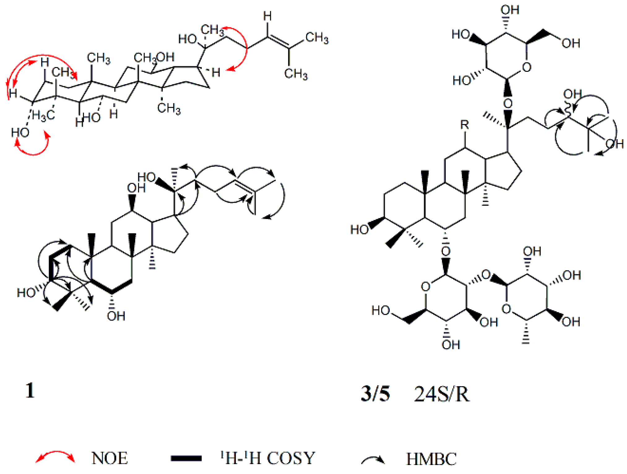
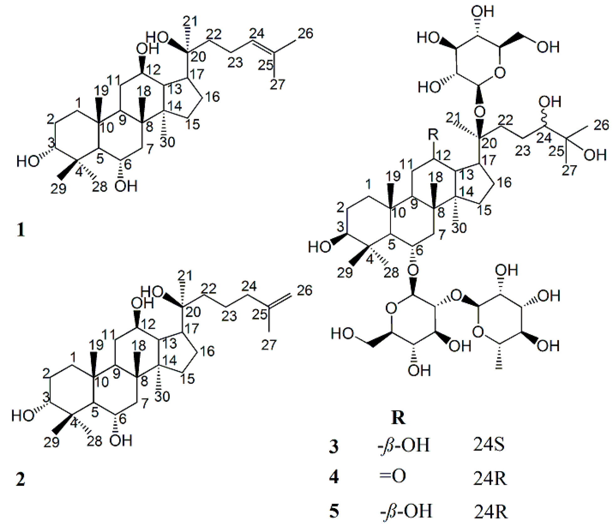
2.1. Structure Elucidation of Compounds 15

Compound 1, white powder: its molecular formula was established as C30H52O4 by HRESIMS. The 1H-NMR spectrum of 1 showed eight methyl singlet signals at δH 0.81, 1.01, 1.11, 1.29, 1.39, 1.62, 1.65, 1.88. The 13C-NMR spectrum of 1 showed signals of eight methyl carbons, six quaternary, eight methane and eight methylene among which two carbon signals (δ 126.6 and 131.1) showed the presence of a C=C bond. The chemical shifts of 1 resembled those of protopanaxatriol except the chemical shifts on A ring [17]. The proton and carbon signals of 1 were fully assigned on the basis of 2D-NMR spectra (Table 1). According to the literature, as compared with its 3β-epimers, the C-1~C-5 chemical shifts of 3α-dammarane-type triperpenes are in relatively higher nuclear magnetic fields, while carbon chemical shifts of C-28~C-29 are in relatively lower nuclear magnetic fields. For example, the C-1~C-5 and C-28~C-29 carbon chemical shift values of two pairs of C-3 epimers were shown as follow: [dammar-20S,24S-epoxy-3α,12β,25-triol: δC 33.5, 25.4, 76.0, 37.5, 49.5, 28.3, 22.0; dammar-20R,24S-epoxy-3β,12β,25-triol: δC 39.0, 27.5, 78.9, 39.0, 55.9, 28.0, 15.4); 3α-ocotillol: δC 34.9, 26.5, 77.4, 39.1, 56.2, 32.6, 22.8; 3β-ocotillol: δC 39.4, 28.1, 78.4, 40.4, 61.9, 31.9, 16.5] [18,19]. Carbon chemical shift values of C-1~C-5 and C-28~C-29 on A ring of 1C 35.0, 26.8, 77.6, 39.3, 56.3, 32.8, 23.1] suggested the existence of α configuration at C-3. NOEs between (i) δ 3.61 (H-3) and 1.29 (H-29) and 2.03 (H-2a), (ii) δ 5.58 (3-OH) and 1.88 (H-28) demonstrated the 3α configuration (Figure 1). Therefore, 1 was elucidated as dammar-24-ene-3α,6α,12β,20S-tetraol (ginsengenin-S1, Figure 2).
Compound 2, white powder: its molecular formula was established as C30H52O4 by HRESIMS. The NMR data of 2 (Table 1) resembled those of 1. The 3α configuration of 2 was determined by its NOEs the same way as 1. Compared with 1, the carbon NMR data of 2 showed the absence of a methyl carbon signal and an additional signal of methylene (δC 39.3) and a pair of typical signals of a terminal double bond (δC 110.4 and 146.8) in place of (δC 126.6 and 131.1), therefore, briefly, the obvious differences between 1 and 2 were the carbon chemical shifts of C-24~C-27. The 1H-NMR data of 2 also showed the absence of a methyl hydrogen signal and two additional hydrogen signals of the terminal double bond (δH 4.80 s, 4.84 s). Thus, 2 was deduced to be dammar-25-ene-3α,6α,12β,20S-tetraol (ginsengenin-S2, Figure 2).
Compounds 3 and 5, obtained as a white powder. Their molecular formulas were identically established as C48H84O20 by HRESIMS. Acidic hydrolysis of 3 and 5 both produced l-rhamnose and d-glucose, which were detected by GC analysis. Nine methyl singlet signals and a methyl doublet signal were detected in the 1H-NMR spectra of both 3 and 5 [3: δH 0.82, 0.89, 1.08, 1.28, 1.46, 1.49, 1.50, 2.00, 1.68 (3H, d, J = 6.1 Hz, H-6″); 5: δH 0.83, 0.91, 1.11, 1.32, 1.53 (6H, overlapped), 1.55, 2.06, 1.73 (3H, d, J = 5.6 Hz, H-6″)], three oxymethine protons [3: δH 3.40 (1H, m, H-3), 3.91 (1H, m, H-12), 3.81 (1H, m, H-24); 5: δH 3.43 (1H, m, H-3), 3.87 (1H, m, H-12), 3.74 (1H, m, H-24)], which were attributed to the aglycone moiety, together with three anomeric protons [3: δH 5.12 (1H, m, H-1‴), 5.13 (1H, m, H-1′), 6.35 (1H, brs, H-1″); 5: δH 5.17 (1H, m, H-1‴), 5.19 (1H, m, H-1′), 6.43 (1H, brs, H-1″)], assignable to one α-l-rhamnopyranosyl and two β-d-glucopyranosyl units. The 13C-NMR data of 3 and 5 showed resemblance with those of ginsenoside Re [20] except the disappearance of C=C bond C-atom signals and the additional appearance of two signals of C-atom [3: δC 79.5 (C-24), 73.1 (C-25); 5: δC 80.1 (C-24), 73.1 (C-25)] which connected the hydroxyl group. The following correlations were found in HMBC of 3: H-26/C-24, C-25, C-27; H-27/C-24, C-25, C-26, which indicated the existence of 2-methylpentane-2,3-diol moiety, and the same phenomenon was observed in 5 (Figure 1). The carbon chemical shifts of glycosyl moiety and the basic parent structure of the aglycone of 5 were consistent with 3 except the side chain (Table 2), which suggested the same nuclear parent of the aglycone and the same type and position of the sugar of 3 and 5, with only slight differences in the carbon chemical shift values of C-22~C-27 [3: δC 33.2, 26.6, 79.5, 73.1, 27.0, 25.2; 5: δC 33.7, 27.2, 80.1, 73.1, 26.9, 25.9]. Therefore, 3 and 5 are a pair of epimers at the 24-position. The carbon chemical shifts of 5 were almost consistent with a saponin isolated from notoginseng through Endophytes biotransformation, of which its structure was identified as 6-O-[α-l-rhamnopyranosyl-(1 2)-β-d-glucopyranosyl]-20-O-β-d-glucopyranosyl-dammarane-12-one-3,6,20,24,25-hexaol [21], however the absolute configuration of C-24 was not mentioned. By our observation, the carbon chemical shifts of C-22~C-24 and C-27 of 3 were in relatively higher nuclear magnetic fields compared with 5, and the configuration of the C-24 (δC 79.5) of 3 was indicated to be 24S and the C-24 (δC 80.1) of 5 was indicated to be 24R according to the literatures [tarecilioside A: δC 80.5 (C-24R); notoginsenoside SFt2: δC 80.1 (C-24R); notoginsenoside ST-6: δC 79.4 (C-24S); notoginsenoside ST-8: δC 79.3 (C-24S)] [22,23,24,25,26]. Thus, 3 and 5 were inferred as 6-O-[α-l-rhamnopyranosyl-(1 2)-β-d-glucopyranosyl]-20-O-β-d-glucopyranosyl-dammarane-3β,6α,12β,20S,24S,25-hexaol (ginsenoside-S3) and 6-O-[α-l-rhamnopyranosyl-(1 2)-β-d-glucopyranosyl]-20-O-β-d-glucopyranosyl-dammarane-3β,6α,12β,20S,24R,25-hexaol, respectively (ginsenoside-S3, Figure 2).
Compounds 4 was obtained as a white amorphous powder. The molecular formula was determined as C48H82O20 by HRESIMS. Compared with 5, the 1H-NMR data of 4 showed the appearance of two different chemical shifts of the proton [H-13 (δH 3.47) and H-17 (δH 2.96)] and the disappearance of H-12 (δH 3.87). The 13C-NMR spectra of 4 resembled those of 5 with the appearance of a 2-methylpentane-2,3-diol moiety, however, differences in the chemical shift values of C-9, C-11~C-14, C-17 [δC 54.3, 40.5, 211.6, 56.8, 56.4, 43.5] suggested the emergence of a carbonyl unit at C-12 in 4. The chemical shifts of 4 were completely assigned using 2D-NMR spectra (Table 2). The configuration of the C-24 (δC 80.2) of 4 was indicated to be 24R according to the C-24 configuration analysis of 3 and 5, thus the structure of 4 was inferred as 6-O-[α-l-rhamnopyranosyl-(1 2)-β-d-glucopyranosyl]-20-O-β-d-glucopyranosyl-dammarane-12-one-3β,6α,20S,24R,25-pentaol (ginsenoside-S4, Figure 2).

2.2. Bioactivity Evaluation

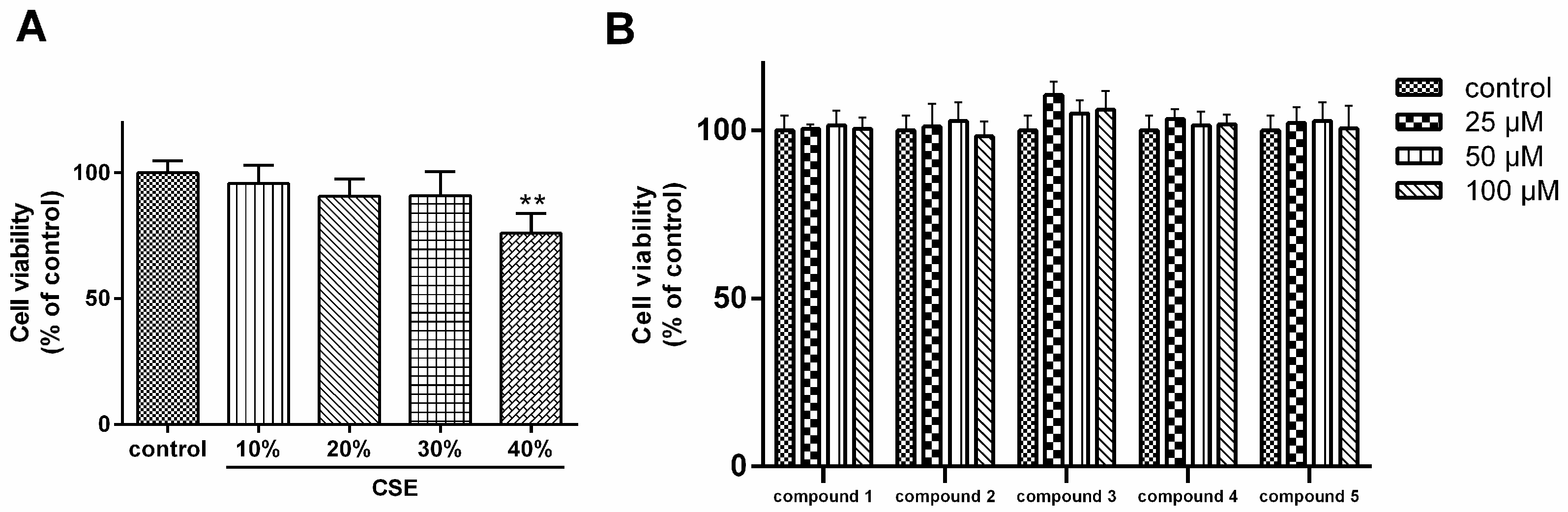
2.2.1. Cytotoxicity of Cigarette Smoke Extract (CSE) and Compounds 15 on A549 Cells

The MTT result showed that cell viability of A549 cells was substantially affected by CSE at a concentration≧40% (Figure 3A). Therefore, 30% CSE was used as stimulation in subsequent experiments. As shown in Figure 3B, the cell viability of A549 cells was not affected by 15. Then we tested the antioxidant effects of 15 on CS-stimulated A549 cells.

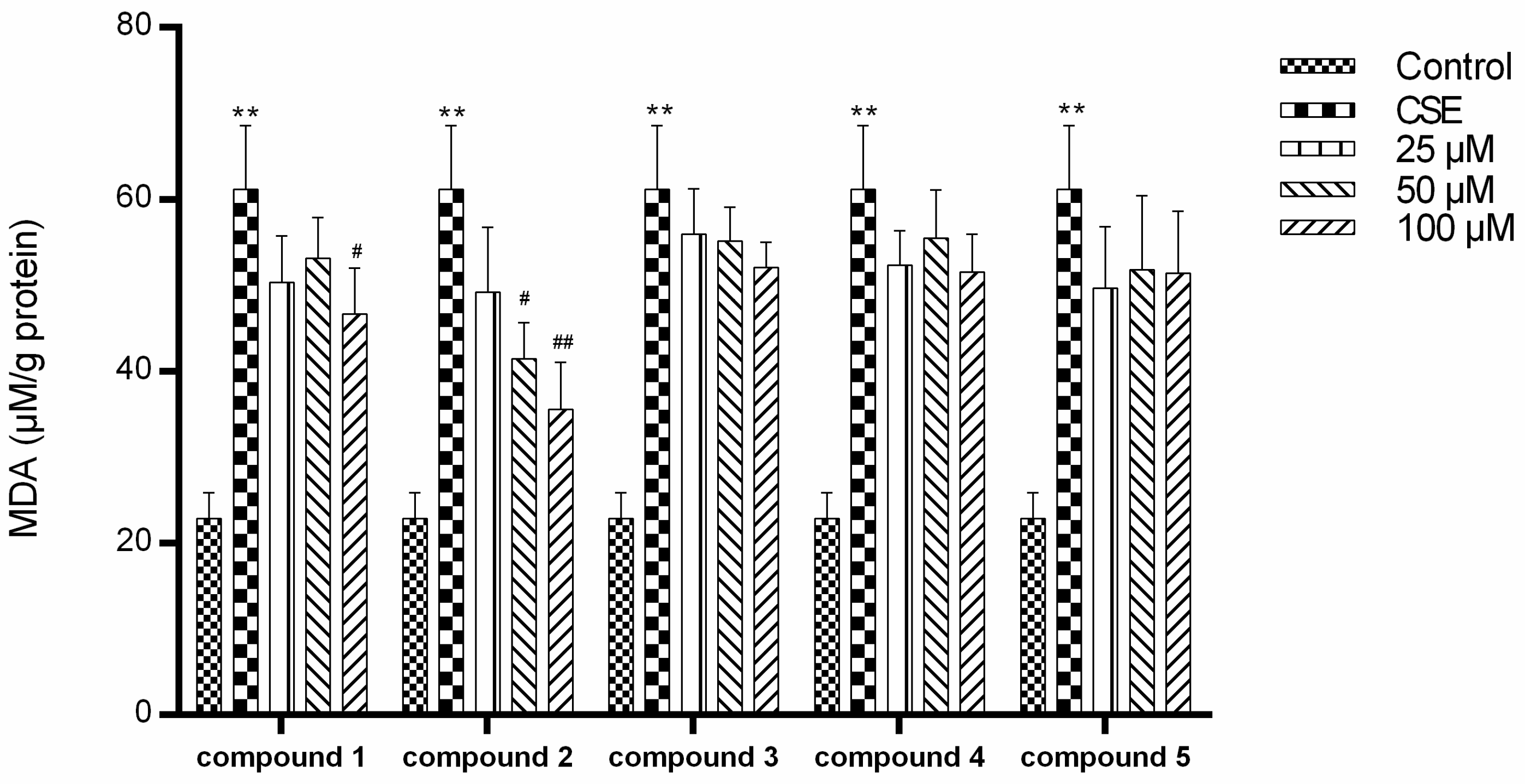
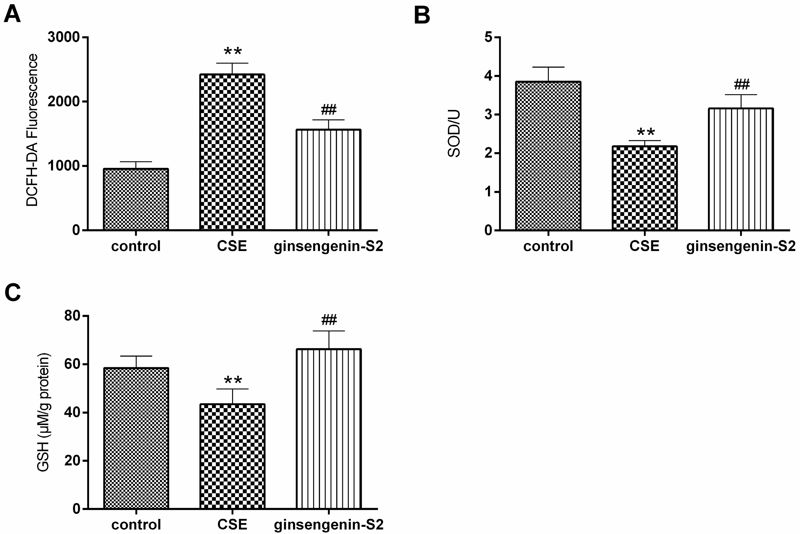
2.2.2. Antioxidant Activity of Compounds 15

As shown in Figure 4, CS exposure significantly increased the intracellular malondialdehyde (MDA) level in human lung epithelial cells. Among these five compounds, ginsengenin-S2 induced a dose-dependent reduction of MDA level.
Tobacco smoke is involved in the increased synthesis of reactive oxygen species (ROS) [27]. In our study, CS exposure induced significantly higher ROS level in A549 cells than the control group (Figure 5A). Interestingly, ginsengenin-S2 down-regulated the ROS level in CS exposed-A549 cells. Then, we investigated the role of ginsengenin-S2 on the activities of two first line defense antioxidants-superoxide dismutase (SOD) and glutathione (GSH) in CS-exposed human lung epithelial cells. As shown in Figure 5B,C, the CS challenge caused the decrease of intracellular GSH content and SOD activity (p < 0.01), with a parallel of increased MDA level which demonstrates that the CS challenge induced oxidative stress in the in vitro cell model. Further, we observed that pretreatment with ginsengenin-S2 could attenuate CS-induced oxidative stress by clearly up-regulating the content of SOD and GSH to a relatively basal level.

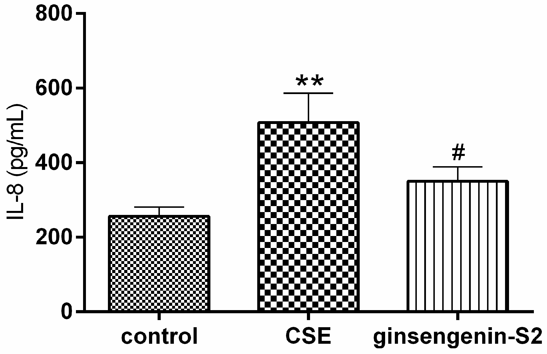
2.2.3. Effect of Ginsengenin-S2 on CS-Induced IL-8 Levels In Vitro

CS is also a leading risk factor for the development of an inflammatory condition characterized by the release of proinflammatory mediators such as interleukin-8 (IL-8). We then investigated whether ginsengenin-S2 inhibited the release of chemokine IL-8 from CS-stimulated lung epithelial cells. As shown in Figure 6, IL-8 production markedly rose in the cell treated with CS (p < 0.01), however pretreatment with ginsengenin-S2 at 100 μM resulted in a decreased IL-8 level (p < 0.05).

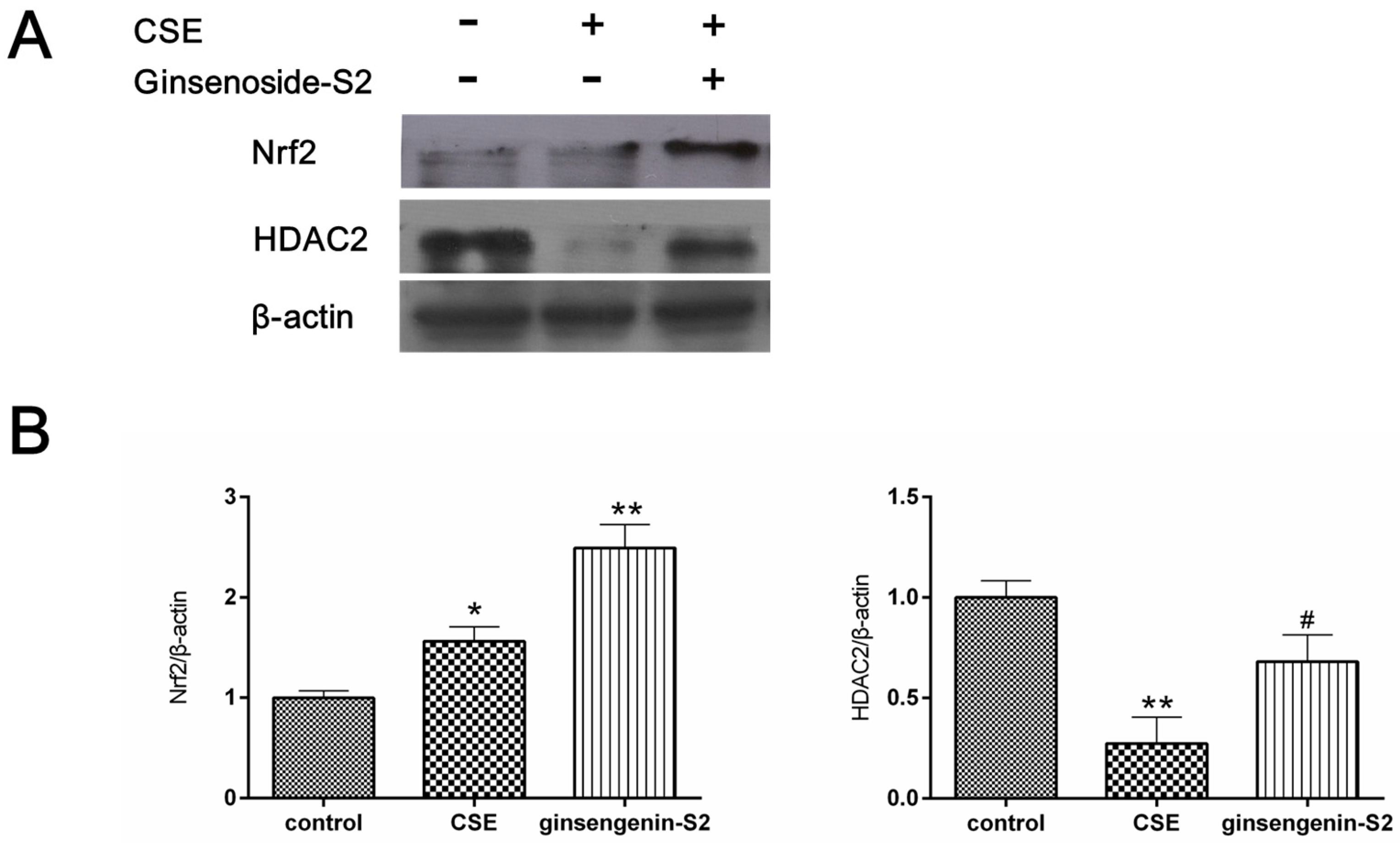
2.2.4. Effect of Ginsengenin-S2 on CS-Mediated Protein Expression of Nrf2 and HDAC2 In Vitro

To explore the underlying mechanism of the antioxidant activity and anti-inflammatory property of ginsengenin-S2, we examined the effect of ginsengenin-S2 on the protein expression of nuclear-related factor 2 (Nrf2) and histone deacetylase 2 (HDAC2) in CS-exposed A549 cells. In our study, we observed that the expression level of Nrf2 increased and the level of HDAC2 decreased after the CS challenge (p < 0.05). Interestingly, treatment with ginsengenin-S2 significantly activated the expression of Nrf2 (p < 0.01) as compared to the control group (Figure 7), which suggested that Nrf2 may participate in the meditative effect of ginsengenin-S2 in CS-induced oxidative stress. In addition, ginsengenin-S2 up-regulated the expression of HDAC2 in CS-exposed lung epithelial cells (p < 0.05).
Oxidative stress is involved in COPD pathogenesis and could cause the enrichment of ROS and even leads to organ tissue damage [28,29]. CS is one of the major risk contributors to COPD which could increase ROS production. As an important role in anti-oxidation, GSH could help reduce the production of lipid peroxide and prevent cell damage [30]. SOD is an enzyme which could scavenge superoxide radicals. It is reported that SOD and GSH-associated enzymes in the lungs of COPD patients are significantly affected by CS [31]. MDA, an important marker for monitoring the process of membrane lipid peroxidation and damage degrees, appears to have a different level between healthy smokers and COPD patients [32]. The current data suggested that ginsengenin-S2, a novel sapogenin, noticeably attenuated CS-induced oxidative damage and inflammatory action via up-regulating the content of intracellular SOD and MDA and inhibiting the level of MDA and IL-8 release. Nrf2, an important antioxidant transcription factor against oxidative stress, is released from the Keap1-Nrf2 complex and translocates from the cytoplasm when cells are exposed to oxidative stress. Increasing studies show that Nrf2 mediates the protective effects of natural products on CS exposure caused oxidative stress and inflammatory responses [16,33]. CS-induced lung inflammation involves the reduction of HDAC2 which is related to steroid resistance in patients with COPD who smoke cigarettes [34]. In this study, we found that ginsengenin-S2 activated the Nrf2 and enhanced the HDAC2 activity, suggesting that the protective effect of ginsengenin-S2 may involve the Nrf2 and HDAC2 pathways. To better utilize the medicinal resource of Panax ginseng Meyer cv. Silvatica, it is necessary to study the protective effect of its root extract against the cigarette smoke-induced COPD or airway fibrosis in vivo in the future.

3. Materials and Methods

3.1. Plant Materials

PGS was provided by the Guandong Green Ecology Ginseng & Pantotrichum Co., Ltd (Fusong, Jilin, China) in 2016 and was identified by Professor Pingya Li of Jilin University. The voucher specimen (No. PGS161220) was deposited at the National and Local United Engineering R&D Center of Ginseng Innovative Drugs, China.

3.2. Apparatus and Chemicals

HRESIMS was performed on Waters Xevo G2-XS QTOF mass spectrometer (Waters Co., Milford, MA, USA). Gas chromatography (GC) was carried out on an Agilent 5979 GC-MS (Agilent, J&W Scientific, Folsom, CA, USA). IR spectra were taken on WGH-30/6 drug-beam infrared spectrophotometer (Gangdong Technology Co. Ltd., Tianjin, China). The NMR spectra were measured on Bruker AV-600 spectrometer (Bruker Co., Karlsruhe, Germany) with tetramethylsilane (Sigma-Aldrich Co., St. Louise, MO, USA) as the internal standard. Column chromatography (CC) was performed with silica gel (200-300 mesh; Anhui Liangchen Silicon Material Co. Ltd., Huoshan, China). Thin-layer chromatography (TLC) was conducted on silica gel GF254 plates (Anhui Liangchen Silicon Material Co. Ltd., Huoshan, China). Preparative HPLC was carried out using a Shodex R1-201H Refractive Index Detector (Kexiao Chemical Equipment Co., Hangzhou, China) and YMC Prep C18 Column (5 μm, 20 mm × 10 mm; YMC Co., Ltd., Kyoto, Japan), 1525 Binary HPLC pump (Waters Co., Milford, MA, USA.). Methanol (MeOH) and acetonitrile (ACN) (HPLC grade, Janssens Pharmaceuticalaan Co., Fair Lawn, NJ, USA), ultrapure water (Wahaha Co., Ltd, Hangzhou, China) and other solvents used were of analytical grade (Tianjin Fine Chemical Works, Tianjin, China).

3.3. Extraction and Isolation

The pearl knots of PGS (1 kg) were completely extracted with 95% ethanol (EtOH) and the ethanolic extracts were concentrated under vacuum. The ethanolic extracts (82 g) were subjected to macro-reticular absorption resin (D101) and began with H2O and then were eluted with 95% EtOH. The EtOH fraction (65 g) was then subjected to silica gel CC (200–300 mesh) and was eluted with CH2Cl2-MeOH (gradient 100:1 to 1:100) to give 41 fractions (Fr 1–41). Fr 7 (1.3 g) was further separated by silica gel CC [petroleum ether (PE)-ethyl acetate (EtOAc), gradient 1:1 to 1:4] to afford three sub-fractions (Fr 7a–7c), Fr 7c (0.6 g) was followed by preparative RP-HPLC (C18 Column, 5 μm, 20 mm × 10 mm) with ACN-H2O (44:56, 4 mL/min) as a mobile phase to afford compounds 6 (30 mg, 24 min), 1 (7 mg, 35 min) and 2 (4 mg, 39 min). Fr 24 (1.8 g) was further separated by silica gel CC (EtOAc-EtOH, 10:1 to 2:1) to obtain four sub-fractions (Fr 24a–24d), compound 7 (0.3 g) was recrystallized from Fr 24a (0.5 g) in MeOH, Fr 24b (0.4 g) was followed by preparative RP-HPLC (C18 Column, 5 μm, 20 mm × 10 mm) with ACN-H2O (20:80, 3 mL/min) as a mobile phase to afford compounds 3 (17 mg, 11 min), 5 (20 mg, 19 min) and 4 (2 mg, 22 min).

3.4. Spectroscopic Data

3.4.1. Ginsengenin-S1 (1)

Dammar-24-ene-3α,6α,12β,20S-tetraol: white amorphous powder; IR νmax 3394, 2960, 1632, 1455, 1383, 1032, 609 cm−1; Libermann-Burchard reaction was positive, suggesting the existence of triterpenoid structure; 1H and 13C-NMR: see Table 1; HRESIMS m/z 521.3880 [M + HCOO] (calculated for C31H53O6, 521.3842); chemical spectra see in the Supplementary Materials.

3.4.2. Ginsengenin-S2 (2)

Dammar-25-ene-3α,6α,2β,20S-tetraol: white amorphous powder; IR νmax 3328, 2964, 1653, 1461, 1386, 1111, 1028, 649 cm−1; Libermann-Burchard reaction was positive, suggesting the existence of triterpenoid structure; 1H and 13C-NMR: see Table 1; HRESIMS m/z 521.3844[M + HCOO] (calculated for C31H53O6, 521.3842); chemical spectra see in the Supplementary Materials.

3.4.3. Ginsenoside-S3 (3)

6-O-[α-l-rhamnopyranosyl-(1 2)-β-d-glucopyranosyl]-20-O-β-d-glucopyranosyl-dammarane-3β,6α,12β,20S,24S,25-hexaol: white amorphous powder; IR νmax 3406, 2944, 1659, 1371, 1032, 597 cm−1; Libermann-Burchard reaction was positive, suggesting the existence of triterpenoid structure; Molish reactions were positive, suggesting the existence of glucoside; 1H and 13C-NMR: see Table 2; HRESIMS m/z 1025.5554 [M + HCOO] calculated for C49H85O22, 1025.5532); chemical spectra see in the Supplementary Materials.

3.4.4. Ginsenoside-S4 (4)

6-O-[α-l-rhamnopyranosyl-(1 2)-β-d-glucopyranosyl]-20-O-β-d-glucopyranosyl-dammarane-12-one-3β,6α,20S,24R,25-pentaol: white amorphous powder; IR νmax 3412, 2944, 1626, 1068,580 cm−1; Libermann-Burchard reaction was positive, suggesting the existence of triterpenoid structure; Molish reactions were positive, suggesting the existence of glucoside; 1H and 13C-NMR: see Table 2; HRESIMS m/z 1023.5424 [M + HCOO] (calculated for C49H83O22, 1023.5376); chemical spectra see in the Supplementary Materials.

3.5. Bioassay

3.5.1. Preparation of Cigarette Smoke Extract

Xiongshi cigarette (China Tobacoo Zhejiang Industrial Co., Ltd, Hangzhou, China; each cigarette contained 0.7 mg of nicotine, 8 mg of tar and 10 mg of carbon monoxide) were used to prepare the cigarette smoke extract (CSE) according to the literature [35]. In brief, one Xiongshi cigarette was bubbled through 25 mL of culture medium to generate 100% concentration CSE, then the solution was filtered through the 0.22 μm pore filter. The CSE (pH 7.2–7.4) was freshly prepared for each experiment and was then diluted to the desired concentration and used within 30 min.

3.5.2. Cell Viability Assay

The triterpenoids were dissolved in DMSO as stock solutions and diluted with DMEM to the needed concentrations. Final DMSO concentration in the sample was less than 0.1%. Cell culture and MTT assay were done as we described previously to evaluate 24 h-incubation of triterpenoids or the 18 h-incubation of CSE on the cell viability of A549 cells [6].

3.5.3. Drug Treatment

A549 cells were cultured in 6-well plates at a density of 5 × 10⁵ cells per 1 mL and were pretreated with triterpenoids for 6 h. After that, the cells were treated with both CSE and triterpenoids for another 18 h of incubation.

3.5.4. Determination of Oxidative Stress

After 18 h of stimulation of CSE, the levels of intracellular MDA, GSH and SOD in the lysates were measured using commercial MDA, GSH and SOD kits (Jiancheng Bioengineering Institute, Nanjing, China) according to the manufacturer’s protocol.

3.5.5. Reactive Oxygen Species (ROS) Assay

A549 cells were cultured in 96-well plates (5 × 10³ cells per well). Drug treatment and CS stimulation were carried out as described in Section 3.5.3 and Section 3.5.4. The cells were washed with warm PBS and the ROS detection was performed with DCFH-DA probe based ROS Assay Kit (Beyotime Biotechnology, Shanghai, China) at 37 °C. The ROS level was expressed as the fluorescence intensity.

3.5.6. Enzyme-Linked Immunosorbent Assay

After 18 h of stimulation of CSE, IL-8 content in the cell culture supernatant was measured using IL-8 ELISA kit (R&D Systems, Minneapolis, MN, USA) according to the manufacturer’s protocol.

3.5.7. Western Blotting

Western blot assay was performed as previous described [6] to study the effects of ginsengenin-S2 on the protein expression of HDAC2 and Nrf2 in CSE-stimulated A549 cells. Anti-HDAC2 (#ab32117), anti-Nrf2 (#ab137550) and anti-β-actin (#ab137550) were from Abcam Company (Cambridge, UK). The HDAC2 and Nrf2 band intensities were adjusted with reference to β-actin control.

3.5.8. Statistical Analysis

Data analysis was performed using GraphPad Prism 6.0 software (GraphPad Software, San Diego, USA). All the data are presented as mean ± S.D. and a two tailed test or a one-way analysis of variance (ANOVA) was used to determine the statistical significance, and p-value <0.05 was considered as significant.

4. Conclusions

In our study, four new dammarane-type triterpenoids, elucidated as ginsengenin-S1 (1), ginsengenin-S2 (2), ginsenoside-S3 (3) and ginsenoside-S4 (4) were isolated from pearl knots of PGS and their antioxidant activities were investigated using CS-stimulated A549 cells. Among these compounds, ginsengenin-S2 exhibited protective activity against CS-induced oxidative damage and ameliorated inflammatory reaction. Further, its protective effect against CS may involve the Nrf2 and HDAC2 pathways. This study revealed the evidence that an active compound from the root of Panax ginseng Meyer cv. Silvatica—ginsengenin-S2 is beneficial to reduce injury from cigarette smoke in vitro and provides some theoretical basis for the development and utilization of the resource Panax ginseng Meyer cv. Silvatica. It reminded us that it is worthwhile to study the in vivo protective effect of the root of Panax ginseng Meyer cv. Silvatica against cigarette smoke using animal models.

Supplementary Materials

The following are available online. 1H-NMR, 13C-NMR, HSQC, HMBC, 1H-1H COSY, NOESY, HRESIMS, Infrared spectrum of compounds 15.

Author Contributions

Formal analysis, X.G.; Funding acquisition, P.L.; Investigation, Z.Q., Z.L., X.G., C.W. and F.W.; Project administration, P.L.; Resources, P.L.; Supervision, J.L.; Writing—original draft, Z.Q. and Z.L.

Funding

This work was financially supported by Jilin Key Scientific & Technological Project (No. 2012ZDGG010); the Talents Team Major Program of Jilin Province of China (JRCBTZ. [2016] No. 3); the National Science and Technology Major Project for New Drug Development in China (No.2010ZX09401-305-26).

Conflicts of Interest

The authors declare no conflicts of interest.

References

  1. Zhou, S.-S.; Auyeung, K.K.-W.; Yip, K.-M.; Ye, R.; Zhao, Z.-Z.; Mao, Q.; Xu, J.; Chen, H.-B.; Li, S.-L. Stronger Anti-obesity Effect of White Ginseng over Red Ginseng and the Potential Mechanisms Involving Chemically Structural/Compositional Specificity to Gut Microbiota. Phytomedicine 2018. [Google Scholar] [CrossRef]
  2. Li, M.; Li, R.J.; Liu, M.Y. Study on the biological nature of ginseng pearl knot. Chin. J. Chin. Mater. Med. 1989, 14, 654–655. [Google Scholar]
  3. Liu, D.; Li, Y.-G.; Xu, H.; Sun, S.-Q.; Wang, Z.-T. Differentiation of the root of Cultivated Ginseng, Mountain Cultivated Ginseng and Mountain Wild Ginseng using FT-IR and two-dimensional correlation IR spectroscopy. J. Mol. Struct. 2008, 883, 228–235. [Google Scholar] [CrossRef]
  4. Xu, X.F.; Xu, S.Y.; Zhang, Y.; Zhang, H.; Liu, M.N.; Liu, H.; Gao, Y.; Xue, X.; Xiong, H.; Lin, R.C.; et al. Chemical Comparison of Two Drying Methods of Mountain Cultivated Ginseng by UPLC-QTOF-MS/MS and Multivariate Statistical Analysis. Molecules 2017, 22, 71. [Google Scholar] [CrossRef]
  5. Hwang, J.W.; Oh, J.H.; Yoo, H.S.; Lee, Y.W.; Cho, C.K.; Kwon, K.R.; Yoon, J.H.; Park, J.; Her, S.; Lee, Z.W.; et al. Mountain ginseng extract exhibits anti-lung cancer activity by inhibiting the nuclear translocation of NF-kappaB. Am. J. Chin. Med. 2012, 40, 187–202. [Google Scholar] [CrossRef] [PubMed]
  6. Qi, Z.; Wang, Z.; Zhou, B.; Fu, S.; Hong, T.; Li, P.; Liu, J. A new ocotillol-type ginsenoside from stems and leaves of Panax quinquefolium L. and its anti-oxidative effect on hydrogen peroxide exposed A549 cells. Nat. Prod. Res. 2019, 1–8. [Google Scholar] [CrossRef]
  7. Qi, Z.; Chen, L.; Li, Z.; Shao, Z.; Qi, Y.; Gao, K.; Liu, S.; Sun, Y.; Li, P.; Liu, J. Immunomodulatory Effects of (24R)-Pseudo-Ginsenoside HQ and (24S)-Pseudo-Ginsenoside HQ on Cyclophosphamide-Induced Immunosuppression and Their Anti-Tumor Effects Study. IJMS 2019, 20, 836. [Google Scholar] [CrossRef]
  8. Xie, C.L.; Li, J.H.; Wang, W.W.; Zheng, G.Q.; Wang, L.X. Neuroprotective effect of ginsenoside-Rg1 on cerebral ischemia/reperfusion injury in rats by downregulating protease-activated receptor-1 expression. Life Sci. 2015, 121, 145–151. [Google Scholar] [CrossRef] [PubMed]
  9. Kang, A.; Xie, T.; Zhu, D.; Shan, J.; Di, L.; Zheng, X. Suppressive Effect of Ginsenoside Rg3 against Lipopolysaccharide-Induced Depression-Like Behavior and Neuroinflammation in Mice. J. Agric. Food Chem. 2017, 65, 6861–6869. [Google Scholar] [CrossRef] [PubMed]
  10. Chen, L.-X.; Qi, Z.; Shao, Z.-J.; Li, S.-S.; Qi, Y.-L.; Gao, K.; Liu, S.-X.; Li, Z.; Sun, Y.-S.; Li, P.-Y. Study on Antidepressant Activity of Pseudo-Ginsenoside HQ on Depression-Like Behavior in Mice. Molecules 2019, 24, 870. [Google Scholar] [CrossRef]
  11. Qi, Z.; Li, W.; Tan, J.; Wang, C.; Lin, H.; Zhou, B.; Liu, J.; Li, P. Effect of ginsenoside Rh2 on renal apoptosis in cisplatin-induced nephrotoxicity in vivo. Phytomedicine 2019, 152862. [Google Scholar] [CrossRef]
  12. Li, Y.; Xie, L.; Xin, S.; Li, K. Values of procalcitonin and C-reactive proteins in the diagnosis and treatment of chronic obstructive pulmonary disease having concomitant bacterial infection. Pak. J. Med. Sci. 2017, 33, 566–569. [Google Scholar] [CrossRef] [PubMed]
  13. Wang, G.; Mohammadtursun, N.; Lv, Y.; Zhang, H.; Sun, J.; Dong, J. Baicalin Exerts Anti-Airway Inflammation and Anti-Remodelling Effects in Severe Stage Rat Model of Chronic Obstructive Pulmonary Disease. Evid. Based Complement Alternat. Med. 2018, 2018, 7591348. [Google Scholar] [CrossRef] [PubMed]
  14. Guan, S.; Liu, Q.; Han, F.; Gu, W.; Song, L.; Zhang, Y.; Guo, X.; Xu, W. Ginsenoside Rg1 Ameliorates Cigarette Smoke-Induced Airway Fibrosis by Suppressing the TGF-beta1/Smad Pathway In Vivo and In Vitro. Biomed Res. Int. 2017, 2017, 6510198. [Google Scholar] [CrossRef] [PubMed]
  15. Guan, S.; Xu, W.; Han, F.; Gu, W.; Song, L.; Ye, W.; Liu, Q.; Guo, X. Ginsenoside Rg1 Attenuates Cigarette Smoke-Induced Pulmonary Epithelial-Mesenchymal Transition via Inhibition of the TGF-beta1/Smad Pathway. Biomed Res. Int. 2017, 2017, 12. [Google Scholar] [CrossRef]
  16. Wang, M.; Chen, X.; Jin, W.; Xu, X.; Li, X.; Sun, L. Ginsenoside Rb3 exerts protective properties against cigarette smoke extract-induced cell injury by inhibiting the p38 MAPK/NF-κB and TGF-β1/VEGF pathways in fibroblasts and epithelial cells. Biomed. Pharmacother. 2018, 108, 1751–1758. [Google Scholar] [CrossRef] [PubMed]
  17. Yu, M.; Zhao, Y. Isolation and identification of a pair of isomers in fruit of Panax ginseng. Chin. Tradit. Herbal Drugs 2002, 33, 404–405. [Google Scholar]
  18. Fujita, S.; Kasai, R.; Ohtani, K.; Yamasaki, K.; Chiu, M.-H.; Nie, R.-L.; Tanaka, O. Dammarane glycosides from aerial part of Neoalsomitra integrifoliola. Phytochemistry 1995, 39, 591–602. [Google Scholar] [CrossRef]
  19. Han, L.; Lin, M.Y.; Zheng, Q.; Liu, H.Y.; Liu, H.Y.; Dong, G.; Liu, J.P.; Li, P.Y. A new epimer of ocotillol from stems and leaves of American ginseng. Nat. Prod. Res. 2014, 28, 935–939. [Google Scholar] [CrossRef]
  20. Han, B.H.; Park, M.H.; Han, Y.N.; Woo, L.K.; Sankawa, U.; Yahara, S.; Tanaka, O. Degradation of ginseng saponins under mild acidic conditions. Planta Med. 1982, 44, 146–149. [Google Scholar] [CrossRef]
  21. Luo, S.L.; Dang, L.Z.; Li, J.F.; Zou, C.G.; Zhang, K.Q.; Li, G.H. Biotransformation of saponins by endophytes isolated from Panax notoginseng. Chem. Biodivers. 2013, 10, 2021–2031. [Google Scholar] [CrossRef] [PubMed]
  22. Zhao, Z.; Matsunami, K.; Otsuka, H.; Shinzato, T.; Takeda, Y. Tareciliosides A–G: Cycloartane glycosides from leaves of Tarenna gracilipes (HAY.) OHWI. Chem. Pharm. Bull (Tokyo) 2008, 56, 1153–1158. [Google Scholar] [CrossRef] [PubMed]
  23. Liu, Q.; Lv, J.-J.; Xu, M.; Wang, D.; Zhu, H.-T.; Yang, C.-R.; Zhang, Y.-J. Dammarane-type saponins from steamed leaves of Panax Notoginseng. Natur. Prod. Bioprosp. 2011, 1, 124–128. [Google Scholar] [CrossRef] [Green Version]
  24. Xu, W.; Liu, X.; Liu, X.L.; Jia, A.L.; Wang, X.W.; Qiu, Z.D. Two new dammarane-type triterpenoid saponins from notoginseng medicinal fungal substance. J. Asian Nat. Prod. Res. 2016, 18, 1138–1142. [Google Scholar] [CrossRef] [PubMed]
  25. Xu, W.; Zhang, J.H.; Liu, X.; Jia, A.L.; Liu, X.L.; Wang, X.W.; Qiu, Z.D. Two new dammarane-type triterpenoid saponins from ginseng medicinal fungal substance. Nat. Prod. Res. 2017, 31, 1107–1112. [Google Scholar] [CrossRef] [PubMed]
  26. Xu, W.; Ding, C.-F.; Wang, X.-W.; Liu, X.-L.; Qiu, Z.-D. Two new triterpenoid saponins from notoginseng medicinal fungal substance. Chin. Chem. Lett. 2015, 26, 1298–1302. [Google Scholar] [CrossRef]
  27. Valavanidis, A.; Vlachogianni, T.; Fiotakis, K. Tobacco smoke: involvement of reactive oxygen species and stable free radicals in mechanisms of oxidative damage, carcinogenesis and synergistic effects with other respirable particles. Int. J. Environ. Res. Public Health 2009, 6, 445–462. [Google Scholar] [CrossRef]
  28. Santus, P.; Corsico, A.; Solidoro, P.; Braido, F.; Di Marco, F.; Scichilone, N. Oxidative stress and respiratory system: pharmacological and clinical reappraisal of N-acetylcysteine. COPD 2014, 11, 705–717. [Google Scholar] [CrossRef]
  29. Zhang, Y.; Wang, Q.; Wang, Y.D.; Sun, B.; Leng, X.W.; Li, Q.; Ren, L.Q. Effect of rutin on cisplatin-induced damage in human mesangial cells via apoptotic pathway. Hum. Exp. Toxicol. 2018, 38, 118–128. [Google Scholar] [CrossRef]
  30. Yan, X.; Song, Y.; Shen, C.; Xu, W.; Chen, L.; Zhang, J.; Liu, H.; Huang, M.; Lai, G.; Qian, G. Mucoactive and antioxidant medicines for COPD: consensus of a group of Chinese pulmonary physicians. Int. J. Chron. Obstruct. Pulmon. Dis. 2017, 12, 803. [Google Scholar] [CrossRef] [PubMed]
  31. Rahman, I.; Kinnula, V.L. Strategies to decrease ongoing oxidant burden in chronic obstructive pulmonary disease. Expert Rev. Clin. Phar. 2012, 5, 293–309. [Google Scholar] [CrossRef] [PubMed] [Green Version]
  32. Domej, W.; Oettl, K.; Renner, W. Oxidative stress and free radicals in COPD–implications and relevance for treatment. Int. J. Chron. Obstruct. Pulmon. Dis. 2014, 9, 1207. [Google Scholar] [CrossRef] [PubMed]
  33. Cui, W.; Zhang, Z.; Zhang, P.; Qu, J.; Zheng, C.; Mo, X.; Zhou, W.; Xu, L.; Yao, H.; Gao, J. Nrf2 attenuates inflammatory response in COPD/emphysema: Crosstalk with Wnt3a/β-catenin and AMPK pathways. J. Cell. Mol. Med. 2018, 22, 3514–3525. [Google Scholar] [CrossRef] [PubMed] [Green Version]
  34. Adenuga, D.; Yao, H.; March, T.H.; Seagrave, J.; Rahman, I. Histone deacetylase 2 is phosphorylated, ubiquitinated, and degraded by cigarette smoke. Am. J. Respir. Cell Mol. Biol. 2009, 40, 464–473. [Google Scholar] [CrossRef]
  35. Chen, L.; Ge, Q.; Tjin, G.; Alkhouri, H.; Deng, L.; Brandsma, C.A.; Adcock, I.; Timens, W.; Postma, D.; Burgess, J.K.; et al. Effects of cigarette smoke extract on human airway smooth muscle cells in COPD. Eur. Respir. J. 2014, 44, 634–646. [Google Scholar] [CrossRef] [PubMed] [Green Version]
Sample Availability: Not available.
Figure 1. The important NOESY, 1H-1H COSY and HMBC correlations of 1; the important HMBC correlations of 3 and 5. 1H-1H COSY, 1H-1H correlation spectroscopy; HMBC, heteronuclear multiple bond correlation; NOESY, nuclear Overhauser effect spectroscopy.
Molecules 24 01159 g001
Figure 2. Chemical structures of 15: ginsengenin-S1 (1); ginsengenin-S2 (2); ginsenoside-S3 (3); ginsenoside-S4 (4); ginsenoside-S5 (5).
Molecules 24 01159 g002
Figure 3. (A) Cytotoxicity of cigarette smoke extract (CSE) on A549 cells; (B) Effect of compound 15 on the cell viability of A549 cells. The results represent the mean ± S.D. (n = 6). ** p < 0.01, compared with control.
Molecules 24 01159 g003
Figure 4. Effect of compounds 15 on the intracellular malondialdehyde (MDA) level in cigarette smoke (CS)-exposed A549 cells. All data were expressed as mean ± S.D., n = 6. ** p < 0.01, compared with control; # p < 0.05, ## p < 0.01, compared with the CSE group.
Molecules 24 01159 g004
Figure 5. Effects of ginsengenin-S2 (100 μM) on (A) the reactive oxygen species (ROS) level, (B) intracellular dismutase (SOD) activity and (C) glutathione (GSH) level in CS-exposed A549 cells. All data were expressed as mean ± S.D., n = 6. ** p < 0.01, compared with control; ## p < 0.01, compared with the CSE group.
Molecules 24 01159 g005
Figure 6. Anti-inflammatory effect of ginsengenin-S2 (100 μM) on the inflammatory cytokine interleukin-8 (IL-8) in CS-exposed A549 cells. All data were expressed as mean ± S.D., n = 4. ** p < 0.01, compared with control; # p < 0.05, compared with the CSE group.
Molecules 24 01159 g006
Figure 7. (A) Effect of ginsengenin-S2 (100 μM) on the protein expression of nuclear-related factor 2 (Nrf2) and histone deacetylase 2 (HDAC2) in the CS-exposed A549 cells was examined by western blot analysis; (B) quantitative analyses of Nrf2 in each group. Data are expressed as mean ± SD (n = 3). * p < 0.05, ** p < 0.01 as compared to the control group; # p < 0.05 as compared to the CSE treated group.
Molecules 24 01159 g007
Table 1. 1H and 13C-NMR (600 MHz, 150 MHz in C5D5N) data for 1 and 2.
Position12Position12
δCδHδCδHδCδHδCδH
1351.85 (1H, m)35.11.88 (1H, m)17552.27 (1H, m)55.12.30 (1H, m)
1.48 (1H, m) 1.49 (1H, m)1817.71.11 (3H, s)17.71.20 (3H, s)
226.82.03 (1H, m)26.92.05 (1H, m)1918.41.01 (3H, s)18.51.05 (3H, s)
1.80 (1H, m) 1.80 (1H, m)2073.3 73.4
377.63.61 (1H, d, 10)77.73.63 (1H, br s)2127.31.39 (3H, s)27.61.39 (3H, s)
439.3 39.4 2236.12.01 (1H, m)35.81.96 (1H, m)
556.31.89 (1H, m)56.41.93 (1H, m) 1.67 (1H, m) 1.62 (1H, m)
667.64.33 (1H, m)67.74.38 (1H, m)2323.32.58 (1H, m)22.82.03 (1H, m)
747.61.94 (1H, m)47.71.97 (1H, m) 2.25 (1H, m) 1.67 (1H, m)
1.88 (1H, m) 1.79 (1H, m)24126.65.30 (1H, t, 6.3)39.32.08 (1H, m)
841.4 41.4 25131.1 146.8
950.31.75 (1H, m)50.41.78 (1H, m)2626.151.65 (3H, s)110.44.84 (1H, s)
1039.5 39.6 4.80 (1H, s)
1132.42.19 (1H, m)32.62.25 (1H, m)27181.62 (3H, s)231.71 (3H, s)
1.57 (1H, m) 1.63 (1H, m)2832.81.88 (3H, s)32.91.91 (3H, s)
1271.43.86 (1H, m)71.43.89 (1H, m)2923.11.29 (3H, s)23.21.32 (3H, s)
1348.52.02 (1H, m)48.62.07 (1H, m)3017.30.81 (3H, s)17.30.83 (3H, s)
1452 52.1 3-OH 5.58 (1H, br s) 5.59 (1H, d, 4.0)
1531.61.58 (1H, m)31.71.62 (1H, m)6-OH 5.15 (1H, br s) 5.17 (1H, d, 6.9)
1.00 (1H, m) 1.03 (1H, m)12-OH 7.22 (1H, s) 7.23 (1H, s)
1627.11.81 (1H, m)27.21.84 (1H, m)20-OH 6.96 (1H, s) 6.96 (1H, s)
1.35 (1H, m) 1.37 (1H, m)
Delta is ppm of chemical shifts which are reported in parts per million (δ), and coupling constants (J) are expressed in Hertz.
Table 2. 1H and 13C-NMR (600 MHz, 150 MHz in C5D5N) data for 35.
Position345
δCδHδCδHδCδH
139.41.59 (1H, m)39.41.39 (1H, m)39.51.62 (1H, m)
0.86 (1H, m) 0.87 (1H, m) 0.89 (1H, m)
227.61.76 (2H, m)27.91.77 (1H, m)27.81.83 (1H, m)
1.81 (1H, m) 1.77 (1H, m)
378.53.40 (1H, m)78.53.46 (1H, m)78.63.43 (1H, m)
439.9 39.7 40.1
560.81.30 (1H, m)60.81.39 (1H, m)61.01.34 (1H, m)
674.54.58 (1H, m)74.54.75 (1H, m)74.74.62 (1H, m)
745.92.12 (1H, m)45.82.34 (1H, m)46.02.19 (1H, m)
1.86 (1H, m) 1.92 (1H, m) 1.91 (1H, m)
841.1 42.1 41.3
949.51.42 (1H, m)54.31.84 (1H, m)49.61.46 (1H, m)
1039.6 40.0 39.8
1130.81.99 (1H, m)40.52.27 (2H, m)31.12.01 (1H, m)
1.38 (1H, m) 1.38 (1H, m)
1270.63.91 (1H, m)211.6 70.73.87 (1H, m)
1348.61.97 (1H, m)56.83.47 (1H, m)48.91.96 (1H, m)
1451.5 56.4 51.7
1530.91.41 (1H, m)32.31.80 (1H, m)31.01.45 (1H, m)
0.74 (1H, m) 0.96 (1H, m) 0.81 (1H, m)
1626.71.63 (1H, m)27.32.44 (1H, m)26.91.67 (1H, m)
1.22 (1H, m) 1.99 (1H, m) 1.24 (1H, m)
1752.82.32 (1H, m)43.52.96 (1H, m)53.02.30 (1H, m)
1817.21.08 (3H, s)17.51.49 (3H, s)17.31.11 (3H, s)
1917.50.89 (3H, s)17.80.98 (3H, s)17.70.91 (3H, s)
2083.9 82.2 83.6
2122.81.49 (3H, s)22.81.53 (3H, s)23.01.53 (3H, overlapped)
2233.22.35 (2H, m)38.52.57 (1H, m)33.72.90 (1H, m)
2.07 (1H, m) 1.85 (1H, m)
2326.62.41 (1H, m)25.22.18 (1H, m)27.22.17 (1H, m)
1.78 (1H, m) 1.85 (1H, m) 2.00 (1H, m)
2479.53.81 (1H, m)80.23.77 (1H, m)80.13.74 (1H, m)
2573.1 73.3 73.1
2627.01.50 (3H, s)26.41.55 (3H, s)26.91.55 (3H, s)
2725.21.46 (3H, s)26.41.54 (3H, s)25.91.53 (3H, overlapped)
2832.12.00 (3H, s)32.52.10 (3H, s)32.32.06 (3H, s)
2917.61.28 (3H, s)17.81.35 (3H, s)17.81.32 (3H, s)
3017.10.82 (3H, s)17.20.85 (3H, s)17.20.83 (3H, s)
6-glc-1′101.85.13 (1H, m)102.15.25 (1H, d, 6.3)102.05.19 (1H, m)
2′78.74.14 (1H, m)78.73.97 (1H, m)78.74.18 (1H, m)
3′78.64.26 (1H, m)78.44.36 (1H, m)79.04.31 (1H, m)
4′72.54.12 (1H, m)72.94.21 (1H, m)72.74.16 (1H, m)
5′78.33.88 (1H, m)79.44.20 (1H, m)78.73.92 (1H, m)
6′63.04.42 (1H, m)63.44.51 (1H, m)63.34.52 (1H, m)
4.16 (1H, m) 4.36 (1H, m) 4.17 (1H, m)
2′-rham-1″101.96.35 (1H, brs)102.36.48 (1H, d, 6.3)102.06.43 (1H, brs)
2″72.34.70 (1H, m)72.84.80 (1H, m)72.54.76 (1H, brs)
3″72.24.57 (1H, m)72.64.67 (1H, m)72.44.64 (1H, m)
4″74.04.25 (1H, m)74.54.30 (1H, m)74.34.30 (1H, m)
5″69.54.83 (1H, td, 9.2, 6.3)69.84.94 (1H, m)69.64.89 (1H, m)
6″18.71.68 (3H, d, 6.1)19.11.76 (3H, d, 5.9)18.91.73 (3H, d, 5.6)
20-glc-1‴98.35.12 (1H, m)98.75.10 (1H, m)98.55.17 (1H, m)
2‴75.23.89 (1H, m)76.03.96 (1H, m)75.63.95 (1H, m)
3‴79.34.25 (1H, m)79.84.36 (1H, m)79.54.30 (1H, m)
4‴71.63.98 (1H, t, 9.1)72.24.14 (1H, m)71.73.98 (1H, m)
5‴78.13.88 (1H, m)78.83.97 (1H, m)78.53.89 (1H, m)
6‴62.94.42 (1H, m)63.44.51 (1H, m)63.24.45 (1H, m)
4.28 (1H, m) 4.29 (1H, m) 4.32 (1H, m)
Delta is ppm of chemical shifts which are reported in parts per million (δ), and coupling constants (J) are expressed in Hertz.

