1. Introduction
Essential oils (EOs) are complex mixtures derived from various parts of plants with strong aromatic components such as terpenes. They are used in many fields such as medicine, cosmetic, and food industry [
1,
2]. The available literature reported that EOs possess, among others, significant antiseptic, antibacterial, antiviral, antioxidant, anti-parasitic, antifungal, and insecticidal activities [
3].
At the moment, Morocco is considered as one of the principal suppliers and producers of some aromatic plants, such as
Artemisia herba-alba Asso,
Mentha pulegium L.,
Lavandula stoechas L., and
Rosmarinus officinalis L. Moreover, these plants produce very high added value products contributing to the economic development of Morocco [
4].
Artemisia herba-alba,
chih in Arabic, belongs to the Asteraceae family; its essential oil is known for its antimicrobial, antioxidant, insecticidal, and antispasmodic activities. It is also used in traditional medicine as an antispasmodic and in treatment of diabetes mellitus [
2,
5].
Origanum majorana L. is a lamiaceous species, known for its antimicrobial, antioxidant, antidiabetic, and antitumoral activities [
6]. In traditional medicine, the plant is used as an antiepileptic and a sedative drug [
7].
The aim of the present study was to identify the components of A. herba-alba and O. majorana EOs from Morocco, and to evaluate their antimicrobial activity, against some Gram-positive and Gram-negative bacteria, and their antifungal efficacy.
3. Discussion
In our A. herba-alba essential oil oxygenated monoterpenes (57.3%) predominated, with cis-thujone (25.5%) and trans-thujone (17.7%) as the main constituents. Vanillyl alcohol (11.5%) and nor-davanone (7.8%) were in appreciable amounts.
These results agree with literature on the essential oil from
A. herba-alba from different countries that evidenced
cis- and/or
trans-thujone as the principal constituents [
8,
9,
10]. On the other hand, other studies showed eucalyptol (32.8%) as the main constituent of the
A. herba-alba EO from Iran, and caryophyllene acetate (10.75%) for a Jordanian EO [
11,
12]. These compounds are totally absent in our essential oil. Camphor is reported as principal component in essential oil from Algeria and Tunisia (ranging between 19.6% and 50.5%) [
13,
14], but it is present in a low percentage in our sample (4.9%).
Moreover, other studies evidenced that davanone is one of the main constituents, with a percent greater than 10% [
15,
16]. In our EO, davanone and its derivative,
cis, threo-davanafuran, accounted for 13.6% of the oil. Instead, this is the first report on the presence of vanillyl alcohol as one of the main constituents of this EO. Other studies reported camphor as the major component of the essential oil (17.8%–50.3%) [
13,
15,
17,
18,
19] that, instead, is absent in our sample or chrysanthenone, present in our EO with its derivative,
iso-chrysanthenyl acetate [
20,
21].
Monoterpenes predominated (91.1%) in the oil of
O. majorana, both hydrocarbons and oxygenated compounds; sesquiterpenes accounted for 6.8%. The main components are terpinen-4-ol (34.1%), α-terpinene (19.2%), and terpineol (8.9%). Our results are in agreement with many studies that reported terpinen-4-ol among the principal constituents of the essential oil of
O. majorana [
22,
23,
24,
25,
26]. Moreover, α-terpinene was present in similar percentage (ranging from 11.08% to 12.72%) also in essential oils from Tunisia and Morocco [
22,
27]. Instead, in the EO from the Venuezelan Andes α-terpinene is reported in lesser percentages (3.6%) [
26].
trans-Sabinene hydrate was reported as one of the principal components in other studies [
28,
29], in our sample its isomer was present in a low quantity (1.3%). Linalool, absent in our essential oil, is the main compound in the EO of
O. majorana from Turkey with a percent of 88.01% [
30]. Moreover, 4-terpinene and γ-terpinene were identified as the main components in
O. majorana from Taiwan and Morocco, respectively [
27,
31].
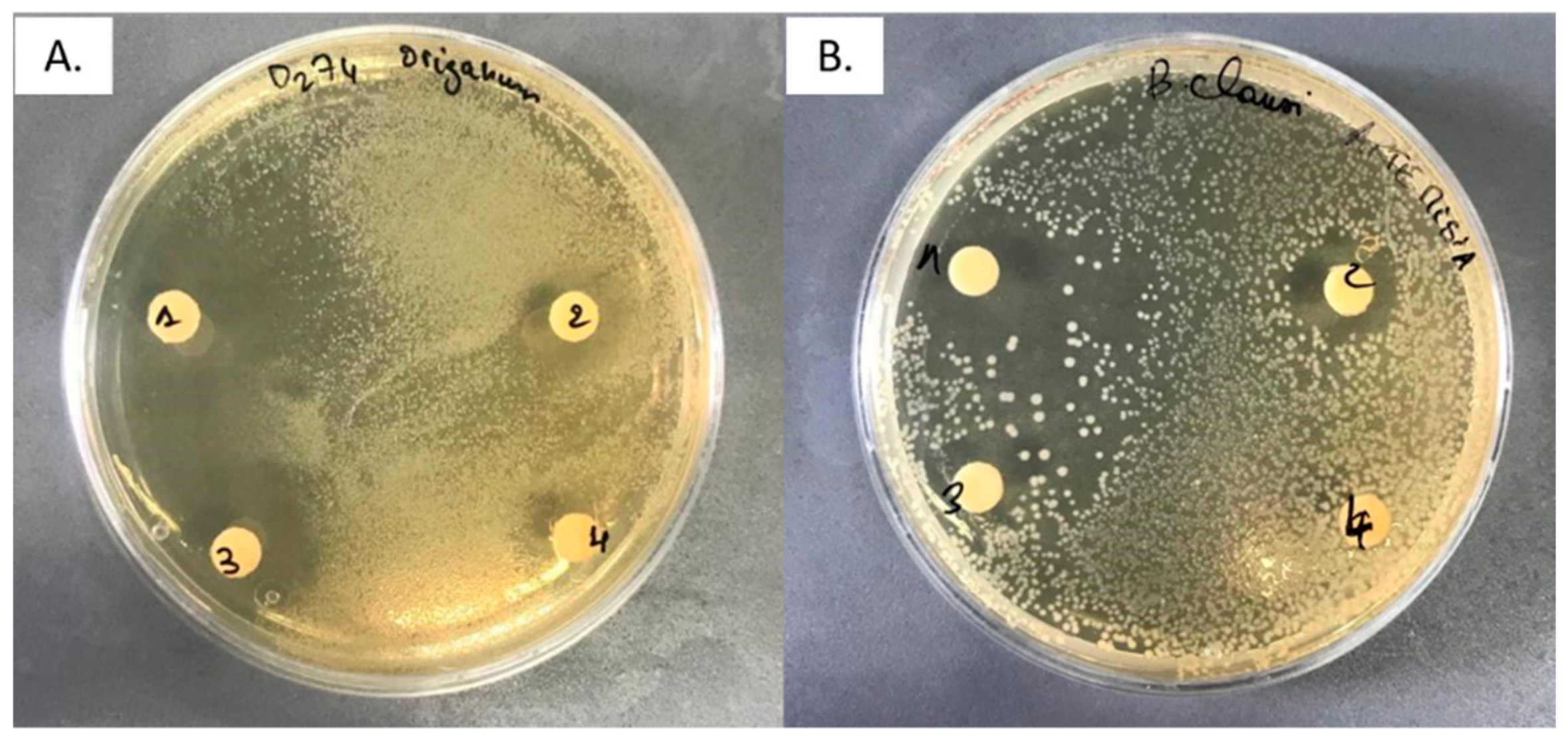
Most microorganisms used in this study were sensitive to both essential oils, with the dose of 20 μL of EO sufficient to stop the growth of almost all tested Gram-positive and Gram-negative strains. In particular, O. majorana EO resulted more active, showing a wide spectrum of activity. On the other hand, the EO of A. herba-alba showed inhibitory effects against 15 bacterial strains.
The available literature reports the antimicrobial activity of
A. herba-alba essential oil against
Staph. aureus,
E. coli, and
B. cereus [
23,
32,
33]. Moreover, several studies showed a great potential of
A. herba-alba EO oil as an antibacterial agent against
Klebsiella pneumoniae,
Listeria monocytogenes,
Vibrio colerae, and
S. Typhimurium [
34,
35,
36]. Our results showed variable antimicrobial and antifungal activity of the essential oil, being the inhibition zones in the range of 10–24 mm. Gram-positive bacteria resulted more sensitive to this EO. The Gram-positive
B. clausii 2226 was the most sensitive tested strain, with the strongest inhibition zone (24.00 ± 1 mm).
B. clausii was used as a model of spore-forming aerobic microorganism and our findings showed that our
A. herba-alba EO is suitable to control the growth of this microorganism. It is well known that spore forming bacteria (also called thermoduric) are the main problem in pasteurized foods, both from the point of view of food spoilage and human intoxication. Gram-negative strains also displayed variable degree of susceptibility to this EO. The maximum activity was showed against the pathogen strain
S. Typhimurium (17.7 ± 0.6), but
C. maltaromaticum F1201,
C. maltaromaticum D1203,
H. alvei 53M,
Ent. faecalis E21, and
Ent. faecalis 226 resulted resistant, since no inhibition zone was observed. Due to the involvement of
S. Typhimurium in the majority of food intoxication across the world, the antimicrobial capability of this EO could be of pivotal importance in the control of this microorganism in foods.
The antimicrobial activity of
O. majorana essential oil appears to be similarly effective against both Gram-positive and Gram-negative microorganisms. These results agree with literature data [
21,
34,
35]. Data of previous research showed that
O. majorana essential oil was active against a large spectrum of different bacteria strains:
E. coli,
Str. agalactiae,
Shigella dysenteriae,
Salmonella Enteritidis,
Staph. aureus,
Ent. faecalis,
E. coli, and
Klebsiella pneumoniae [
26,
37].
In our study, all tested strains were sensitive to this essential oil, with the Gram-positive S. aureus the most sensitive with the greatest inhibition zone (32.2 ± 2.5 mm); the more sensitive Gram-negative was S. Typhimurium, with an inhibition zone of 29.7 ± 0.6 mm.
The antimicrobial activity of both essential oils could be related to their content in oxygenated monoterpenes, which constitute about 57.3% and 53.0% of the EOs of
A. herba alba and
O. majorana, respectively. Similar findings have been already previously reported [
38,
39].
The major components of
O. majorana EO, e.g., terpinen-4-ol, α-terpinol and α-pinene, have been reported for their antimicrobial and antifungal properties [
21]. Additionally, the main constituents of the EO of
A. herba-alba, cis- and
trans-thujone and vanillyl alcohol, have been reported for their antimicrobial, anti-inflammatory, and antioxidant activities [
40,
41]. Oxygenated monoterpenes exhibit high antimicrobial activity on whole cell and possess antifungal effects. These compounds diffuse into and damage cell membrane structures [
42].
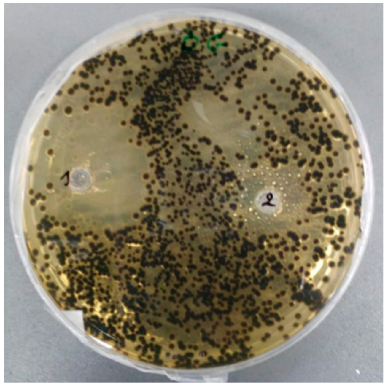
Our results showed high antifungal activity for both essential oils, with the highest inhibitory activity shown by the EO of
A. herba-alba against
Aspergillus niger (inhibition zone 23.6 ± 1.5 mm). These results are consistent with data previously reported [
29,
43].
4. Materials and Methods
4.1. Plant Material
The aerial parts of A. herba-alba and O. majorana were collected in the Azzemour region, South West Morocco, in June 2018, in flowering stage, and dried in the shade. The plants were identified by Prof. V. De Feo. A voucher specimen of each plant is stored in Department of Agricultural Sciences, University of Naples Federico II.
4.2. Essential Oil Extraction
One kilogram of
A. herba-alba and
O. majorana aerial parts was subjected to hydrodistillation for 3 h, according to the standard procedure described in the European Pharmacopoeia [
44]. The oils were solubilized in
n-hexane, filtered over anhydrous sodium sulphate and stored under N
2 at +4 °C in the dark, until tested and analyzed.
4.3. GC-FID Analysis
Analytical gas chromatography was carried out on a Perkin-Elmer Sigma-115 gas-chromatograph (Perkin Elmer, Waltham, MA, USA) equipped with an FID and a data handling processor. The separation was achieved using a HP-5 MS fused-silica capillary column (30 m × 0.25 mm i.d., 0.25 µm film thickness). Column temperature: 40 °C, with 5 min initial hold, and then to 270 °C at 2 °C/min, 270 °C (20 min); injection mode splitless (1 µL of a 1:1000 n-hexane solution). Injector and detector temperatures were 250 °C and 290 °C, respectively. Analysis was also run by using a fused silica HP Innowax polyethylene glycol capillary column (50 m × 0.20 mm i.d., 0.25 µm film thickness). In both cases, helium was used as carrier gas (1.0 mL/min).
4.4. GC/MS Analysis
Analysis was performed on an Agilent 6850 Ser. II apparatus (Agilent, Roma, Italy), fitted with a fused silica DB-5 capillary column (30 m × 0.25 mm i.d., 0.33 µm film thickness), coupled to an Agilent Mass Selective Detector MSD 5973 (Agilent); ionization energy voltage 70 eV; electron multiplier voltage energy 2000 V. Mass spectra were scanned in the range 40–500 amu, scan time 5 scans/s. Gas chromatographic conditions were as reported in the previous paragraph; transfer line temperature, 295 °C.
4.5. Identification of the Essential Oil Components
Most constituents were identified by comparison of their Kovats retention indices (Ri) (determined relative to the tR of
n-alkanes (C10–C35), with either those of the literature [
45,
46,
47] and mass spectra on both columns or those of authentic compounds available in our laboratories by means of NIST 02 and Wiley 275 libraries [
48]. The components relative concentrations were obtained by peak area normalization.
4.6. Antibacterial Assay
The antibacterial activity was evaluated in vitro, by means of the agar diffusion test on the plate. The activity of the essential oils was tested on the 20 microorganisms reported in
Table 6. All of them belong to the collection of the Department of Agricultural Sciences, University of Naples Federico II.
Microbial strains were previously grown in TSB tryptone soya broth for 24 h. A volume of 0.1 mL of the microbial suspensions (about 1 × 10⁸ CFU/mL) was uniformly distributed on Nutrient agar plates in sterile conditions. Different amounts of essential oils were spotted on the inoculated plates: 50, 40, 20, 15, 10, and 5 µL for O. majorana and 20, 15, 10, and 5 µL for A. herba-alba essential oils. After 10 min, under sterile conditions, plates were then incubated at optimal growth condition culture of each strain. The antimicrobial activity was evidenced by measuring the diameter (in mm) of the zone of inhibition. Ethanol was used as the negative control; tetracycline (10 µg) and gentamycin (10 µg) were used as positive controls. Each experiment was carried out in three independent replicates and result is the average with standard deviation.
4.7. Antifungal Activity
The antifungal activity was evaluated in vitro, using the agar well diffusion method on the plates. The activity was tested against Aspergillus niger isolated from a spoiled butter sample and identified by phenotypic characteristics. The fungus was previously grown in TSA agar plates at 28 °C until spore formation. Then, 1 mL of a spore suspension in quarter strength Ringer solution, containing about 1 × 10⁸ spores per mL, was uniformly distributed on Nutrient agar plates in sterile conditions, then a hole was punched with sterile cork and 20 µL of each EO was introduced into the well; the plates were incubated at 28 °C for 4–5 days. Ethanol was used as the negative control. The antifungal activity was evaluated by measuring diameter of the inhibition area. Each experiment was carried out in three independent replicates and result is the average with standard deviation.
4.8. Statistical Analysis
Data of each experiment were statistically analyzed using GraphPad Prism 6.0 software (GraphPad Software Inc., San Diego, CA, USA), followed by comparison of means (one-way ANOVA) using Dunnett’s multiple comparisons test, at the significance level of p < 0.05.


 ,
, 

