New and Potent Quinuclidine-Based Antimicrobial Agents

 and
and
Abstract
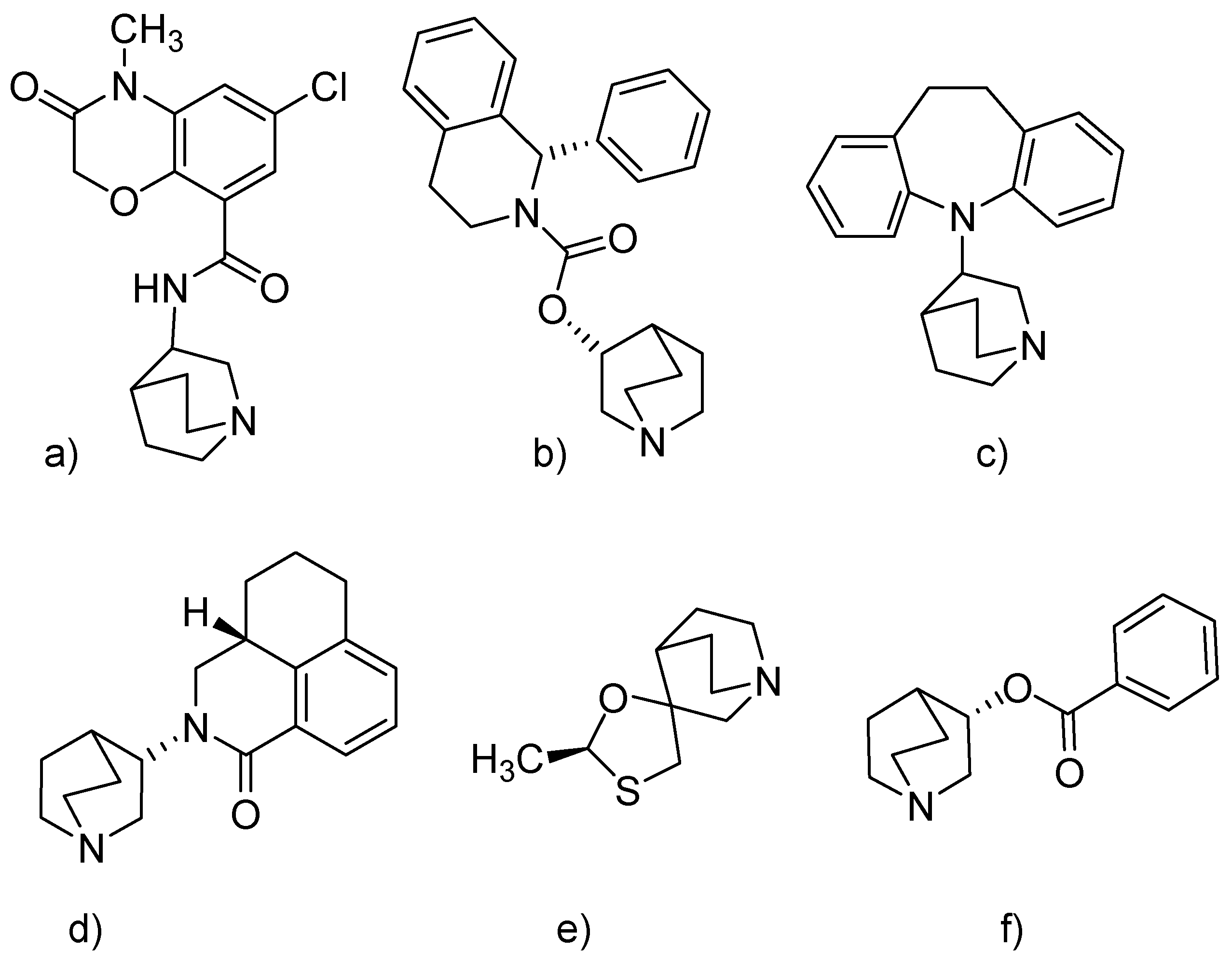
:1. Introduction
2. Results and Discussion
2.1. Synthesis of Compounds
2.2. Antimicrobial Activity
2.3. MTT Assay
2.4. Effects of the Compounds on Cellular Reactive Oxygen Species and Antioxidative Defence
2.5. Multi-Way Analysis
3. Materials and Methods
3.1. Synthesis of Compounds
General Procedure for the Synthesis of N-quaternary 3-hydroxyiminoquinuclidinium bromides 1–10
3.2. Antimicrobial Evaluation
3.2.1. Disc Diffusion Assay
3.2.2. Minimum Inhibitory Concentration Assay
3.3. MTT Assay
ROS
3.4. Antioxidant Measurements
3.5. Statistical Analysis
3.6. Computational Methods
4. Conclusions
Author Contributions
Funding
Acknowledgments
Conflicts of Interest
References
- Fauci, A.S. Infectious Diseases: Considerations for the 21st Century. Clin. Infect. Dis. 2001, 32, 675–685. [Google Scholar] [CrossRef] [PubMed] [Green Version]
- Boucher, H.W.; Talbot, G.H.; Bradley, J.S.; Edwards, J.E.; Gilbert, D.; Rice, L.B.; Scheld, M.; Spellberg, B.; Bartlett, J. Bad bugs, no drugs: No ESKAPE! An update from the Infectious Diseases Society of America. Clin. Infect. Dis. 2009, 48, 1–12. [Google Scholar] [CrossRef] [PubMed]
- Pendleton, J.N.; Gorman, S.P.; Gilmore, B.F. Clinical relevance of the ESKAPE pathogens. Expert. Rev. Anti. Infect. Ther. 2013, 11, 297–308. [Google Scholar] [CrossRef] [PubMed]
- World Health Organization. Antimicrobial Resistance: Global Report on Surveillance; World Health Organization: Geneva, Switzerland, 2014. [Google Scholar]
- Gomtsyan, A. Heterocycles in drugs and drug discovery. Chem. Heterocycl. Compd. 2012, 48, 7–10. [Google Scholar] [CrossRef]
- Mittal, A. Synthetic Nitroimidazoles: Biological Activities and Mutagenicity Relationships. Sci. Pharm. 2009, 77, 497–520. [Google Scholar] [CrossRef] [Green Version]
- Nagalakshmi, G. Synthesis, antimicrobial and antiinflammatory activity of 2,5-disubstituted-1,3,4-oxadiazoles. Indian J. Pharm. Sci. 2008, 70, 49–55. [Google Scholar] [CrossRef] [PubMed] [Green Version]
- Joule, J.A.; Mills, K. Heterocyclic Chemistry, 4th ed.; Blackwell Publishing: Hoboken, NJ, USA, 2000; p. 369. [Google Scholar]
- Nekrasov, D.D. Biological Activity of 5-and 6-Membered Azaheterocycles and Their Synthesis from 5-Aryl-2,3-Dihydrofuran-2,3-diones. Chem. Heterocycl. Compd. 2001, 37, 263–275. [Google Scholar] [CrossRef]
- Sperry, J.B.; Wright, D.L. Furans, thiophenes and related heterocycles in drug discovery. Curr. Opin. Drug Discovery Dev. 2005, 8, 723–740. [Google Scholar]
- Polshettiwar, V.; Varma, R.S. Greener and expeditious synthesis of bioactive heterocycles using microwave irradiation. Pure Appl. Chem. 2008, 80, 777–790. [Google Scholar] [CrossRef]
- Katritzky, A.R. Heterocyclic chemistry: An academic subject of immense industrial importance. Chem. Heterocycl. Compd. 1992, 28, 241–259. [Google Scholar] [CrossRef]
- Song, C.E. An Overview of Cinchona Alkaloids in Chemistry; Wiley: Hoboken, NJ, USA, 2009. [Google Scholar]
- Gralla, R.; Lichinitser, M.; Van der Vegt, S.; Sleeboom, H.; Mezger, J.; Peschel, C.; Tonini, G.; Labianca, R.; Macciocchi, A.; Aapro, M. Palonosetron improves prevention of chemotherapy-induced nausea and vomiting following moderately emetogenic chemotherapy: Results of a double-blind randomized phase III trial comparing single doses of palonosetron with ondansetron. Ann. Oncol. 2003, 14, 1570–1577. [Google Scholar] [CrossRef] [PubMed]
- Bazina, L.; Maravić, A.; Krce, L.; Soldo, B.; Odžak, R.; Bučević Popović, V.; Aviani, I.; Primožič, I.; Šprung, M. Discovery of novel quaternary ammonium compounds based on quinuclidin-3-ol as new potential antimicrobial candidates. Eur. J. Med. Chem. 2019, 163, 626–635. [Google Scholar] [CrossRef] [PubMed]
- Odžak, R.; Šprung, M.; Soldo, B.; Skočibušić, M.; Gudelj, M.; Muić, A.; Primožič, I. Quaternary salts derived from 3-substituted quinuclidine as potential antioxidative and antimicrobial agents. Open Chem. 2017, 15, 320–331. [Google Scholar] [CrossRef]
- Skočibušić, M.; Odžak, R.; Štefanić, Z.; Križić, I.; Krišto, L.; Jović, O.; Hrenar, T.; Primožič, I.; Jurašin, D. Structure-Property Relationship of Quinuclidinium Surfactants—Towards Multifunctional Biologically Active Molecules. Colloids Surf. B Biointerfaces 2016, 140, 548–559. [Google Scholar] [CrossRef]
- Gerba, C.P. Quaternary Ammonium Biocides: Efficacy in Application. Appl. Environ. Microbiol. 2015, 81, 464–469. [Google Scholar] [CrossRef] [Green Version]
- Grob, C.A.; Renk, E. 3-Chinuclidincarbonsaure. Helv. Chim. Acta 1954, 37, 1689–1698. [Google Scholar] [CrossRef]
- Sternbach, L.H.; Kaiser, S. Antispasmodics. I. Bicyclic Basic Alcohols. J. Am. Chem. Soc. 1952, 74, 2215–2218. [Google Scholar] [CrossRef]
- World Health Organization. Critically Important Antimicrobials for Human Medicine: Ranking of Antimicrobial Agents for Risk Management of Antimicrobial Resistance Due to Non-Human Use; World Health Organization: Geneva, Switzerland, 2017. [Google Scholar]
- Tam, V.H.; Kabbara, S.; Vo, G.; Schilling, A.N.; Coyle, E.A. Comparative pharmacodynamics of gentamicin against Staphylococcus aureus and Pseudomonas aeruginosa. Antimicrob. Agents Chemother. 2006, 50, 2626–2631. [Google Scholar] [CrossRef]
- Hrenar, T.; Primožič, I.; Fijan, D.; Majerić Elenkov, M. Conformational analysis of spiro-epoxides by principal component analysis of molecular dynamics trajectories. Phys. Chem. Chem. Phys. 2017, 19, 31706–31713. [Google Scholar] [CrossRef]
- Maravić, A.; Skočibušić, M.; Cvjetan, S.; Šamanić, I.; Fredotović, Ž.; Puizina, J. Prevalence and diversity of extended-spectrum-β-lactamase-producing Enterobacteriaceae from marine beach waters. Mar. Pollut. Bull. 2015, 90, 60–67. [Google Scholar] [CrossRef]
- Clinical and Laboratory Standards Institute. Performance Standards for Antimicrobial Susceptibility Testing; eighteenth informational supplement, CLSI document M100-18; Clinical and Laboratory Standards Institute: Wayne, PA, USA, 2008. [Google Scholar]
- Góth, L.A. Simple method for determination of serum catalase activity and revision of reference range. Clin. Chim. Acta 1991, 196, 143–151. [Google Scholar] [CrossRef]
- Campbell, M.K.; Farrell, S.O. Biochemistry; Brooks/Cole: Belmont, CA, USA; Cengage Learning: Boston, MA, USA, 2012. [Google Scholar]
- Tietze, F. Enzymic method for quantitative determination of nanogram amounts of total and oxidized glutathione: Applications to mammalian blood and other tissues. Anal. Biochem. 1969, 27, 502–522. [Google Scholar] [CrossRef]
- Stewart, J.J.P. Optimization of parameters for semiempirical methods VI: More modifications to the NDDO approximations and re-optimization of parameters. J. Mol. Model. 2013, 19, 1–32. [Google Scholar] [CrossRef] [PubMed]
- Stewart, J.J.P. Stewart Computational Chemistry; MOPAC2016: Colorado Springs, CO, USA, 2016. [Google Scholar]
- Hrenar, T. QCC, Quantum Chemistry Code; Revision 0.6826; Zagreb, Croatia, 2019; Available online: https://masongroup.lab.uiowa.edu/qcc-everyone (accessed on 1 June 2019).
- Primožič, I.; Hrenar, T.; Baumann, K.; Krišto, L.; Križić, I.; Tomić, S. Mechanochemical and Conformational Study of N-heterocyclic Carbonyl-Oxime Transformations. Croat. Chem. Acta 2014, 87, 155–162. [Google Scholar] [CrossRef]
- Hrenar, T. moonee, Code for Manipulation and Analysis of Multi- and Univariate Data; Revision 0.6826; University of Zagreb: Zagreb, Croatia, 2019. [Google Scholar]
- Jović, O.; Smolić, T.; Jurišić, Z.; Meić, Z.; Hrenar, T. Chemometric Analysis of Croatian Extra Virgin Olive Oils from Central Dalmatia Region. Croat. Chem. Acta 2013, 86, 335–344. [Google Scholar] [CrossRef]
- Novak, P.; Kišić, A.; Hrenar, T.; Jednačak, T.; Miljanić, S.; Verbanec, G. In-line reaction monitoring of entacapone synthesis by Raman spectroscopy and multivariate analysis. J. Pharm. Biomed. Anal. 2011, 54, 660–666. [Google Scholar] [CrossRef] [PubMed]
- Frisch, M.J.; Trucks, G.W.; Schlegel, H.B.; Scuseria, G.E.; Robb, M.A.; Cheeseman, J.R.; Scalmani, G.; Barone, V.; Petersson, G.A.; Nakatsuji, H.; et al. Gaussian 16, Revision A.03; Gaussian, Inc.: Wallingford, CT, USA, 2016. [Google Scholar]
- Gnuplot 5.0. Available online: http://gnuplot.info/ (accessed on 1 June 2019).
- Tucker, L. Some mathematical notes on three-mode factor analysis. Psychometrika 1966, 31, 279–311. [Google Scholar] [CrossRef]
Sample Availability: Samples of the compounds 1–10 are available from the authors. |


| Comp. | Diameters of the Inhibition Zone (mm) | |||||
|---|---|---|---|---|---|---|
| Gram-Positive Bacteria | Gram-Negative Bacteria | |||||
| Bacillus cereus | Enterococcus faecalis | Staphylococcus aureus | Escherichia coli | Klebsiella pneumoniae | Pseudomonas aeruginosa | |
| qox | 8.2 ± 0.9 | 7.7 ± 1.4 | 10.4 ± 1.7 | 8.6 ± 1.9 | 7.6 ± 1.1 | 10.8 ± 1.4 |
| 1 | 9.9 ± 1.4 | 6.2 ± 1.3 | 9.3 ± 0.8 | 9.6 ± 1.1 | 8.4 ± 1.1 | 12.1 ± 1.8 |
| 2 | 12.1 ± 1.8 | 14.2 ± 2.1 | 15.7 ± 1.7 | 14.8 ± 1.2 | 16.1 ± 0.9 | 19.2 ± 1.4 |
| 3 | 10.4 ± 1.5 | 12.7 ± 1.4 | 13.5 ± 1.7 | 15.1 ± 1.2 | 13.7 ± 1.8 | 16.3 ± 1.5 |
| 4 | 16.1 ± 0.4 | 15.4 ± 1.2 | 18.1 ± 1.1 | 17.7 ± 0.9 | 14.7 ± 0.4 | 18.7 ± 0.9 |
| 5 | 18.2 ± 1.2 | 19.4 ± 1.8 | 20.8 ± 1.1 | 20.8 ± 2.1 | 19.8 ± 0.4 | 24.8 ± 1.7 |
| 6 | 16.8 ± 0.8 | 17.4 ± 1.7 | 17.2 ± 0.3 | 19.1 ± 1.5 | 18.1 ± 1.8 | 20.1 ± 2.1 |
| 7 | 10.8 ± 1.7 | 6.4 ± 2.1 | 9.5 ± 1.3 | 8.1 ± 1.2 | 7.5 ± 1.4 | 11.0 ± 0.8 |
| 8 | 7.8 ± 0.3 | 9.6 ± 1.2 | 11.4 ± 0.7 | 6.6 ± 1.1 | 8.6 ± 1.1 | 10.6 ± 1.1 |
| 9 | 23.5 ± 1.3 | 17.3 ± 2.4 | 27.2 ± 1.3 | 22.7 ± 0.8 | 24.7 ± 1.3 | 27.6 ± 1.7 |
| 10 | 24.0 ± 1.2 | 23.0 ± 1.4 | 21.0 ± 0.8 | 22.4 ± 0.9 | 25.1 ± 0.6 | 29.8 ± 1.3 |
| Gen | 18.2 ± 0.7 | 13.2 ± 0.6 | 23.9 ± 0.4 | 11.5 ± 0.9 | 16.9 ± 0.3 | 9.4 ± 0.6 |
| Comp. | MIC (µg/mL) | |||||
|---|---|---|---|---|---|---|
| Gram-Positive Bacteria | Gram-Negative Bacteria | |||||
| B. cereus | E. faecalis | S. aureus | E. coli | K. pneumoniae | P. aeruginosa | |
| 1 | 128.00 | 256.00 | 128.00 | 128.00 | 256.00 | 128.00 |
| 2 | 64.00 | 64.00 | 32.00 | 16.00 | 16.00 | 8.00 |
| 3 | 64.00 | 32.00 | 32.00 | 16.00 | 64.00 | 32.00 |
| 4 | 32.00 | 16.00 | 16.00 | 32.00 | 32.00 | 16.00 |
| 5 | 4.00 | 2.00 | 1.00 | 1.00 | 2.00 | 0.25 |
| 6 | 8.00 | 4.00 | 4.00 | 4.00 | 8.00 | 2.00 |
| 7 | 64.00 | 128.00 | 64.00 | 128.00 | 256.00 | 32.00 |
| 8 | 128.00 | 64.00 | 64.00 | 256.00 | 128.00 | 128.00 |
| 9 | 1.00 | 8.00 | 12.50 | 25.00 | 50.00 | 0.50 |
| 10 | 1.00 | 4.00 | 2.00 | 1.00 | 0.50 | 0.25 |
| Gentamicin | 4.00 | 4.00 | 1.00 | 32.00 | 8.00 | 64.00 |
| Cefotaxime | 0.25 | 0.50 | 0.50 | 0.50 | 0.50 | 16.00 |
© 2019 by the authors. Licensee MDPI, Basel, Switzerland. This article is an open access article distributed under the terms and conditions of the Creative Commons Attribution (CC BY) license (http://creativecommons.org/licenses/by/4.0/).
Share and Cite
Radman Kastelic, A.; Odžak, R.; Pezdirc, I.; Sović, K.; Hrenar, T.; Čipak Gašparović, A.; Skočibušić, M.; Primožič, I. New and Potent Quinuclidine-Based Antimicrobial Agents. Molecules 2019, 24, 2675. https://doi.org/10.3390/molecules24142675
Radman Kastelic A, Odžak R, Pezdirc I, Sović K, Hrenar T, Čipak Gašparović A, Skočibušić M, Primožič I. New and Potent Quinuclidine-Based Antimicrobial Agents. Molecules. 2019; 24(14):2675. https://doi.org/10.3390/molecules24142675
Chicago/Turabian StyleRadman Kastelic, Andreja, Renata Odžak, Iskra Pezdirc, Karlo Sović, Tomica Hrenar, Ana Čipak Gašparović, Mirjana Skočibušić, and Ines Primožič. 2019. "New and Potent Quinuclidine-Based Antimicrobial Agents" Molecules 24, no. 14: 2675. https://doi.org/10.3390/molecules24142675