Article

Melatonin-Mediated Development of Ovine Cumulus Cells, Perhaps by Regulation of DNA Methylation

1
Animal Husbandry and Veterinary Research Institute of Tianjin, Tianjin 300381, China
2
Jilin Provincial Key Laboratory of Grassland Farming, Northeast Institute of Geography and Agoecology, Chinese Academy of Sciences, Changchun, Jilin 130062, China
3
State Key Laboratory of Stem Cell and Reproduction Biology, Institute of Zoology, Chinese Academy of Science, Beijing 100101, China
*
Authors to whom correspondence should be addressed.
These authors contributed equally to this paper.
Molecules 2018, 23(2), 494; https://doi.org/10.3390/molecules23020494
Submission received: 19 January 2018 / Revised: 13 February 2018 / Accepted: 16 February 2018 / Published: 23 February 2018

Abstract

:
Cumulus cells of pre-pubertal domestic animals are dysfunctional, perhaps due to age-specific epigenetic events. This study was designed to determine effects of melatonin treatment of donors on methylation modification of pre-pubertal cumulus cells. Cumulus cells from germinal vesicle stage cumulus oocyte complexes (COCs) were collected from eighteen lambs which were randomly divided into control group (C) and melatonin group given an 18 mg melatonin implant subcutaneous (M). Compared to the C group, the M group had higher concentrations of melatonin in plasma and follicular fluid (p < 0.05), greater superovulation, a higher proportion of fully expanded COCs, and a lower proportion of apoptotic cumulus cells (p < 0.05). Real-time PCR results showed that melatonin up-regulated expression of genes MT1, Bcl2, DNMT1, DNMT3a and DNMT3b, but down-regulated expression of genes p53, Caspase 3 and Bax (p < 0.05). Furthermore, melatonin increased FI of FITC (global methylation level) on cumulus cells (p < 0.05). To understand the regulation mechanism, the DNMTs promoter methylation sequence were analyzed. Compared to the C group, although there was less methylation at two CpG sites of DNMT1 (p < 0.05) and higher methylation at two CpG sites of DNMT3a (p < 0.05), there were no significant differences in methylation of the detected DNMT1 and DNMT3a promoter regions. However, there were lower methylation levels at five CpG sites of DNMT3b, which decreased methylation of detected DNMT3b promoter region on M group (p < 0.05). In conclusion, alterations of methylation regulated by melatonin may mediate development of cumulus cells in lambs.

1. Introduction

Evaluation of the reproductive potential of pre-pubertal animal can solve the lack of embryonic origin for the commercialization of embryo transfer, significantly reduce the cost of embryo production and greatly improve the breeding and production efficiency [1]. However, developmental capacity of oocytes from pre-pubertal animals is reduced compared to their adult counterparts. Besides incomplete or deficient maturation of cytoplasm [2], altered protein synthesis [3], and impaired metabolism [4], cumulus cell dysfunction also contribute.
Cumulus cells are considered to have important roles in oocyte maturation by: (1) keeping the oocyte under meiotic arrest; (2) participating in the induction of meiotic resumption; (3) supporting cytoplasmic maturation. These key cumulus cell functions during oocyte maturation are attributable to their elaborate gap junctional network and to their specific metabolic capacities [5,6]. Pre-pubertal cumulus cells have a prominent nucleus and limited cytoplasm with high transcriptional activity, communicating with each other by a few short processes, with limited processes reaching the oocyte [7,8]. Young donors had disturbed DNA methylation processes due to insufficient methyltransferases during oocyte maturation and embryo development [9,10,11], although methylation characteristics of the cumulus cells remain unclear. It was, therefore, hypothesized that pre-pubertal cumulus cells were epigenetically immature because DNA methylation modifications lacked adult-like cumulus cell methylation patterns.
In animals that exhibit seasonal reproduction, such as sheep, photoperiodic information is conveyed to the reproductive neuroendocrine system by circadian secretion of melatonin from the pineal gland [12]. A night-time increase in plasma melatonin concentrations has been reported to occur within 1–6 week-old mammals after birth [13]. In addition to being an antioxidant, melatonin is likely an epigenetic regulator, as it and its metabolites have similar structures and hypothetically could regulate DNA methyltransferases (DNMTs), either by masking target sequences or by blocking the active site of the enzyme [14,15]. Melatonin is a highly lipophilic and somewhat hydrophilic molecule that easily crosses cell membranes, reaching intracellular organelles including the nucleus [16] to accumulate in the nucleus and it interacts with specific nuclear binding sites [17]. So-called nuclear receptors for melatonin have been identified and some studies have linked them to melatonin control of cell growth and differentiation [18]. Furthermore, melatonin binding sites were identified not only in granulosa cells from preovulatory follicles [19,20], but also in cumulus and granulosa cells [21]. Two distinct receptor subtypes MT1 and MT2 genes have been cloned and mapped in several animal species [22,23,24]. In mammals, MT1 seemed to be involved more in regulation of reproductive activity than MT2 [25]. Although, addition of melatonin during in vitro maturation (IVM) protected cumulus cells from DNA damage [26], little information is available about effects of melatonin on pre-pubertal cumulus cells in vivo, especially with regards to epigenetic modification.
The objective was to investigate potential epigenetic mechanisms improving cumulus cells quality in prepubertal lambs by determining whether melatonin treatment altered gene expression of key enzymes and methylation modification.

2. Results

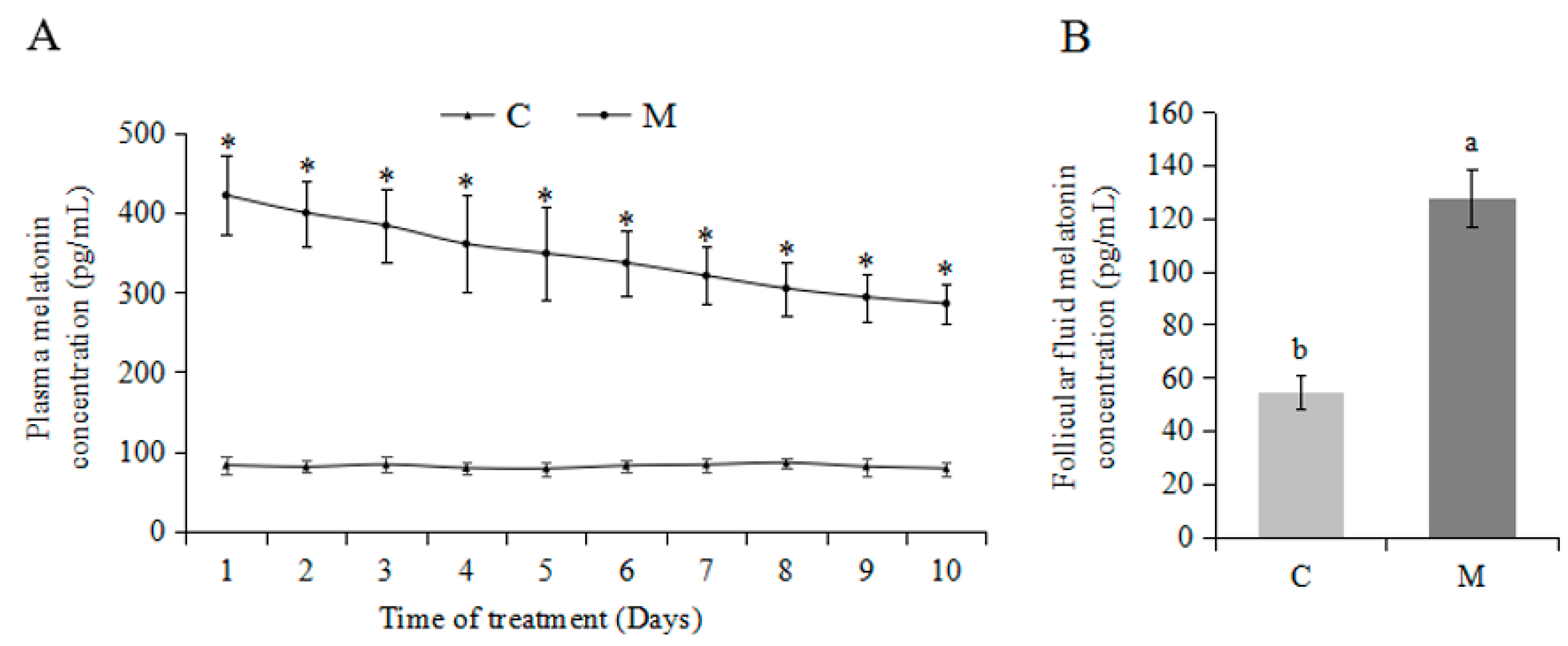
2.1. Effects of Exogenous Melatonin on Plasma and Follicular Fluid Melatonin Concentrations

Compared to the C group, the M group had higher concentrations (p < 0.05) of melatonin in both plasma and follicular fluid (Figure 1).

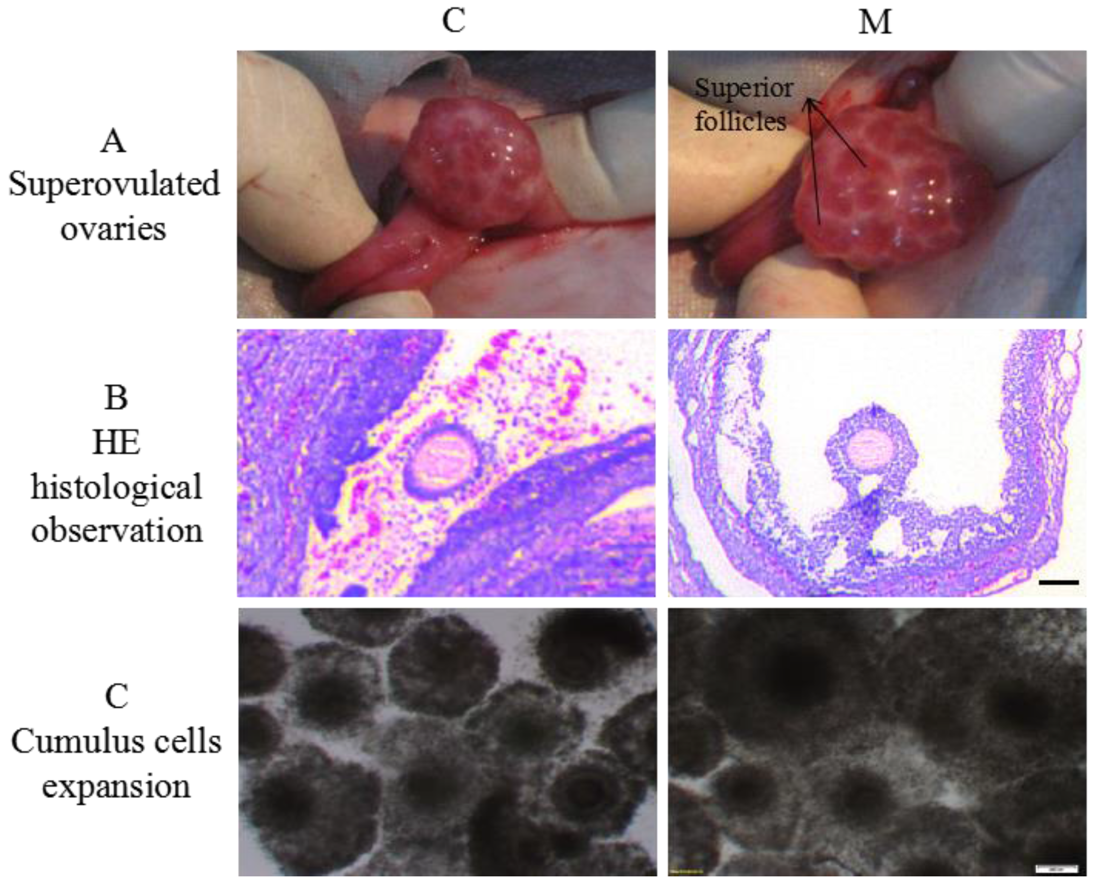
2.2. Effects of Melatonin on Superovulaton and Cumulus Cells Expansion

Compared to the C group, the M group had a better superovulatory response (p < 0.05; Table 1; Figure 2A,B), lower proportions of not expanded and partially expanded cumulus oocyte complexes (COCs), and higher proportion of fully expanded COCs (p < 0.05; Table 2; Figure 2C).

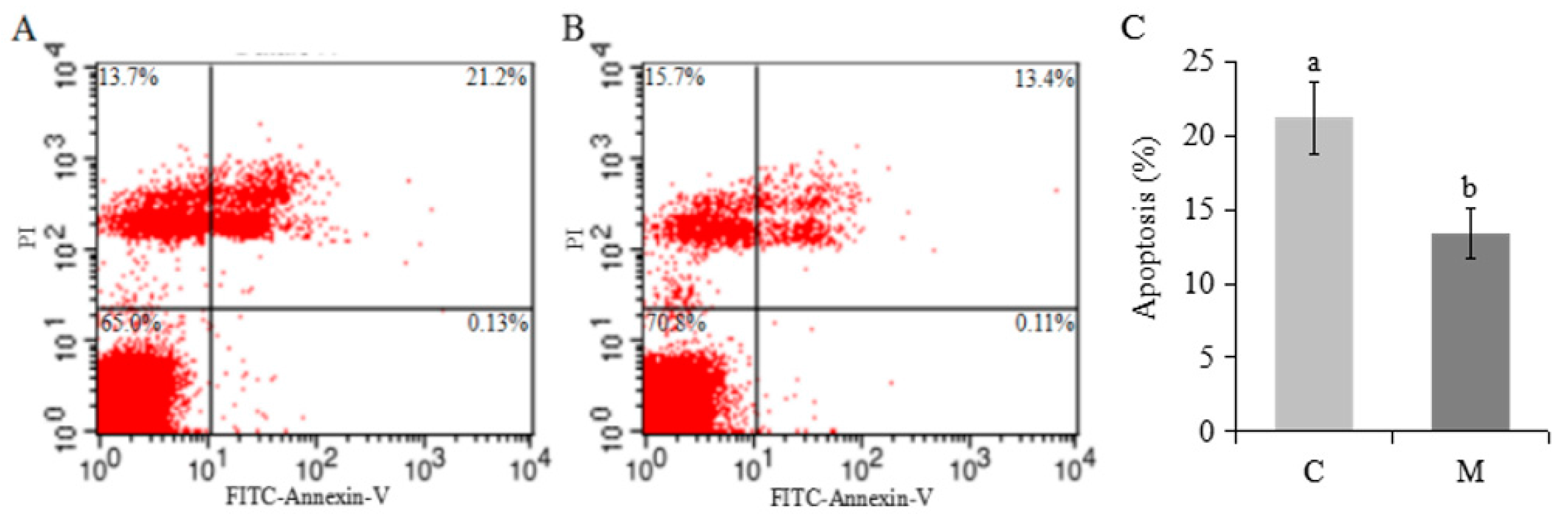
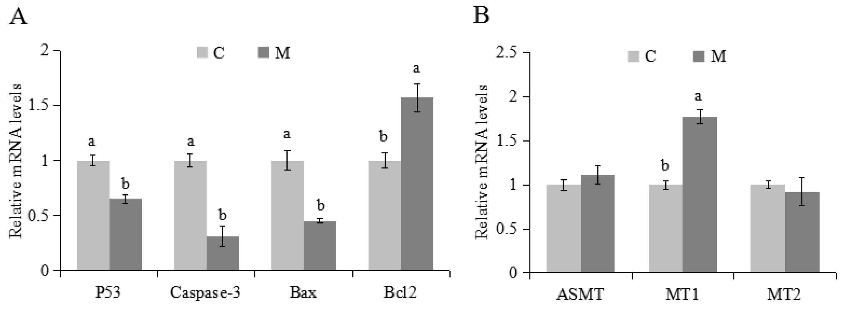
2.3. Effects of Melatonin on Apoptosis and Expression of Related Genes in Cumulus Cells

There was a lower proportion of apoptotic cumulus cells in the M versus C groups (p < 0.05; Figure 3A–C). Melatonin increased mRNA expression of Bcl2 and MT1, but decreased P53, Caspase 3 and Bax (p < 0.05; Figure 4A,B). Melatonin had no effect on ASMT or MT2 (Figure 4B).

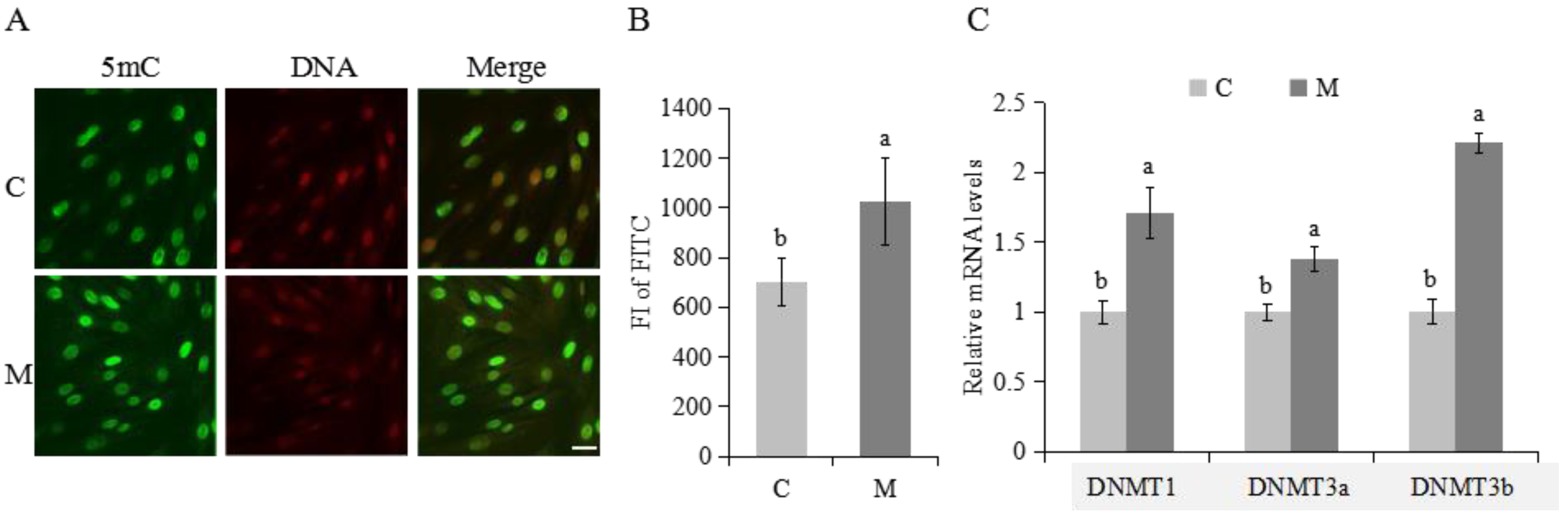
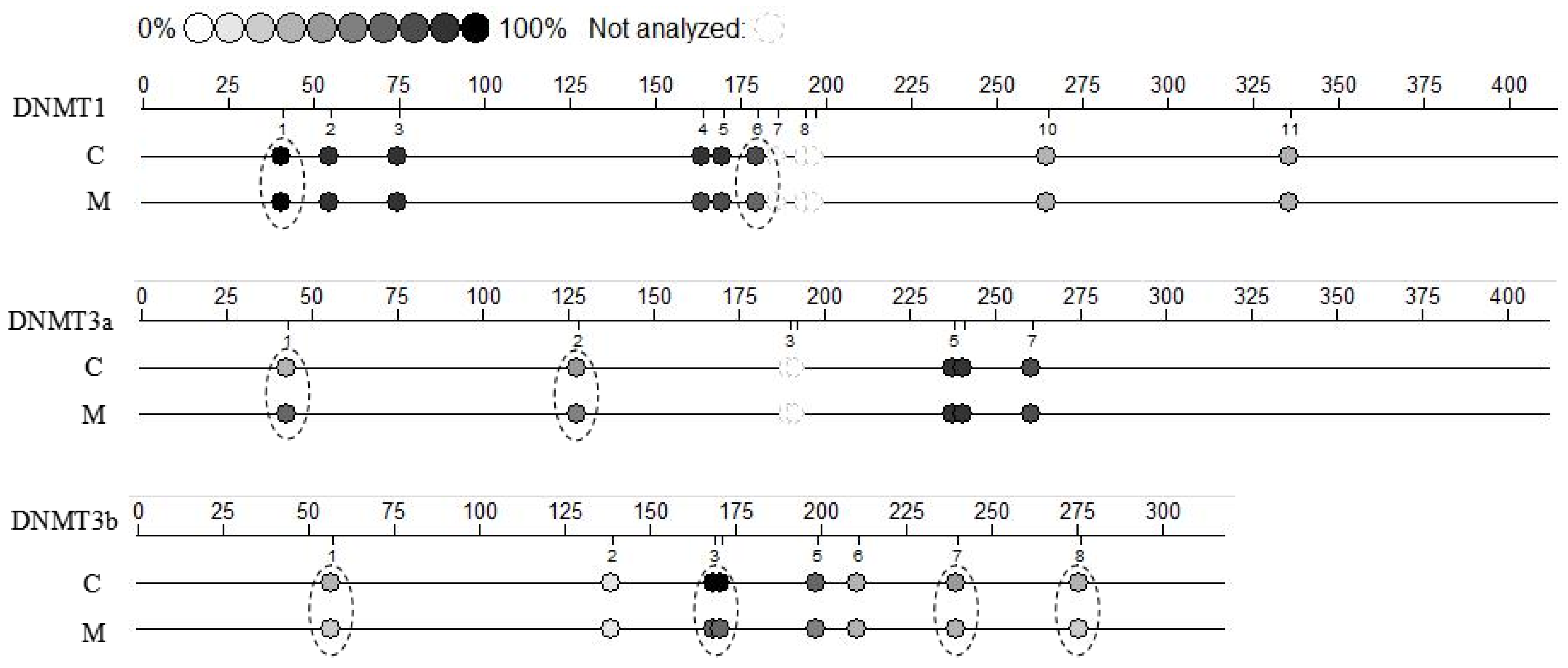
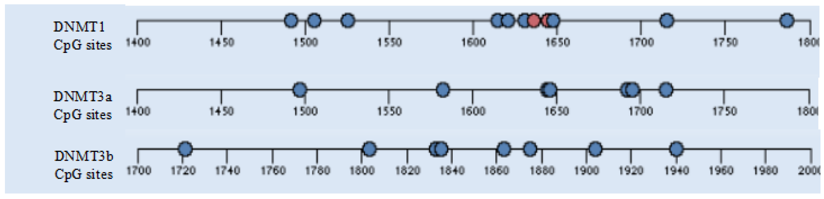
2.4. Methylation Modifications in Cumulus Cells

Melatonin increased FI of FITC (methylation marker) and increased mRNA expression of DNMT1, DNMT3a and DNMT3b on cumulus cells (p < 0.05; Figure 5A–C).
Compared to the C group, there were no significant differences in methylation of DNMT1 and DNMT3a promoter CpG region, and decreased methylation of DNMT3b promoter CpG region on melatonin group (p < 0.05; Appendix: Table A2, Table A3 and Table A4). There were lower methylation levels at two CpG sites (CpG40, CpG179) of DNMT1 (p < 0.05; Figure 6, Appendix: Table A2) and at five CpG sites (CpG56, CpG168:170, CpG239, CpG275) of DNMT3b (p < 0.05; Figure 6, Appendix: Table A4), and higher methylation levels at two CpG sites (CpG42, CpG127) of DNMT3a (p < 0.05; Figure 6, Appendix: Table A3) in the M group.

3. Discussion

In addition to its antioxidant properties, melatonin regulates gene expression [15] and therefore has epigenetic effects. In the present study in prepubertal lambs, melatonin promoted cumulus cells development, and regulated related genes expression, perhaps due to alterations of methylation modification.
In 1-mo-old superovulated lambs, exogenous melatonin significantly increased melatonin concentrations in both plasma and follicular fluid, enhanced superovulation and cumulus cells expansion. Melatonin has improved the quality of human [27], porcine [28,29], bovine [24,26,30,31], and murine [32,33] oocytes (with or without cumulus cells), embryos, and cumulus cells by reducing cytoplasmic ROS concentrations or DNA damage. Melatonin concentrations in the follicular fluid vary with follicle size, increasing with an increased follicular diameter in humans [34]. High melatonin concentrations in follicular fluid help to prevent atresia, enabling full development of preovulatory follicles and protecting adjacent cells and the oocyte from free radicals [35,36]. Perhaps melatonin modulates the follicular response to LH by elevating mRNA expression of LH receptors in granulosa and cumulus cells [37]. Additionally, melatonin promotes follicular development by increasing production of insulin-like growth factor I which stimulates mitogenic growth of cumulus cells [38]. Although previous in vivo studies demonstrated exogenous FSH may mask effects of melatonin on follicular development, exogenous melatonin tended to increase the number of developing follicles [39]. Perhaps the role of melatonin in regulation of germ cell development is species- or age-specific.
Mammalian cumulus cells have very important roles during oocyte growth and maturation. They supply nutrients [40] and/or messenger molecules for oocyte development [41,42], and to mediate effects of hormones on oocytes [43]. Moreover, cumulus cells expansion is considered an important marker for oocyte maturation and is essential for fertilization, subsequent cleavage, and blastocyst development [44]. Our results confirmed that melatonin had a potentially significant effect on the degree of lamb cumulus cells expansion. The same promoting effects of melatonin on cumulus cell expansion were reported in bovine [24] and porcine oocytes [45].
It is well established that melatonin induces apoptosis in cancer-like cells [46,47], whereas in normal cells it prevents apoptosis [48]. A lower proportion of apoptotic cumulus cells in this study indicated that melatonin had a beneficial effect, confirmed by decreasing expression of genes P53, Caspase 3 and Bax, and increasing expression of Bcl2 genes. In general, it is believed that the balance between Bax and Bcl-2 determines the propensity of cells to respond to a given insult by apoptosis or survival [49]. Melatonin shifts the balance towards a protective state by suppressing pro-apoptotic Bax and inducing expression of Bcl-2 [50,51].
MT1 and MT2, expressed in follicular cumulus, mural granulosa cells and oocytes [52], contribute to regulation of follicular development, proliferation, and influence hormone signaling [53]. In the present study, up-regulation of MT1 gene expression in lambs indicated that the positive effect of melatonin on cumulus cells may be mediated by MT1. Therefore, we speculated that some effects of melatonin on lamb cumulus cells were likely to be directly mediated by receptor mechanisms. Moreover, the unchanged expression of MT2 in cumulus cells supported previous reports suggesting that MT1 was more important than MT2 for regulation of reproduction [25]. In the absence of alterations of gene expression of ASMT, we inferred that there was production of melatonin secretion as early as 1 month of age in lambs and consequently, exogenous melatonin treatment may not affect endogenous melatonin secretion on cumulus cells.
Pre-pubertal germ cells are epigenetically immature, and epigenetics has been proposed to be involved in acquisition of full developmental or proliferative competence. Methylation sequence changes are associated with donor age [10], with less DNA methylation in pre-pubertal germ cells [6,11]. The positive effect of melatonin on cumulus cells in this study may be related to increasing global methylation. There is direct evidence of epigenetic actions for melatonin, including significant increases in mRNA expression for various HDAC isoforms and elevated histone H3 acetylation in neural stem cell lines [54]. Methylation actions of melatonin suggest epigenetic regulation at a co-regulator level rather than selective enzymatic inhibition or activation [54]. Several studies indicated that melatonin inhibited COX-2 and iNOS transcriptional activation, suggested to be an epigenetic action [55]. This had significant benefits in treatment of experimental hyperglycemia mediated by epigenetic regulation [56]. Furthermore, this also involved anti-inflammatory actions, controlled by activation of the NF-κB and AP-1 family, which induces various epigenetic processes by altering chromatin structure [57,58].
The higher level of global methylation in this study may due to overexpression of DNMT1, DNMT3a and DNMT3b genes. Despite uncertainty regarding how melatonin can regulate a variety of genes such as DNMTs, MT1, apoptosis genes but always in the right direction (some inhibition, some activation), these actions may be due to an epigenetic mechanism [59,60]. Most genes are either active or inactive under steady-state conditions and in order to change their status (e.g., from on to off and vice versa), several epigenetic modifications such as histone modifications and DNA methylations are required. Accumulating data in the turning on/off of genes and gene regulation by melatonin have converged with discoveries of epigenetic mechanisms. Although there were lower methylation level at two CpG sites (CpG40, CpG179) of DNMT1 and higher methylation level at two CpG sites (CpG42, CpG127) of DNMT3a after melatonin treatment, there were no differences in methylation of detected promoter region of DNMT1 and DNMT3a. Melatonin decreased methylation of the DNMT3b promoter CpG region, reflected at five CpG sites (CpG56, CpG168:170, CpG239, CpG275) after melatonin treatment. Melatonin regulated DNMTs gene induction by methylation and also seemed to recruit basal transcriptional machinery to the promoter region of DNMT-related genes. In conclusion, some of the positive effects of melatonin on cumulus cells was attributed to regulation of methylation events.

4. Materials and Methods

All procedures involving animals were approved by the Chinese Academy of Science Animal Care and Use Committee (Grant No. 20120208). All chemicals were purchased from Sigma Aldrich (St. Louis, MO, USA), unless otherwise indicated.

4.1. Animals

Eighteen 4-wk-old Hu-sheep lambs were obtained from the Institute of Animal Husbandry and Veterinary Medicine (Tianjin, China). Lambs were housed in a 12L:12D light-dark cycle, in a temperature-controlled (25 °C) room, with feed and water available ad libitum. Lambs were randomly and equally allocated into two groups, the control group (C) and the melatonin implantation group (M). Lambs in the M group had an implant containing 18 mg melatonin (Melovine®, CEVA Salud Animal, Barcelona, Spain), placed subcutaneous at the base of the left ear.

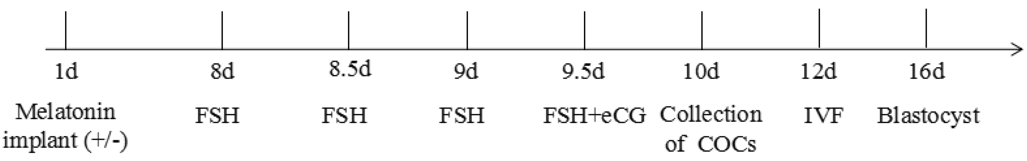
4.2. Donor Superovulation

All lambs were given 125 IU of follicle-stimulating hormone (FSH, Sansheng, Ningbo, China), injected im on two occasions, 24 h apart. In the M group, the first dose of FSH was given 7 days after placement of the melatonin implant. In addition, 250 IU of equine chorionic gonadotrophin (eCG, Sansheng, Ningbo, China) was given im at the time of the last FSH treatment (Appendix, Figure A1. Superovulation scheme).

4.3. Blood Sampling and Hormone Assay

Every day during the superovulation period, blood samples (3 mL) were collected for ten consecutive days (Appendix, Figure A1. Superovulation scheme) by jugular venipuncture using heparinized vacutainers (Beyotime, Jiangsu, China) to detect plasma melatonin concentration. To minimize effects of circadian rhythms, sample collection was consistently done at night (2:0) in resting conditions, always by the same operator. Blood was placed into chilled tubes (Beyotime) containing 0.125 M EDTA and 0.025 M o-phenanthroline, centrifuged (1000× g at 4 °C for 15 min), and plasma immediately extracted and stored at −20 °C. A commercial enzyme-linked immunosorbent assay for melatonin (EIA 1431, sensitivity 0.977 mIU/mL; DRG Diagnostics, Marburg, Germany), was done according to manufacturer's instructions. Intra- and inter-assay coefficients of variations were 4.0 and 13.7%, respectively.

4.4. The Cumulus Oocyte Complexes Collection

At 14 h after eCG, COCs were collected by aspiration. Briefly, lambs were anaesthetized with acepromazine maleate (0.05 mg/kg body weight) and sodium pentothal (10 mg/kg body weight), the abdomen opened on the left or right lower 5–7 cm of the breast with 3–4 cm incision. Ovaries were exposed and COCs were aspirated from visible follicles (2–5 mm in diameter) using a 10-mL syringe equipped with an 18 gauge needle and containing HEPES-buffered DMEM/F-12. Following aspiration, the ovaries and tips of the uterine horns were washed extensively with saline to minimize adhesions. The aspirated fluid was examined under a phase-contrast microscope (Olympus BX60, Olympus, Japan) and only COCs with homogeneous granular cytoplasm and at least three or four layers of compact cumulus cells were selected. The COCs from nine sheep of each group were collected together, then were randomly selected for following experiments (Appendix, Table A1). Melatonin concentrations in follicular fluids were determined by ELISA (same assay used for plasma).

4.5. Histology Staining

After COCs being collected, eight ovaries were removed from four donors in each group. Samples were placed overnight in 4% buffered formaldehyde (37% formaldehyde, Merck, Darmstadt, Germany). Fixed tissues were embedded in paraffin blocks and the whole ovary was sectioned serially at 4 μm thickness. Three sequential sections were put on each slide. Every second or third slide was stained with hematoxylin and eosin.

4.6. In Vitro Maturation (IVM)

The maturation medium contained 20% (v/v) heat-inactivated estrous sheep serum, 10 μg/mL FSH, 10 μg/mL luteinizing hormone, 10 ng/mL epidermal growth factor, and 1 μg/mL estradiol-17β in TCM199 medium. Each drop contained 50 μL in vitro maturation medium that was equilibrated in a CO2 incubator (Thermo, Waltham, MA, USA) for 2 h before the COCs were placed in the medium. Each group of 15 oocytes was cultured in a 50 μL drop of maturation medium in humidified air with 5% CO2 at 39 °C for 24 h. The degree of cumulus cells expansion was subjectively assessed under a phase-contrast microscope (Olympus BX60, Tokyo, Japan) after 22 h of IVM; COCs were classified as not expanded, partially expanded (outer layer of cells was loosened), or fully expanded (all cumulus cells were loosened), as described [61].

4.7. In Vitro Fertilization (IVF) and In Vitro Culture

The COCs were incubated with 0.1% hyaluronidase to dissociate cumulus cells after IVM, then the oocytes were fertilized with the same ram fresh sperm in synthetic oviduct fluid medium containing 20% (v/v) estrous sheep serum and 10 μg/mL heparin (in IVF medium) in the incubator. The IVF drops were prepared and equilibrated in an incubator for 2 h before insemination. The volume of each drop was 40 μL. Groups of up to five oocytes were transferred into the IVF drops. Sperm concentration was calculated using a hemocytometer (SDM1, Minitube, Diefenbach, Germany) and diluted to 1 × 10⁶ cells/mL with IVF medium. Subsequently, there was 10 μL of the sperm suspension added to the 40 μL IVF drops. The gametes were co-incubated at 39 °C in a 5% CO2 humidified air atmosphere for 22 h.
At approximately 22 h following the addition of semen to the culture medium, presumptive zygotes were washed in synthetic oviductal fluid (SOF) medium to remove sperm before being transferred to 50 μL culture droplets of SOF supplemented with 1% (v/v) Basal Medium Eagle-essential amino acids, 1% (v/v) Modified Eagle Medium-nonessential amino acids, 1 mM glutamine, and 6 mg/mL fatty acid-free BSA under mineral oil. The contents of the dishes were incubated at 39 °C in a 5% CO2, 5% O2, 90% N2 humidified atmosphere. Cleavage and hatching rates were recorded at 48 h and 7 d post-IVF, respectively.

4.8. Cumulus Cell Apoptosis

A portion of COCs were washed two or three times in HEPES-buffered DMEM/F-12, cumulus cells dissociated by incubation with 0.1% hyaluronidase for 5 min and continuous pipetting to isolate cumulus cells from oocytes. Thereafter, the cumulus cells suspension was centrifuged at 1000× g for 5 min and the supernatant decanted. This was repeated two or three times, using phosphate-buffered saline (PBS) to wash the cellular pellet.
Apoptosis was assessed using a commercial kit (FITC Annexin V Apoptosis Detection Kit I; Becton Dickinson, Sunnyvale, CA, USA), in accordance with manufacturer’s instructions. Cells were washed with PBS and centrifuged three times at 800× g for 5 min. Then, 100 µL of PBS was added into each centrifuge tube, followed by addition of 5 µL of FITC solution and 5 μL of propidium iodide (20 μg/mL). Cells were incubated at room temperature in the dark for 15 min. Samples were assayed within 2 h using a flow cytometer (Becton Dickinson). Data for 20,000 cells per sample were stored in the list mode using FACS Analyzer flow cytometry software 3.0 (Becton Dickinson); thereafter, these data were passed through a Hewlett Packard Consort 30 system (Palo Alto, CA, USA) and analyzed by SuperCyt Analyst 3 software (Sierra Cytometry, Reno, NV, USA). Apoptotic and dead cells were distinguished by staining with propidium iodide and FITC. All experiments were performed in biological triplicates, and data were representative of at least three independent experiments.

4.9. Global Methylation Analysis

After cumulus cells were isolated, the suspension was placed in a cell culture plate, DMEM/F-12 (penicillin & streptomycin (Hyclone, Logan, Utah, USA) and 10% fetal calf serum (BI, Tel Aviv, Israel)) was added to the plate, and the plate incubated at 38.5 °C with 5% CO2 in humidified air. Cumulus cells used for immunocytochemical staining were permeabilized with 1% Triton X-100 (Beyotime) in phosphate-buffered saline (PBS) for 30 min and then treated in 2 M HCl (Beyotime) for 30 min at 25 °C. Non-specific binding was inhibited with 0.1% BSA (Beyotime) for 30 min at room temperature, and then cells were incubated with anti-5meC antibodies (1:500; Epigentek Group Inc., Farmingdale, NY, USA) at 4 °C overnight. Cumulus cells were extensively washed and then probed with fluorescein isothiocyanate-conjugated anti-mouse IgG (1:100; Santa Cruz Biotechnology, Inc., Dallas, TX, USA) for 1.5 h at 37 °C. The DNA was visualized by counterstaining with 10 μg/mL propidium iodide for 10 min. After extensive washing, cumulus cells were incubated in PBS containing 10% triethylenediamine. Fluorescence was detected with an Olympus BX40 spectral confocal scanning microscope at excitation wavelengths of 488 and 543 nm. System settings were held constant for all examinations. Fluorescence intensity was quantified using FV10-ASW 3.0 Free Viewer software (Olympus). The mean fluorescence pixel value was measured from at least 100 cells per plate and five plates per sample.

4.10. RNA Purification and qRT-PCR

Total RNA was extracted using TRIzol reagent (Invitrogen, Carlsbad, CA, USA), and RNase-free DNase was used to remove genomic DNA. Integrity and concentration of RNA were determined by measuring absorbance at 260 nm. Total RNA (1.0 µg) from each sample was re-suspended in a 20 µL final volume of reaction buffer, containing 25 mM Tris-HCl, 37.5 mM KCl, 10 mM dithiothreitol, 1.5 mM MgCl2, 10 mM of each dNTP, and 0.5 mg oligo (dT)15 primers to synthesize the cDNA. After the reaction mixture reached 42 °C, 20 units of reverse transcriptase was added to each tube, and the sample incubated for 1 h at 42 °C. Reverse transcription was stopped by denaturing the enzyme at 95 °C. The final PCR mixture contained 2.5 µL cDNA, 1× PCR buffer, 1.5 mM MgCl2, 200 µM dNTP mixture, 1 U of Taq DNA polymerase, 1 µM sense and antisense primers, and 5.0 µL sterile water. The qRT-PCR was conducted using the CFX96TM Real-Time PCR Detection System (Bio-Rad, Hercules, CA, USA) under standard conditions. Transcripts were quantified in triplicate for each sample, and β-actin was used as a reference. Expression levels were calculated using the comparative Ct (2−ddCt) method [62]. Primers used are listed in Table 3.

4.11. DNA Methylation Sequence

An aliquot (1 µm) of DNA was treated with bisulfite using an EpiTect Bisulfite Kit (Qiagen, Hilden, Germany). Then, DNA methylation of CpG sites (Figure 7) in the promoter region of the DNMT1, DNMT3a, DNMT3b gene were analyzed using EpiTYPER (MassARRAY system; Agena Biosciences, Santiago, CA, USA) according to the manufacturer’s instructions. DNMT1 forward (aggaagagagTGTAAGGTAAGGGTTTAATTTTATTTTT) and reverse (cagtaatacgactcact atagggagaaggctCCAACCTCAATTTCCTCATCTATAA) primers corresponding to product 394 size, coverage 9/11 CpG, DNMT3a forward (aggaagagagTTAGAGGGTGTTTTGGAAAGGGTAA) and reverse (cagtaatacgactcactatagggagaaggctAACAAAAACAAATATTTCCTATATACACC) primers corresponding to product 392 size, coverage 7/7 CpG, DNMT3b forward (aggaagagagGTTGTTATGGAGAGGAGAGAAGTTG) and reverse (cagtaatacgactcactatagggaga aggctACCAACACCCAAAACAAAAAAA) primers corresponding to product 298 size, coverage 8/8 CpG, were designed using EpiDesigner (Agena Bioscience), and spectrum characteristics were validated with RSeqMeth (Figure 7). Cycling conditions were: denaturation (94 °C for 4 min) then 45 cycles of amplification (94 °C for 20 s, 56 °C for 30 s, and 72 °C for 1 min) and a final extension step at 72 °C for 3 min. Samples were electrophoresed using 2% (w/v) agarose gel to confirm amplification. The CpG sites were unambiguously interrogated, and their genomic locations detailed (Figure 7). Mass spectra methylation ratios were generated using EpiTYPER v1.2 (Agena Biosciences). Finally, reliability of the methylation assay was confirmed using Epitect unmethylated (0%) and methylated (100%) DNA samples (Qiagen) as positive controls. For each participant, average DNA methylation values were calculated by averaging across a total of CpG cites. All experiments were performed in biological triplicates, and data were representative of three independent experiments.

4.12. Statistical Analyses

All data (except those in Table 2 and maturation rate, cleavage rate, blastocyst rate in Table 1) were presented as mean ± SEM and analyzed with one-way ANOVA, followed by Duncan’s test, using SPSS 18.0 statistical software (SPSS Inc., Chicago, IL, USA). Values in Table 2 and maturation rate, cleavage rate, blastocyst rate in Table 1 were analyzed using a Chi-square test. For all analyses, p < 0.05 was considered significant. Data are expressed as mean ± SEM.

Acknowledgments

This study was financially supported by the Tianjin Science and Technology Project (Grant No. 17ZXZYNC00040), the Youth Innovation Promotion Association of the Chinese Academy of Sciences (2015184).

Author Contributions

Conceived and designed the experiments: Yi Fang and Xiaosheng Zhang. Performed the experiments: Yi Fang, Shoulong Deng, Jinlong Zhang, Haijun Liu, Yihai Li and Xiaosheng Zhang. Analyzed the data: Yi Fang and Xiaosheng Zhang. Contributed reagents/materials/analysis tools: Jinlong Zhang and Xiaosheng Zhang. Wrote the paper: Yi Fang and Yixun Liu.

Conflicts of Interest

The authors declare no conflict of interest.

Appendix

Table A1. Number of COCs used per group for different tests (n = 9).
GroupsSum No. COCsNo. COCs Examined
ExpansionApoptosisRT-PCRMethylation
C4972166981131
M7773669288231
Table A2. Mean ± SEM effect of melatonin on methylation sequence of DNMT1.
GroupCpG Position (%)Sum (%)
405474163:169179185193196264335
C100 ± 5.55 a84.2 ± 9.1388.5 ± 8.0277.1 ± 5.7380.2 ± 9.09 aNANANA35.4 ± 4.2435.1 ± 3.5170.6 ± 8.71
M79.7 ± 9.01 b96.6 ± 11.681.9 ± 6.3380.6 ± 9.1162.4 ± 5.83 bNANANA35.9 ± 2.8935.6 ± 3.7767.6 ± 7.49
The experiment was repeated three times; data presented as mean ± SEM; a,b within a column, means without a common superscript differed (p < 0.05).
Table A3. Mean ± SEM effect of melatonin on methylation sequence of DNMT3a.
GroupCpG Position (%)Sum (%)
42127189:191237:240260
C47.5 ± 4.02 b44.3 ± 5.05 bNA85.4 ± 8.0877.4 ± 6.4863.3 ± 7.34
M64.1 ± 7.17 a53.2 ± 4.13 aNA81.9 ± 6.9179.7 ± 7.0569.3 ± 5.77
The experiment was repeated three times; data presented as mean ± SEM; a,b within a column, means without a common superscript differed (p < 0.05).
Table A4. Mean ± SEM effect of melatonin on methylation sequence of DNMT3b.
GroupCpG Position (%)Sum (%)
56138168:170198210239275
C36.1 ± 4.55 a15.4 ± 2.1392.9 ± 8.81 a66.6 ± 5.3737.2 ± 4.3350.6 ± 4.22 a31.4 ± 3.25 a46.7 ± 5.79 a
M27.6 ± 3.01 b14.7 ± 1.6468.4 ± 5.19 b51.4 ± 4.4234.8 ± 3.4738.7 ± 4.09 b20.6 ± 1.68 b36.0 ± 4.82 b
The experiment was repeated three times; data presented as mean ± SEM; a,b within a column, means without a common superscript differed (p < 0.05).
Figure A1. Superovulation scheme (n = 9).
Molecules 23 00494 g0a1

References

  1. Zhang, X.S.; Zhan, J.L.; Cui, M.S.; Li, Y.H.; Feng, X.Z. The study on the effect factors of superovulation on lamb. Chin. Herbiv. Sci. 2013, 33, 67–70. (In Chinese) [Google Scholar]
  2. Kelly, J.M.; Kleemann, D.O.; Walker, S.K. Enhanced efficiency in the production of offspring from 4- to 8-week-old lambs. Theriogenology 2005, 63, 1876–1890. [Google Scholar] [CrossRef] [PubMed]
  3. Kochhar, H.P.; Wu, B.; Morris, L.; Buckrell, B.C.; Pollard, J.W. Maturation status, protein synthesis and developmental competence of oocytes derived from lambs and ewes. Reprod. Domest. Anim. 2002, 37, 19–25. [Google Scholar] [CrossRef] [PubMed]
  4. O’Brien, J.K.; Dwarte, D.; Ryan, J.P.; Maxwell, W.M.; Evans, G. Developmental capacity, energy metabolism and ultrastructure of mature oocytes from prepubertal and adult sheep. Reprod. Fertil. Dev. 1996, 8, 1029–1037. [Google Scholar] [CrossRef] [PubMed]
  5. Zhou, C.J.; Wu, S.N.; Shen, J.P.; Wang, D.H.; Kong, X.W. The beneficial effects of cumulus cells and oocyte-cumulus cell gap junctions depends on oocyte maturation and fertilization methods in mice. PeerJ 2016, 4, 1761. [Google Scholar] [CrossRef] [PubMed]
  6. Ptak, P.L.; Dattena, M.; Tischner, M.; Cappai, P. Offspring from one-month-old lambs: studies on the developmental capability of prepubertal oocytes. Biol. Reprod. 1999, 61, 1568–1574. [Google Scholar] [CrossRef] [PubMed]
  7. Haenisch-Woehl, A.; Kölle, S.; Neumüller, C.; Sinowatz, F.; Braun, J. Morphology of canine cumulus-oocyte complexes in pre-pubertal bitches. Anat. Histol. Embryol. 2003, 32, 373–377. [Google Scholar] [CrossRef] [PubMed]
  8. Anguita, B.; Paramio, M.T.; Morató, R.; Romaguera, R.; Jiménez-Macedo, A.R. Effect of the apoptosis rate observed in oocytes and cumulus cells on embryo development in prepubertal goats. Anim. Reprod. Sci. 2009, 116, 95–106. [Google Scholar] [CrossRef] [PubMed]
  9. Fang, Y.; Zhang, X.S.; Zhang, J.L.; Zhong, R.Z.; Zhou, D.W. Global DNA methylation and related mRNA profiles in sheep oocytes and early embryos derived from pre-pubertal and adult donors. Anim. Reprod. Sci. 2016, 164, 144–151. [Google Scholar] [CrossRef] [PubMed]
  10. Diederich, M.; Hansmann, T.; Heinzmann, J.; Bargkues, B.; Herrmann, D. DNA methylation and mRNA expression profiles in bovine oocytes derived from prepubertal and adult donors. Reproduction 2012, 144, 319–330. [Google Scholar] [CrossRef] [PubMed]
  11. Ptak, G.; Matsukawa, K.; Palmieri, C.; Salda, L.D.; Scapolo, P.A.; Loi, P. Developmental and functional evidence of nuclear immaturity in prepubertal oocytes. Hum. Reprod. 2006, 21, 2228–2237. [Google Scholar] [CrossRef] [PubMed]
  12. Biian, E.L.; Karsch, F.J.; Hopkins, J.W. Role of the pineal gland in ovine photoperiodism:regulation of seasonal breeding and negative feedback effects of estradiol upon luteinizinghormone secretion. Endocrinology 1983, 113, 329–336. [Google Scholar]
  13. Claypool, L.E.; Wood, R.I.; Yellon, S.M.; Foster, D.L. The ontogeny of melatonin secretion in the lamb. Endocrinology 1989, 124, 2135–2143. [Google Scholar] [CrossRef] [PubMed]
  14. Korkmaz, A.; Topal, T.; Oter, S.; Tan, D.X.; Reiter, R.J. Hyperglycemia-related pathophysiologic mechanisms and potential beneficial actions of melatonin. Mini Rev. Med. Chem. 2008, 8, 1144–1153. [Google Scholar] [CrossRef] [PubMed]
  15. Korkmaz, A.; Rosales-Corral, S.; Reiter, R.J. Gene regulation by melatonin linked to epigenetic phenomena. Gene 2012, 503, 1–11. [Google Scholar] [CrossRef] [PubMed]
  16. Menendez-Pelaez, A.; Poeggeler, B.; Reiter, R.J.; Barlow, W.L.; Pablos, M.I.; Tan, D.X. Nuclear localization ofmelatonin in different mammalian tissues: Immunocytochemical and radioimmunoassay evidence. J. Cell. Biochem. 1993, 53, 373–382. [Google Scholar] [CrossRef] [PubMed]
  17. Tomas-Zapico, C.; Coto-Montes, A. A proposed mechanismto explain the stimulatory effect of melatonin on antioxidativeenzymes. J. Pineal Res. 2005, 39, 99–104. [Google Scholar] [CrossRef] [PubMed]
  18. Karasek, M.; Pawlikowski, M. Antiproliferative effects of melatonin and CGP 52608. Biol. Signals Recept. 1999, 8, 75–78. [Google Scholar] [CrossRef] [PubMed]
  19. Yie, S.M.; Niles, L.P.; Younglai, E.V. Melatonin receptors on human granulosa cell membranes. J. Clin. Endocrinol. Metab. 1995, 80, 1747–1749. [Google Scholar] [PubMed]
  20. Niles, L.P.; Wang, J.; Shen, L.; Lobb, D.K.; Younglai, E.V. Melatonin receptor mRNA expression in human granulosa cells. Mol. Cell. Endocrinol. 1999, 156, 107–110. [Google Scholar] [CrossRef]
  21. Kang, J.T.; Koo, O.J.; Kwon, D.K.; Park, H.J.; Jang, G.; Kang, S.K.; Lee, B.C. Effects of melatonin on in vitro maturation of porcine oocyte and expression of melatonin receptor RNA in cumulus and granulosa cells. J. Pineal Res. 2009, 46, 22–28. [Google Scholar] [CrossRef] [PubMed]
  22. Peyrot, F.; Ducrocq, C. Potential role of tryptophan derivatives in stress responses characterized by the generation of reactive oxygen and nitrogen species. J. Pineal Res. 2008, 45, 235–246. [Google Scholar] [CrossRef] [PubMed]
  23. Carcangiu, V.; Vacca, G.M.; Mura, M.C.; Dettori, M.L.; Pazzola, M.; Luridiana, S.; Bini, P.P. Relationship between MTNR1A melatonin receptor gene polymorphism and seasonal reproduction in different goat breeds. Anim. Reprod. Sci. 2009, 110, 71–78. [Google Scholar] [CrossRef] [PubMed]
  24. El-Raey, M.; Geshi, M.; Somfai, T.; Kaneda, M.; Hirako, M.; Abdel-Ghaffar, A.E.; Sosa, G.A.; El-Roos, M.E.; Nagai, T. Evidence of melatonin synthesis in the cumulus oocyte complexes and its role in enhancing oocyte maturation in vitro in cattle. Mol. Reprod. Dev. 2011, 78, 250–262. [Google Scholar] [CrossRef] [PubMed]
  25. Weaver, D.R.; Liu, C.; Reppert, S.M. Nature’s knockout: the Mel1b receptor is not necessary for reproductive and circadian responses to melatonin in Siberian hamsters. Mol. Endocrinol. 1996, 10, 1478–1487. [Google Scholar] [PubMed]
  26. Takada, L.; Junior, A.M.; Mingoti, G.Z.; Balieiro, J.C.; Cipolla-Neto, J.; Coelho, L.A. Effect of melatonin on DNA damage of bovine cumulus cells during in vitro maturation (IVM) and on in vitro embryo development. Res. Vet. Sci. 2012, 92, 124–127. [Google Scholar] [CrossRef] [PubMed]
  27. Brzezinski, A.; Seibel, M.M.; Lynch, H.J.; Deng, M.H.; Wurtman, R.J. Melatonin in human preovulatory follicular fluid. J. Clin. Endocrinol. Metab. 1987, 64, 865–867. [Google Scholar] [CrossRef] [PubMed]
  28. Shi, J.M.; Tian, X.Z.; Zhou, G.B.; Wang, L.; Gao, C.; Zhu, S.E.; Zeng, S.M.; Tian, J.H.; Liu, G.S. Melatonin exists in porcine follicular fluid and improves in vitro maturation and parthenogenetic development of porcine oocytes. J. Pineal Res. 2009, 47, 318–323. [Google Scholar] [CrossRef] [PubMed]
  29. Rodriguez-Osorio, N.; Kim, I.J.; Wang, H.; Kaya, A.; Memili, E. Melatonin increases cleavage rate of porcine preimplantation embryos in vitro. J. Pineal Res. 2007, 43, 283–288. [Google Scholar] [CrossRef] [PubMed]
  30. Zhao, X.M.; Hao, H.S.; Du, W.H.; Zhao, S.J.; Wang, H.Y.; Wang, N.; Wang, D.; Liu, Y.; Qin, T.; Zhu, H.B. Melatonin inhibits apoptosis and improves the developmental potential of vitrified bovine oocytes. J. Pineal Res. 2016, 60, 132–141. [Google Scholar] [CrossRef] [PubMed]
  31. Pang, Y.W.; Sun, Y.Q.; Jiang, X.L.; Huang, Z.Q.; Zhao, S.J.; Du, W.H.; Hao, H.S.; Zhao, X.M.; Zhu, H.B. Protective effects of melatonin on bovine sperm characteristics and subsequent in vitro embryo development. Mol. Reprod. Dev. 2016, 83, 993–1002. [Google Scholar] [CrossRef] [PubMed]
  32. Coelho, L.A.; Peres, R.; Amaral, F.G.; Reiter, R.J.; Cipolla-Neto, J. Daily differential expression of melatonin-related genes and clock genes in rat cumulus-oocyte complex: Changes after pinealectomy. J. Pineal Res. 2015, 58, 490–499. [Google Scholar] [CrossRef] [PubMed]
  33. Tamura, H.; Takasaki, A.; Miwa, I.; Taniguchi, K.; Maekawa, R.; Asada, H.; Taketani, T.; Matsuoka, A.; Yamagata, Y.; Shimamura, K.; et al. Oxidative stress impairs oocyte quality and melatonin protects oocytes from free radical damage and improves fertilization rate. J. Pineal Res. 2008, 44, 280–287. [Google Scholar] [CrossRef] [PubMed]
  34. Nakamura, Y.; Tamura, H.; Takayama, H.; Kato, H. Increased endogenous level of melatonin in preovulatory human follicles does not directly influence progesterone production. Fertil. Steril. 2003, 80, 1012–1016. [Google Scholar] [CrossRef]
  35. Tamura, H.; Nakamura, Y.; Korkmaz, A.; Manchester, L.C.; Tan, D.X.; Sugino, N.; Reiter, R.J. Melatonin and the ovary: Physiological and pathophysiological implications. Fertil. Steril. 2009, 92, 328–343. [Google Scholar] [CrossRef] [PubMed]
  36. Reiter, R.J.; Tan, D.X.; Manchester, L.C.; Paredes, S.D.; Mayo, J.C.; Sainz, R.M. Melatonin and reproduction revisited. Biol. Reprod. 2009, 81, 445–456. [Google Scholar] [CrossRef] [PubMed]
  37. Woo, M.M.; Tai, C.J.; Kang, S.K.; Nathwani, P.S.; Pang, S.F.; Leung, P.C. Direct action of melatonin in human granulosa-luteal cells. J. Clin. Endocrinol. Metab. 2001, 86, 4789–4797. [Google Scholar] [CrossRef] [PubMed]
  38. Picinato, M.C.; Hirata, A.E.; Cipolla-Neto, J.; Curi, R.; Carvalho, C.R.; Anhe, G.F.; Carpinelli, A.R. Activation of insulin and IGF-1 signaling pathways by melatonin through MT1 receptor in isolated rat pancreatic islets. J. Pineal Res. 2008, 44, 88–94. [Google Scholar] [CrossRef] [PubMed]
  39. PTsiligianni, T.; Valasi, I.; Cseh, S.; Vainas, E.; Faigl, V. Effects of melatonin treatment on follicular development and oocyte quality in Chios ewes-short communication. Acta Vet. Hung. 2009, 57, 331–335. [Google Scholar] [CrossRef] [PubMed]
  40. Haghighat, N.; Van Winkle, L.J. Developmental change in follicular cell-enhanced amino acid uptake into mouse oocytes that depends on intact gap junctions and transport system Gly. J. Exp. Zool. 1990, 253, 71–82. [Google Scholar] [CrossRef] [PubMed]
  41. Buccione, R.; Schroeder, A.C.; Eppig, J.J. Interactions between somatic cells and germ cells throughout mammalian oogenesis. Biol. Reprod. 1990, 43, 543–547. [Google Scholar] [CrossRef] [PubMed]
  42. Thibault, C.; Szollosi, D.; Gerard, M. Mammalian oocyte maturation. Reprod. Nutr. Dev. 1987, 27, 865–896. [Google Scholar] [CrossRef] [PubMed]
  43. Zuelke, K.A.; Brackett, B.G. Luteinizing hormone-enhanced in vitro maturation of bovine oocytes with and without protein supplementation. Biol. Reprod. 1990, 43, 784–787. [Google Scholar] [CrossRef] [PubMed]
  44. Gutnisky, C.; Dalvit, G.C.; Pintos, L.N.; Thompson, J.G.; Beconi, M.T.; Cetica, P.D. Influence of hyaluronic acid synthesis and cumulus mucification on bovine oocyte in vitro maturation, fertilisation and embryo development. Reprod. Fertil. Dev. 2007, 19, 488–497. [Google Scholar] [CrossRef] [PubMed]
  45. Choi, J.; Park, S.M.; Lee, E.; Kim, J.H.; Jeong, Y.I.; Lee, J.Y.; Park, S.W.; Kim, H.S.; Hossein, M.S.; Jeong, Y.W.; et al. Anti-apoptotic effect of melatonin on preimplantation development of porcine parthenogenetic embryos. Mol. Reprod. Dev. 2008, 75, 1127–1135. [Google Scholar] [CrossRef] [PubMed]
  46. Joo, S.S.; Yoo, Y.M. Melatonin induces apoptotic death in LNCaP cells via p38 and JNK pathways: Therapeutic implications for prostate cancer. J. Pineal Res. 2009, 47, 8–14. [Google Scholar] [CrossRef] [PubMed]
  47. Leja-Szpak, A.; Jaworek, J.; Pierzchalski, P.; Reiter, R.J. Melatonin induces pro-apoptotic signaling pathway in human pancreatic carcinoma cells (PANC-1). J. Pineal Res. 2010, 49, 248–255. [Google Scholar] [CrossRef] [PubMed]
  48. Jung, K.H.; Hong, S.W.; Zheng, H.M.; Lee, H.S.; Lee, H.; Lee, D.H.; Lee, S.Y.; Hong, S.S. Melatonin ameliorates cerulein-induced pancreatitis by the modulation of nuclear erythroid 2-related factor 2 and nuclear factor-kappaB in rats. J. Pineal Res. 2010, 48, 239–250. [Google Scholar] [CrossRef] [PubMed]
  49. Wong, A.C.; Ryan, A.F. Mechanisms of sensorineural celldamage, death and survival in the Cochlea. Front. Aging Neurosci. 2015, 7, 58. [Google Scholar] [CrossRef] [PubMed]
  50. Juknat, A.A.; Mendez, M.V.; Quaglino, A.; Fameli, C.I.; Mena, M.; Kotler, M.L. Melatonin prevents hydrogen peroxide-induced Bax expression in cultured rat astrocytes. J. Pineal Res. 2005, 38, 84–92. [Google Scholar] [CrossRef] [PubMed]
  51. Maity, P.; Bindu, S.; Dey, S.; Goyal, M.; Alam, A.; Pal, C.; Reiter, R.; Bandyopadhyay, U. Melatonin reduces indomethacin-induced gastric mucosal cell apoptosis by preventing mitochondrial oxidative stress and the activation of mitochondrial pathway of apoptosis. J. Pineal Res. 2009, 46, 314–323. [Google Scholar] [CrossRef] [PubMed]
  52. Tian, X.; Wang, F.; He, C.; Zhang, L.; Tan, D.; Reiter, R.J.; Xu, J.; Ji, P.; Liu, G. Beneficial effects of melatonin on bovine oocytes maturation: a mechanistic approach. J. Pineal Res. 2014, 57, 239–247. [Google Scholar] [CrossRef] [PubMed]
  53. Talpur, H.S.; Worku, T.; Rehman, Z.U.; Dad, R.; Bhattarai, D.; Bano, I.; Farmanullah; Liang, A.; He, C.; Yang, L. Knockdown of melatonin receptor 1 and induction of follicle-stimulating hormone on the regulation of mouse granulosa cell function. Reprod. Biol. 2017, 17, 380–388. [Google Scholar] [CrossRef] [PubMed]
  54. Sharma, R.; Ottenhof, T.; Rzeczkowska, P.A.; Niles, L.P. Epigenetic targets for melatonin: Induction of histone H3 hyperacetylation and gene expression in C17.2 neural stem cells. J. Pineal Res. 2008, 45, 277–284. [Google Scholar] [CrossRef] [PubMed]
  55. Deng, W.G.; Tang, S.T.; Tseng, H.P.; Wu, K.K. Melatonin suppresses macrophage cyclooxygenase-2 and inducible nitric oxide synthase expression by inhibiting p52 acetylation and binding. Blood 2006, 108, 518–524. [Google Scholar] [CrossRef] [PubMed]
  56. Tain, Y.L.; Huang, L.T.; Chan, J.Y. Transcriptional regulation of programmed hypertension by melatonin: An epigenetic perspective. Int. J. Mol. Sci. 2014, 15, 18484–18495. [Google Scholar] [CrossRef] [PubMed]
  57. Adcock, I.M.; Cosio, B.; Tsaprouni, L.; Barnes, P.J.; Ito, K. Redox regulation of histone deacetylases and glucocorticoid-mediated inhibition of the inflammatory response. Antioxid. Redox Signal. 2005, 7, 144–152. [Google Scholar] [CrossRef] [PubMed]
  58. Glass, C.K.; Ogawa, S. Combinatorial roles of nuclear receptors in inflammation and immunity. Nat. Rev. Immunol. 2006, 6, 44–55. [Google Scholar] [CrossRef] [PubMed]
  59. Korkmaz, A.; Reiter, R.J. Epigenetic regulation: A new research area for melatonin? J. Pineal Res. 2008, 44, 41–44. [Google Scholar] [CrossRef] [PubMed]
  60. Crosio, C.; Cermakian, N.; Allis, C.D.; Sassone-Corsi, P. Light induces chromatin modification in cells of the mammalian circadian clock. Nat. Neurosci. 2000, 3, 1241–1247. [Google Scholar] [CrossRef] [PubMed]
  61. Marei, W.F.; Wathes, D.C.; Fouladi-Nashta, A.A. Impact of linoleic acid on bovine oocyte maturation and embryo development. Reproduction 2010, 139, 979–988. [Google Scholar] [CrossRef] [PubMed]
  62. Livak, K.J.; Schmittgen, T.D. Analysis of relative gene expression data using real-time quantitative PCR and the 2(-Delta Delta C(T)) Method. Methods 2001, 25, 402–408. [Google Scholar] [CrossRef] [PubMed]
Sample Availability: Samples of the compounds are not available from the authors.
Figure 1. Melatonin concentrations in plasma and follicular fluid. (n = 9). * p < 0.05 in Figure 1A; a,b in Figure 1B, for columns, means without a common superscript differed (p < 0.05). (A) Melatonin concentrations of plasma, (B) melatonin concentrations of follicular fluid.
Molecules 23 00494 g001
Figure 2. (A) Ovaries after superovulation, (B) HE histological appearance (bar = 500 μm) and (C) expansion of cumulus cell after IVM (×10).
Molecules 23 00494 g002
Figure 3. Effects of melatonin on cumulus cells apoptosis. Flow cytometry (A: C group; B: M group); mean ± SEM cumulus cells apoptosis (C). The experiment was repeated three times; Data presented as mean ± SEM; a,b in Figure 3C, for columns, means without a common superscript differed (p < 0.05).
Molecules 23 00494 g003
Figure 4. Effects of melatonin on expression of related genes in cumulus cells. Expression of related apoptosis genes in cumulus cells (A); expression of melatonin synthetase and receptors genes (B). The experiment was repeated three times; data presented as mean ± SEM; a,b for adjacent columns, means without a common superscript differed (p < 0.05).
Molecules 23 00494 g004
Figure 5. Abundance of 5-methylcytosine (5mC) and gene expression of DNMTs in cumulus cells collected from C and M groups; (A) Representative immunofluorescence images of 5mC (green), and propidium iodide-stained nuclei (red). Scale bars, 20 μm. (B) global 5mC DNA in cumulus cells. (C) relative mRNA levels of DNMTs. The experiments were repeated three times; data presented as mean ± SEM; a,b in Figure 5B,C, for adjacent columns, means without a common superscript differed (p < 0.05).
Molecules 23 00494 g005
Figure 6. Methylation sequencing in cumulus cells; the experiment was repeated three times; loci within a circle differed (p < 0.05).
Molecules 23 00494 g006
Figure 7. Gene structures of detected promoter region of DNMT1, DNMT3a and DNMT3b. Locations of CpG sites in this study indicated in blue were analyzed, whereas those indicated in red were either not uniquely discriminated in the spectra or had low call rates.
Molecules 23 00494 g007
Table 1. Effect of melatonin on lamb superovulation.
GroupsMean ± SEM
No. Follicles ≥ 2 mm (n = 9)
Mean ± SEM
No. Oocytes (n = 9)
IVF
Maturation Rate (%)
(No. Mature/No. Total)
Cleavage Rate (%)
(No. Cleavage/No. Mature)
Blastocyst Rate (%)
(No. Blastocyst/No. Cleavage)
C79.4 ± 9.22 b55.2 ± 4.29 b71.8 (155/216) b52.3(81/155) b7.41(6/81) b
M106 ± 14.9 a86.3 ± 7.77 a79.5 (291/366) a68.4(199/291) a14.1(28/199) a
a,b Within a column, means without a common superscript differed (p < 0.05).
Table 2. Effect of melatonin on cumulus cells expansion.
GroupsNo. COCs ExaminedNo. (%) Oocytes with Cumulus Cells
Not ExpandedPartially ExpandedFully Expanded
C21639 (18.0) a67 (31.0) a110 (51.0) b
M36629 (8.00) b59 (16.1) b278 (76.0) a
a,b Within a column, means without a common superscript differed (p < 0.05).
Table 3. Primer sequences and conditions.
GenePrimer SequenceT annealing (°C) × No. CyclesFragment Size (bp)
β-Actin5′GTCATCACCATCGGCAATGA
3′CGTGAATGCCGCAGGATT
60 × 35159
ASMT5′TCATTTTCCTGAGTGCGTTG
3′CTCCCAGGTTCTCTTTGCTG
58 × 35205
MT15′GGAGGGTGAAACCTGACGAC
3′CCCAGCAAATGGCAAAGAGG
57 × 3599
MT25′GGCTCCGTCTTCAACATCACC
3′GCAGAAGGACCAGCAGGGTG
60 × 35145
P535′GAAGACCTACCCTGGCAATTAC
3′AGAACAGCTTGTTAAGGGAAGG
60 × 35103
Bax5′GGCTGGACATTGGACTTCCTTC
3′TGGTCACTGTCTGCCATGTGG
57 × 35112
Caspase 35′CAGACAGTGGTGCTGAGGATGA
3′GCTACCTTTCGGTTAACCCGA
56 × 35211
Bcl25′GACTGACACTGAGTTTGGCTACG
3′GAGTCCTTTCCACTTCGTCCTG
58 × 35152
DNMT15′CGCATGGGCTACCAGTGCACCTT
3′GGGCTCCCCGTTGTATGAAATCT
58 × 35158
DNMT3a5′CAACGGAGAAGCCTAAGGTCAA
3′TTGAGGCTCCCACAAGAGATG
60 × 35244
DNMT3b5′GACTCATTGGAGGACCAGCTGAAGC
3′CAGCACCTCCAGGCACTCCACACAG
59 × 35130

