Article

Bioactive Compounds from the Stems of Clausena lansium

1
State Key Laboratory of Bioactive Substance and Function of Natural Medicines, Institute of Materia Medica, Chinese Academy of Medical Sciences and Peking Union Medical College, Beijing 100050, China
2
Beijing Research Institute of Chinese Medicine, Beijing University of Chinese Medicine, Beijing 100029, China
*
Author to whom correspondence should be addressed.
Molecules 2017, 22(12), 2226; https://doi.org/10.3390/molecules22122226
Submission received: 9 November 2017 / Revised: 9 December 2017 / Accepted: 11 December 2017 / Published: 14 December 2017
(This article belongs to the Collection Bioactive Compounds)

Abstract

:
In view of the significant neuroprotective effect of Clausena lansium, we continued to separate the n-butanol and the water extracts from the stems of C. lansium in order to find the leading compounds with significant activity. Two new phenolic glycosides, Clausenolside A–B (12), one new pair of phenolic enantiomers (3a, 3b), and two new monoterpenoids, clausenapene A–B (45), together with twelve known analogues (617) were isolated from the stems of C. lansium. Compounds 117 were obtained from C. lansium for the first time. Compounds 3a, 3b, 4, 16, and 17 showed strong or moderate potential neuroprotective effects on inhibited PC12 cell injury induced by okadaic acid, and compound 9 exhibited strong potential hepatoprotective activities. Their structures were elucidated on the basis of spectroscopic analyses, including UV, IR, NMR experiments, and electronic circular dichroism (ECD) spectra.

1. Introduction

Clausena lansium (Lour.) Skeels (syn. Clausena wampi (Blanco) Oliv.; Clausena punctate (Sonn.) Rehd. & Wils.; Cookia punctate Sonn.; Cookia wampi Blanco; Quinaria lansium Lour.) is a minor member of the Rutaceae. It is an attractive shrub or small tree with somewhat grapelike fruit, similar to the citrus fruits and commonly called Wampee, False or Fool’s Curry [1]. It grows in the southern area of mainland China and is cultivated in Taiwan, Fujian, Guangdong, Guangxi, Hainan, etc. It also occurs in Vietnam, the Philippines, Malaysia, Singapore, Miami, etc. [2]. In traditional Chinese medicine, the leaves and roots of C. lansium were used to treat coughs, asthma, dermatological diseases, viral hepatitis, and gastro-intestinal diseases. The fruit were used to treat digestive disorders and the seeds were used to treat acute and chronic gastro-intestinal inflammation, ulcers, and so on [3].
Various bioactive constituents including coumarins, carbazole alkaloids, and amide alkaloids have been isolated and identified from this plant [4,5,6]. Our research group has previously characterized a variety of new carbazole alkaloids, new amide glycosides, new coumarins, and new megastigmane glucoside from the leaves and stems of C. lansium, and several of these compounds showed selective neuroprotective and hepatoprotective effects [7,8,9,10,11,12]. However, the n-BuOH and the water extracts from the stems of C. lansium have not been investigated in detail. Herein, this paper reports on a further investigation of the water and n-BuOH extracts from the stems of C. lansium, which led to the isolation and characterization of two new phenolic glycosides (12), one new pair of phenolic enantiomers (3a and 3b), two new monoterpenoids (45), together with twelve known analogues (617) (Figure 1). They were obtained from C. lansium for the first time. The determination of their absolute configurations occurred through spectroscopic analysis and electronic circular dichroism (ECD) experiments. Moreover, compounds 14 and 617 were assayed for their in vitro hepatoprotective and neuroprotective effects.

2. Results and Discussion

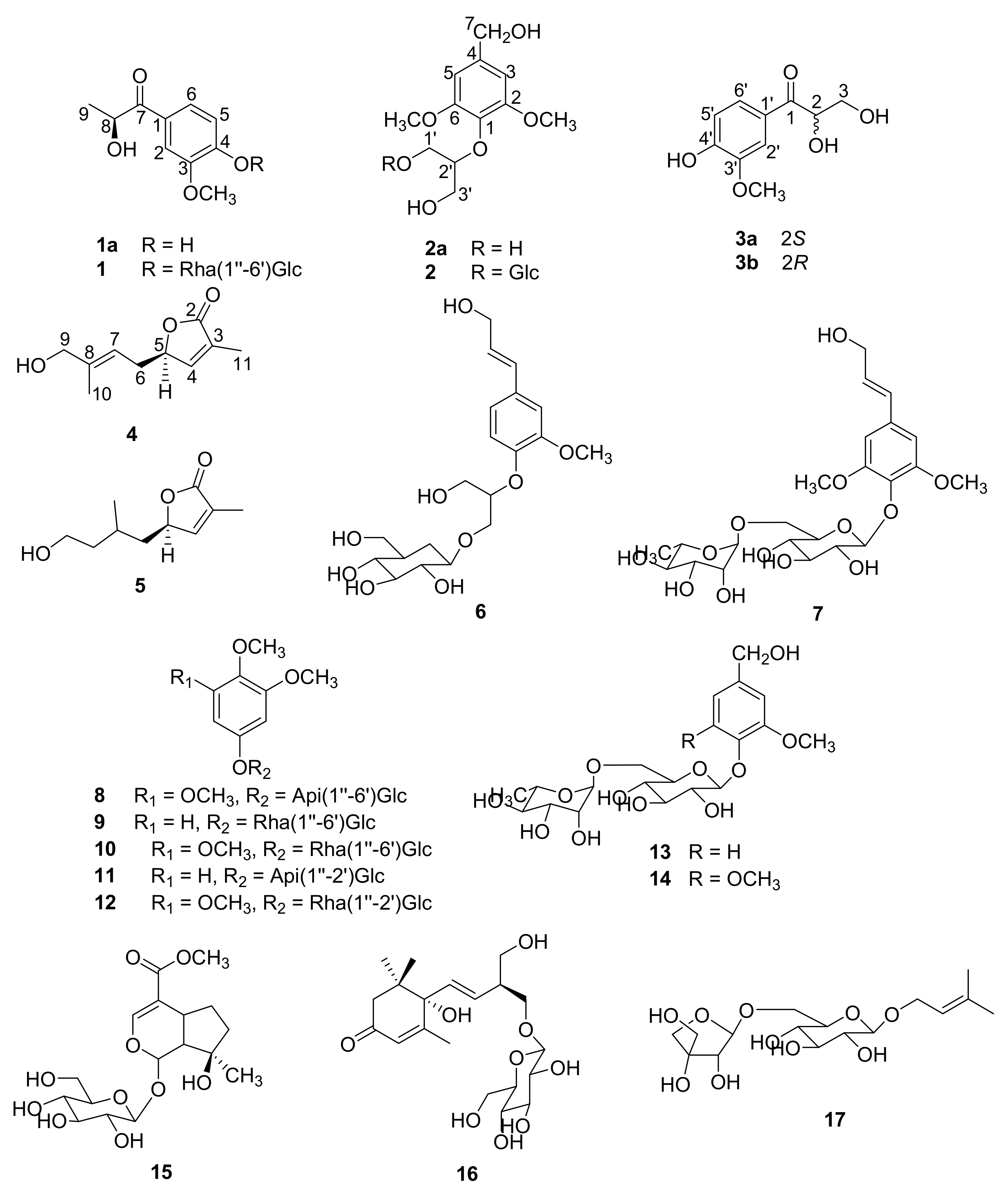
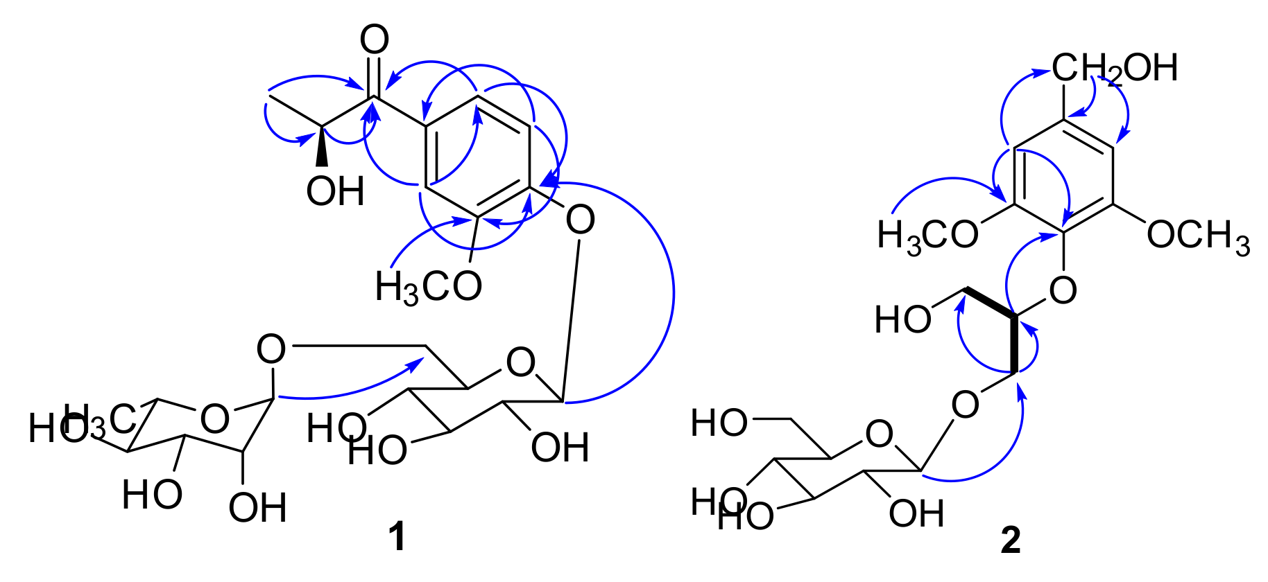
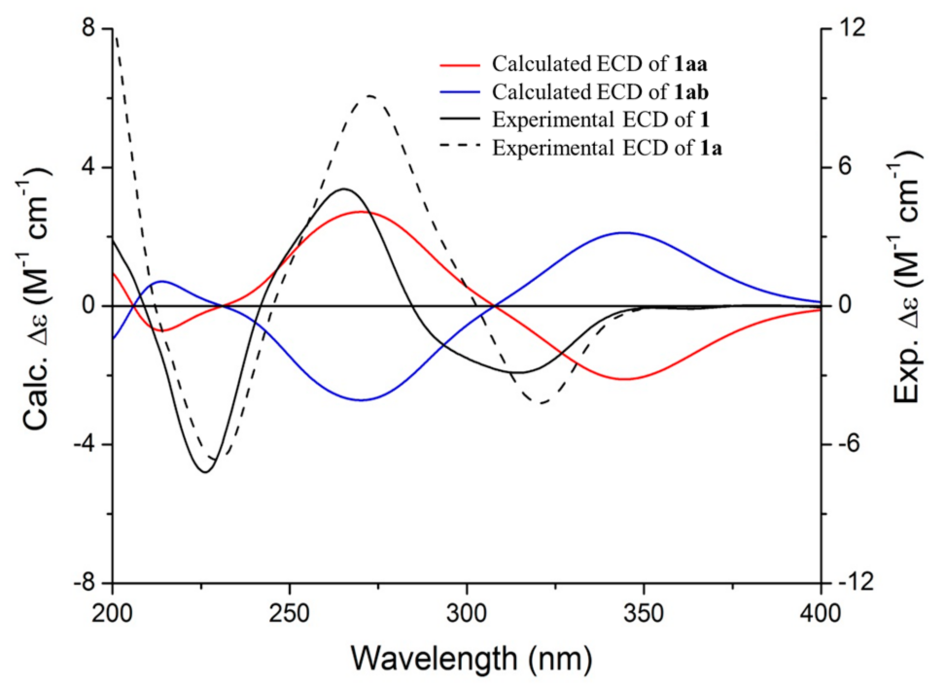
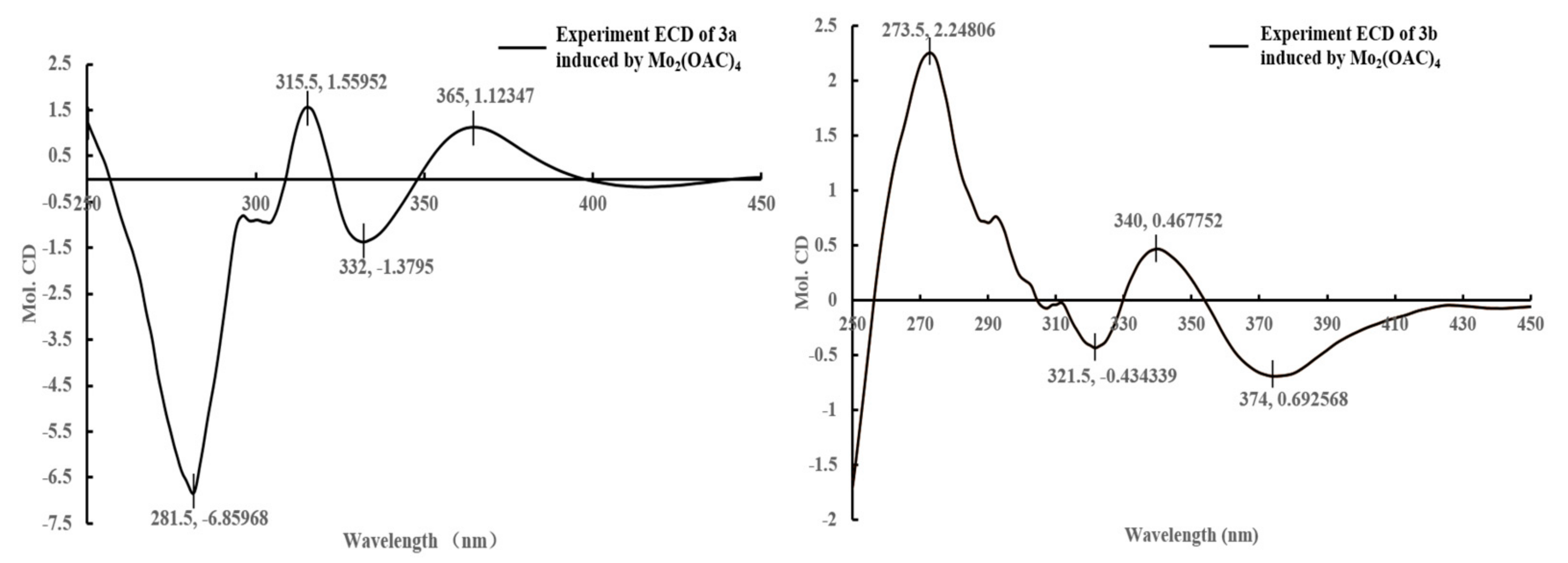
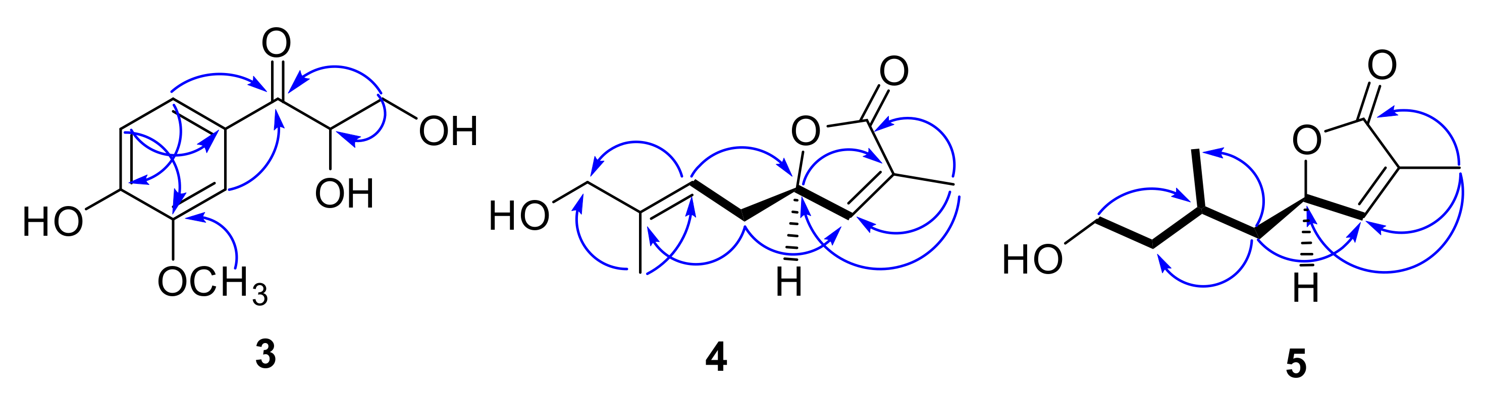
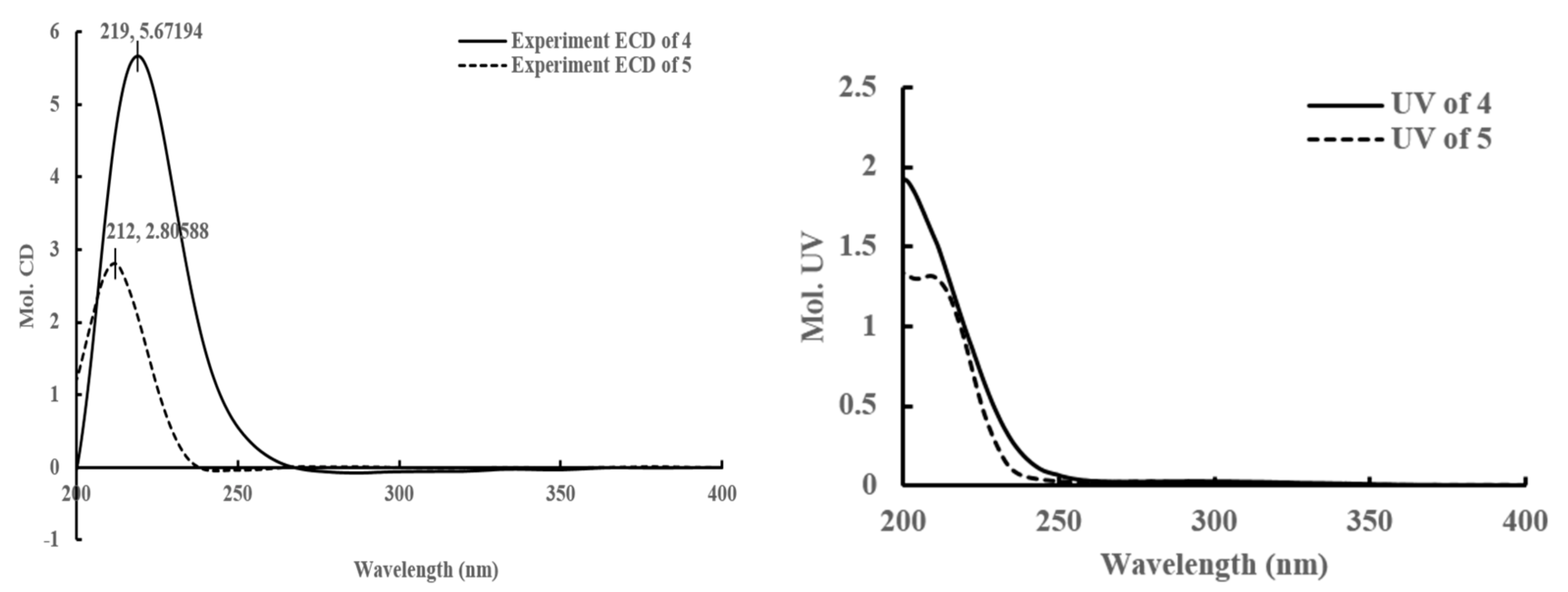
2.1. Purification and Characterization

Clausenolside A (1) was obtained as a white, amorphous solid. Its molecular formula was deduced as C22H32O13 on the basis of its 13C-NMR and HRESIMS at m/z 527.1731 [M + Na]+, calculated as C22H32NaO13, 527.1735, implying seven indices of hydrogen deficiency. The 1H-NMR spectrum (Table 1) revealed three aromatic protons [δH 7.51 (1H, d, J = 2.0 Hz, H-2), 7.62 (1H, dd, J = 8.5, 2.0 Hz, H-6), 7.13 (1H, d, J = 8.5 Hz, H-5)], an oxygenated methine group δH 5.08 (1H, m, H-8), a methoxyl group δH 3.83 (3H, s, 3-OCH3), two methyl groups [δH 1.27 (3H, d, J = 6.7 Hz, H-9), 1.09 (3H, d, J = 6.2 Hz, H-6″)], and a set of protons for two glycosyl moieties, including two anomeric protons [δH 5.01 (1H, d, J = 6.1 Hz, H-1′), 4.52 (1H, br s, H-1″)]. The 13C-NMR and distortionless enhancement by polarization transfer (DEPT) spectra (Figure S3) along with the heteronuclear singular quantum correlation (HSQC) correlations (Figure S4) exhibited the presence of a benzene ring, a keto-carbonyl group, a methoxyl group, an oxygenated methine group, a methyl group, a glucosyl group, and a rhamnosyl group. On the basis of the NMR data analysis (Table 1), compound 1 was identified as a phenolic glycoside. In the heteronuclear multiple bond correlation (HMBC) spectrum (Figure S5), Correlations from H-8 to C-7 and from H-9 to C-7 and C-8 indicated that the oxygenated methine group was attached to C-7 and C-9. The correlations of H-2/C-4 (δC 150.6), C-6 (δC 122.7), C-7 (δC 200.3); H-6/C-7 (δC 200.3), C-4 (δC 150.6); H-5/C-1 (δC 128.5), C-3 (δC 148.7); and 3-OCH3/ C-3 (δC 148.7) demonstrated that the carbonyl was attached to C-1 and the methoxyl group was resonated at C-3. Correlations from H-1′ to C-4 and from H-1″ to C-6′ indicated that the rhamnosyl group was linked with C-6′ and the glucosyl group was linked with C-4 (Figure 2). The aglycone (1a) and sugar moieties were produced by acid hydrolysis of 1. Sugar moieties were confirmed to be d-glucose and l-rhamnose by silylation followed with gas chromatography (GC) analysis. The absolute configuration of 1a was defined as 8S by comparison of the experimental ECD spectra and the calculated ECD data using the time-dependent density functional theory (TDDFT) method at the B3LYP/6-31G (d) level [13]. The calculated ECD spectrum of (8S) 1a (Figure 3) matched the experimental spectrum of 1a and 1 very well, which indicated that the structure of 1a had not changed in the process of acid hydrolysis and the absolute configuration of 1 was elucidated as 8S. Thus, the structure of 1 was assigned as depicted.
Clausenolside B (2) was obtained as an amorphous white powder. Its molecular formula was assigned as C18H28O11 based on the 13C-NMR spectroscopic data and HRESIMS (m/z 443.1532 [M + Na]+, calculated as C18H28NaO11 443.1524), implying five indices of hydrogen deficiency. The NMR spectra (Table 1) of 2 were generally similar to those of compound 6 [14], except that the methoxyl group and the hydroxymethyl group of 2 replaced the hydrogen proton of C-6 and the propen-2-en-1-ol of C-4 replaced 6. In the HMBC spectrum (Figure 2), the correlations from H-3 and H-5 to C-1 (δC 133.9), C-2 (δC 152.7), and C-7 (δC 63.0); from H-7 to C-3 (δC 103.5), C-4 (δC 138.2), and C-5 (δC 103.5) showed that the hydroxymethyl group was linked to C-4; the correlations from H-1′ to C-1 (δC 133.9), C-2′ (δC 67.6); from H-2′ to C-1′ (δC 81.4), C-3′ (δC 60.1); from H-1″ to C-2′ (δC 67.6) indicated that the propanetriol group was linked to C-1 and the β-glucopyranosyl unit was linked to C-2′. The correlations from OCH3 to C-2 (δC 152.7), C-6 (δC 152.7) demonstrated that the methoxyl group had attached to C-2 and C-6. The aglycone (2a) and sugar moiety were produced by an acid hydrolysis of 2. Sugar moiety was confirmed to be d-glucose by silylation followed with GC analysis. Hence, the structure of 2 was assigned as shown.
Compound 3 (3a/3b) was obtained as a white powder. Its molecular formula C10H12O5 was deduced from the HRESIMS (m/z 235.0573 [M + Na]+, calculated as C10H12NaO5, 235.0577) and the 13C-NMR spectroscopic data, corresponding with five indices of hydrogen deficiency. The IR spectrum displayed absorptions characteristic of amino (3394 cm−1), amide (1667 cm−1), and of aromatic ring (1591, 1517, and 1465 cm−1) groups. According to 1H and 13C-NMR (Table 2), the plane structure of 3 was the same as 2,3-dihydroxy-1-(4-hydroxy-3-methoxyphenyl)-propan-1-one [15].
The specific rotation of 3 approached zero, and no Cotton effect was found in the ECD spectrum of 3, indicating a racemic mixture. The subsequent chiral resolution of 3 afforded the anticipated enantiomers 3a and 3b, which showed mirror image-like ECD curves (Figure 4) and specific rotations {3a: [ α ] D 20 +3.6 (c 0.43, MeOH); 3b: [ α ] D 20 −3.7 (c 0.66, MeOH)}. In order to confirm the absolute configuration of 3a and 3b, the 3′,4′-diol moiety of 3a and 3b was determined using induced circular dichroism (CD) spectra by Snatzke’s method [16,17]. A positive Cotton effect at 315 nm (Figure 5) in the induced CD spectrum indicated the 2R configuration for 3a by means of the empirical helicity rule. Meanwhile, a negative Cotton effect at 321 nm (Figure 5) in the induced CD spectrum indicated the 2S configuration for 3b by means of the empirical helicity rule. According to the above information, the absolute configuration of 3a was 2R, and then the absolute configuration of 3b was 2S. Therefore, compounds 3a and 3b were was assigned as (+)-(R)-2,3-dihydroxy-1-(4-hydroxy-3-methoxyphenyl)propan-1-one and (−)-(S)-2,3-dihydroxy-1-(4-hydroxy-3-methoxyphenyl)propan-1-one.
Compound 4 was obtained as colorless oil. Its molecular formula was assigned as C10H14O3 based on the 13C-NMR spectroscopic data and the HRESIMS (m/z 205.0835 [M + Na]+, calculated as C10H14NaO3, 205.0835), implying four indices of hydrogen deficiency. The IR spectrum displayed characteristic absorptions of a five-membered ring unsaturated lactone group (1755 cm−1). The 1H-NMR (Table 3) spectrum showed a set of signals for two olefinic protons at δH 7.40 (1H, m, H-4), 5.37 (1H, m, H-7), one methine at δH 5.10 (1H, m, H-5), and two methyl groups at δH 1.64 (3H, s, H-10), δH 1.80 (3H, s, H-11). 13C-NMR (Table 3) and HSQC spectra (Figure S36) exhibited one carbonyl at δC 173.7, two double bonds at δC 128.1, 129.0, 130.8, 150.5, one oxymethine at δC 79.5, two methylenes at δC 42.7, 57.5, and two methyl groups at δC 16.4, 10.2. The 1H and 13C-NMR of 4 displayed signals characteristic of 3-substituted furanomonoterpene. The 1H, 1H- COSY (Figure 6) showed correlations between H-5 and H-4, H-6, as well as between H-8 and H-9. In the HMBC spectrum (Figure 6), the cross-peaks between H-4/C-2, C-3, C-5, C-11, H-5/C-3, C-7, C-8, H-9/C-6, C-7, and 11-CH3/C-2, C-4, C-5 were observed. A positive Cotton effect at 219 nm (Figure 7) in the CD spectrum indicated the 5R configuration for 4 by means of the octant rule of lactones [18,19]. According to above information, the plane structure of 4 was elucidated as (5R,E)-5-(4-hydroxy-3-methylbut-2-en-1-yl)-3-methylfuran-2(5H)-one and was given the trivial name clausenapene A.
Compound 5 was also obtained as a colorless oil. Its molecular formula was assigned as C10H16O3 based on the 13C-NMR spectroscopic data and the HRESIMS (m/z 207.0993 [M + Na]+, calculated as C10H16NaO3, 207.0992), implying three indices of hydrogen deficiency. The IR spectrum displayed absorptions that are characteristic of a five-membered ring unsaturated lactone group (1750 cm−1). A comparison of the 1H and the 13C-NMR (Table 3) of 5 with 4 suggested that their structures are closely similar, except, that the prenyl at C-5 of 4 were replaced by the isopentyl of 5. The 1H and the 1H-COSY showed correlations (Figure 6) between H-5 and H-4, H-6; between H-6 and H-5, H-7; between H-7 and H-6, H-8, H-10; as well as between H-9 and H-8. In the HMBC spectrum, the cross-peaks between H-4/C-2, C-3, C-5, C-11, H-6/C-4, C-8, C-10, H-9/C-7, and 11-CH3/C-2, C-4, C-5 were observed. Compound 5 also exhibited a positive Cotton effect at 212 nm (Figure 7) in the CD spectrum, which indicated that the absolute configuration for 5 was R, as well as 4 by means of the octant rule of lactones [18,19]. According to the above information, the plane structure of 5 was elucidated as (5R)-5-(4-hydroxy-2-methylbutyl)-3-methylfuran-2(5H)-one and was given the trivial name clausenapene B.

2.2. Structure Identification of the Known Compounds

The nine known phenolic glycosides were identified as -O-β-d-glucopyranosyl-2-{2-methoxy-4-[1-(E)-propen-2-ol] phenoxyl}-ropane-3-ol (6) [14], 2,6-dimethoxy-4-(3-hydroxy-propen-1-yl)phenyl-4-O-α-l-rhamnopyranosyl-(1″-6′)-β-d-glucopyranoside (7) [20], 3,4,5-trimethoxyphenol 1-O-β-d-apiofuranosyl-(1″-6′)-β-d-glucopyranoside (8) [21], 3,4-dimethoxyphenyl-6-O-α-l-rhamnopyranosyl-(1″-6′)-β-d-glucopyranoside (9) [22], 12-O-(α-l-rhamnopyranosyl-(1″-6′)-β-d-glucopyranosyloxy)-3,4,5-trimethoxybenzene (10) [23], 3,4-dimethoxyphenol-O-β-d-apiofuranosyl-(1″-2′)-β-d-glucopyranoside (11) [24], khaephuoside A (12) [25], rhyncoside D (13) [26], 2-methoxy-4-hydroxymethylphenol-1-O-α-l-rhamnopyranosyl-(1″-6′)-β-d-glucopy-ranoside (14) [27]. Three terpene glycosides were identified as mussaenoside (15) [28], canangaionooside (16) [29], and isopentenol 1-O-β-d-apiofuranosyl-(1″-6′)-β-d-glucopyranoside (17) [30] on the basis of the analysis and the comparison of their spectroscopic data with literature values.

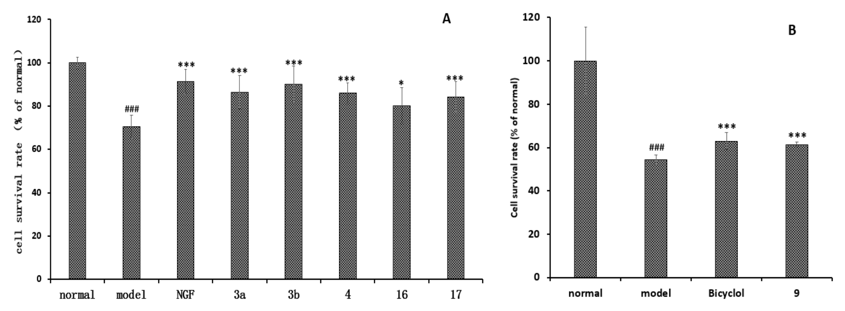
2.3. Neuroprotective Effect and Hepatoprotective Effect of Compounds 14 and 617

Compounds 14 and 617 were evaluated for their neuroprotective effect on PC12 cells induced by okadaic acid (OKA) in vitro using the MTT method. As shown in Figure 8, at 10 μM, 3a, 3b, 4, 16, 17 increased the cell survival rate of the okadaic acid-treated group, while other compounds were inactive. Compounds 14 and 617 were also tested for hepatoprotective activities against N-acetyl-p-aminophenol (APAP)-induced toxicity in HepG2 (human hepatocellular liver carcinoma cell line) cells, using the hepatoprotective activity drug bicyclol as the positive control. As shown in Figure 8, compound 9 exhibited hepatoprotective activity, while other compounds were inactive.

2.4. Discussion

The Rutaceae have 150 genres and 1700 species and are distributed worldwide, although mainly in the tropics and subtropics. Their chemical composition mainly includes essential oils, alkaloids (phenylalanine anthranilic acid, carbazole, imidazolalkaloide, indolalkaloide, etc.), amides, cumarines (umbelliferonderivates, aesculetinderivates, daphnetinderivates, 5,7-dihydroxycumarins, isopropyldihydrofurocumarines, etc.), flavonoids, lignanes, phenolics, tetracyclic triterpenes and limonoids, diterpenes, pentacyclic triterpenes and saponins, etc. [31].
Clausena lansium (Lour.) Skeels, which belongs to the Rutaceae, has been cultivated in southern China and other warm areas of the world. Many chemical components, including carbazole alkaloids, coumarins, acyclic amides, cyclic amides, quinolones, phenyl glycosides, lactams and oxyneolignan were characterized from the stems, roots and leaves of C. lansium, and showed various biological activities. Some of the carbazole alkaloids, alkaloid glycosides, amides, and coumarins have exhibited potential anti-inflammatory activity, neuroprotective, hepatoprotective, and cytotoxicity activities [5,7,9,11,32,33,34,35,36]. However, as for the constituents from the fruit, seeds and peels, the references were very few. Few alkaloids, amides, and monoterpenes were isolated from the seeds of C. lansium [37,38]. The 8-hydroxypsoralen which showed antioxidant and cytotoxic activities, and the two new monoterpenoid coumarins (clauslactone V-W) which showed α-glucosidase inhibitory activity were obtained from the peels of C. lansium [6]. Some monoterpenoid coumarins and seven carbazole alkaloids were also isolated from the peels of Clausena lansium (Lour.) Skeels, and claulansine J exhibited moderate antibacterial activity against Staphylococcus aureus [39,40]. Three new jasmonoid glucosides, two new sesquiterpenes, two new coumarins, and others were isolated from the fruit of C. lansium. One coumarin was active against S. aureus and S. dysenteriae, and also exhibited moderate antioxidant activity, while one sesquiterpene, (+)-(E)-a-santalen-12-oic-acid, showed an inhibitory effect on B. cereus [41].
In this paper, the stems of C. lansium were collected in the Liuzhou commercial cultivation in Guangxi, China. Liuzhou, located in northern Guangxi, is a subtropical monsoon climate. Light, temperature, and water are very rich in Liuzhou. Therefore, the chemical components of C. lansium collected in Liuzhou could vary and be rich. Compounds 117 were obtained from the BuOH and the water extracts from the stems of C. lansium for the first time. The results are basically the same as those reported in the genus Rutaceae and for C. lansium. Some A,D-seco-limonoids have been characterized from the stems of Clausena emarginata [42] and we think that the two new monoterpenoids, clausenapenes A and B, may be decomposition products from the limonoides. In our studies, compounds 14 and 617 were assayed for the hepatoprotective and neuroprotective effects in vitro, in order to discover potential lead compounds. Herein, compounds 3a, 3b, 4, 16, and 17 showed strong or moderate potential neuroprotective effects by inhibiting PC12 cell injury induced by okadaic acid, and compound 9 exhibited strong potential hepatoprotective activities. It indicated that it is worth studying the chemical compositions of the BuOH and the water extracts of the stems of C. lansium to find more lead compounds.

3. Materials and Methods

3.1. General Experimental Procedures

Optical rotations were measured on a JASCO P2000 automatic digital polarimeter (Jasco Corporation, Tokyo, Japan). UV spectra were recorded on a JASCO V-650 spectrophotometer (Jasco Corporation, Tokyo, Japan), CD spectra were measured on a JASCO J-815 spectropolarimeter (Jasco Corporation, Tokyo, Japan). IR spectra were recorded on a Nicolet 5700 spectrometer (Thermo Nicolet Corporation, Madison, SD, USA) using an FT-IR microscope transmission method. NMR spectra were acquired with Bruker AVIIIHD 600 (Bruker Corporation, Karlsruhe, Germany), Varian 600 and 400 (Varian Medical Systems, Inc., Palo Alto, CA, USA) in DMSO-d6. HRESIMS spectra were collected on UHPLC-Q Tof-MS (Agilent Technologies, Santa Clara, CA, USA). The MPLC system was composed of two C-605 pumps (Büchi, Flawil, Swizerland), a C-635 UV detector (Büchi), a C-660 fraction collector (Büchi), and an Octadecylsilyl (ODS) column (450 mm × 60 mm, 50 μm, 400 g; YMC, London, UK). Semi-preparative HPLC was conducted using a Shimadzu LC-6AD instrument (Shimadzu, Kyoto, Japan) with an SPD-20A detector and a Daicel Chiralpak AD-H column (250 mm × 10 mm, 5 μm). Preparative HPLC was also performed on a Shimadzu LC-6AD instrument (Shimadzu, Kyoto, Japan) with an YMC-Pack ODS-A column (250 mm × 20 mm, 5 μm). Column chromatography (CC) was performed with silica gel (200–300 mesh, Qingdao Marine Chemical Inc., Qingdao, China), MCI Gel (CHP20/P120, Mitsubishi chemical, Tokyo, Japan), SF-PRP 512A (100–200 mesh, Beijing Sunflower and Technology Development Co., Beijing, China), ODS (50 μm, YMC, Japan), and Sephadex LH-20 (GE, Upsala, Sweden). The TLC analysis was carried out on glass precoated silica gel GF254 plates. Spots were visualized under UV light or by spraying with 10% sulfuric acid in EtOH, followed by heating.

3.2. Cell Lines, Chemicals and Biochemical

PC12 cells (adrenal gland; pheochromocytoma) were purchased from the American Type Culture Collection. Human HepG2 hepatoma cells were purchased from the Cell Culture Centre at the Institute of Basic Medical Sciences, Chinese Academy of Medical Sciences. Dimethyl Sulphoxide (DMSO), Bicyclol, Okadaic Acid, 3-(3,4-dimehylthiazol-2-yl)-2,5-diphenyl-tetrazolium bromide (MTT) were obtained from Sigma (St. Louis, MO, USA). Dulbecco’s Modified Eagle’s Medium (DMEM), fetal bovine serum (FBS), and equine serum were purchased from Gibco BRL (New York, NY, USA). All other chemicals were of analytical grade and were commercially available.

3.3. Plant Materials

The stems of C. lansium were collected in Liuzhou, Guangxi, China, in March 2013, and were from commercial cultivation. C. lansium was identified by Engineer Guangri Long, Forestry of Liuzhou. A voucher specimen has been deposited at the Herbarium of Institute of Materia Medica, Chinese Academy of Medical Sciences and Peking Union Medical College (ID-S-2320). The 95% ethanol extract from the stems of C. lansium was stored in a refrigerator at −80 °C.

3.4. Extraction and Isolation

Air-dried, powdered stems of C. lansium (200 kg) were extracted with 95% ethanol (1000 L × 2 h × 3). The residue was suspended in water and then partitioned with EtOAc (3 × 40 L), and n-BuOH (3 × 40 L), successively.
After removing the solvent, the n-BuOH-soluble portion (850 g) was fractionated via a macroporous adsorbent resin (HPD-100) column with H2O, 30% EtOH, 60% EtOH, and 95% EtOH to yield four corresponding fractions A–D. Fraction B (420 g) was fractionated via silica gel column chromatography, eluting with CHCl3–MeOH–H2O (9:1:0.1, 8:2.5:0.3, 7:3:0.5, 6:4:0.4) to afford fifteen fractions B1–B15 on the basis of TLC analysis. Fraction B1 (39.6 g) was further separated by PRP-512A, silica gel column chromatography and preparative HPLC (detection at 210 nm, 60% CH3OH, 8 mL/min) to yield 4 (30 mg) and 5 (2 mg).
The water-soluble portion (25 L) was fractionated via a macroporous absorbent resin (HPD-100) column with H2O, 20% EtOH, 40% EtOH, and 95% EtOH to yield four corresponding fractions a–d. Fraction b (296 g) was further separated by silica gel column chromatography with CHCl3–MeOH–H2O (7:3:0.5, 6:4:0.4) to afford eight fractions B1–B8 on the basis of TLC analysis. Fraction B1 (10 g) was further separated by silica gel column chromatography, Sephadex LH-20, preparative HPLC (detection at 210 nm, 20% CH3CN, 8 mL/min) to yield 3 (29 mg). Compound 3 was further separated by semipreparative chiral HPLC (n-hexane–2-propanol, 2:1, 3 mL/min) to give 3a (2.8 mg) and 3b (2.1 mg). Fraction B2 (40 g) was further separated by MCI gel, and preparative HPLC (detection at 210 nm, 12% CH3CN, 8 mL/min) to yield 15 (29 mg). Fraction B3 (39 g) was further separated by PRP-512A with 10% EtOH, 15% EtOH, 20% EtOH, 30% EtOH, and 95% EtOH to yield fourteen fractions B3-1–B3-14. Fractions B3-2 was further separated by Sephadex LH-20, silica gel column chromatography, and preparative HPLC to yield 1 (18 mg), 2 (6 mg), 9 (4 mg), 13 (13 mg), and 14 (29 mg). Fraction B3-7 was further separated by Sephadex LH-20 and preparative HPLC to yield 6 (15 mg), 7 (11 mg), 8 (14 mg), 10 (118 mg), 11 (15 mg), 12 (6 mg), 16 (8 mg), 17 (17 mg).

3.5. Characterization

Compound 1: white powder; [ α ] D 20 +61.9 (c 1.0 MeOH); UV (MeOH) λmax (log ε) 206.4 (4.08), 223.6 (4.04), 269.6 (3.86), 304 (3.63) nm; IR (microscope) νmax 3389, 2921, 1677, 1592, 1511, 1455, 1419, 1268, 1119, 1069 cm−1; 1H-NMR (DMSO-d6, 400 MHz) and 13C-NMR (DMSO-d6, 100 MHz), see Table 1; HRESIMS m/z 527.1731 [M + Na]+ (calculated for C22H32NaO13, 527.1735).
Compound 2: white powder; [ α ] D 20 −6.16 (c 0.37 MeOH); UV (MeOH) λmax (log ε) 207.0 (4.37), 271.2 (3.05) nm; IR (microscope) νmax 3390, 2921, 2849, 1646, 1595, 1504, 1465, 1422, 1124, 1039 cm−1; 1H-NMR (DMSO-d6, 400 MHz) and 13C-NMR (DMSO-d6, 100 MHz), see Table 1; HRESIMS m/z 443.1532 [M + Na]+ (calculated for C18H28NaO11, 443.1524).
Compound 3a: white powder; [ α ] D 20 +3.6 (c 0.1 MeOH); UV (MeOH) λmax (log ε) 206.0 (4.41), 230.4 (4.29), 278.8 (4.14), 306.4 (4.10) nm; IR (microscope) νmax 3394, 2921, 2849, 1667, 1591, 1517, 1465, 1427, 1286, 1107, 779 cm−1; 1H-NMR (DMSO-d6, 400 MHz) and 13C-NMR (DMSO-d6, 100 MHz), see Table 2; HRESIMS m/z 235.0573 [M + Na]+ (calculated for C10H12NaO5, 235.0577).
Compound 3b: white powder; [ α ] D 20 −3.7 (c 0.1 MeOH); UV (MeOH) λmax (log ε) 206.2 (4.38), 230.6 (4.24), 278.4 (4.10), 306.0 (4.06) nm; IR (microscope) νmax 3394, 2921, 2849, 1647, 1591, 1517, 1468, 1420, 1285, 1107, 779 cm−1; 1H-NMR (DMSO-d6, 400 MHz) and 13C-NMR (DMSO-d6, 100 MHz), see Table 2; HRESIMS m/z 235.0573 [M + Na]+ (calculated for C10H12NaO5, 235.0577).
Compound 4: colourless oil; [ α ] D 20 −50.1 (c 0.1, MeOH); UV (MeOH) λmax (log ε) 203.6 (4.39) nm; CD (c 0.33, MeOH) λmax 219 (5.67) nm; IR (microscope) νmax 3418, 2925, 1755, 1659, 1441, 1384, 1261, 1101, 998 cm−1; 1H-NMR (DMSO-d6, 400 MHz) and 13C-NMR (DMSO-d6, 100 MHz), see Table 3; HRESIMS m/z 205.0835 [M + Na]+ (calculated for C10H14NaO3, 205.0835).
Compound 5: colourless oil; [ α ] D 20 −58.5 (c 0.1 MeOH); UV (MeOH) λmax (log ε) 201.4 (3.76) nm; CD (c 0.5, MeOH) λmax 212 (2.80) nm; IR (microscope) νmax 3394, 2921, 1750, 1646, 1468, 1117, 998 cm−1; 1H-NMR (DMSO-d6, 600 MHz) and 13C-NMR (DMSO-d6, 150 MHz), see Table 3; HRESIMS m/z 207.0993 [M + Na]+ (calculated for C10H16NaO3, 207.0992).

3.6. Acid Hydrolysis and GC Analysis of Compounds 1 and 2

Compound 2 (2 mg) was dissolved in 2 mol HCl-H2O (2 mL) and was then heated to 90 °C for 15 h. The reaction mixture was extracted with EtOAc. The aqueous layer was evaporated under vacuum, diluted repeatedly with H2O, and evaporated in vacuo to furnish a neutral residue. The residue was dissolved in anhydrous pyridine (1 mL), to which 2 mg of l-cysteine methyl ester hydrochloride was added. The mixture was stirred at 60 °C for 2 h, and after evaporation in vacuo to create dryness, 0.2 mL of N-trimethylsilylimidazole was added. The mixture was kept at 60 °C for another 2 h.
The reaction mixture was partitioned between n-hexane and H2O (2 mL each), and then the n-hexane extract was analyzed by GC under the following conditions: capillary column, HP-5 (30 m × 0.25 mm, with a 0.25 μm film; Dikma, Beijing, China); detection, FID; detector temperature, 280 °C; injection temperature, 250 °C; initial temperature 200 °C, then raised to 280 at 5 °C/min, final temperature maintained for 10 min; carrier, N2 gas. From the acid hydrolysate of 2, d-glucofuranurono-6, 3-lactone was confirmed by comparison of the retention time of its derivative, with that of an authentic sugar derivatized in a similar way, which showed a retention time of 18.4 min. The constituent sugar of compound 1 was identified by the same method as 2. Retention times of authentic sample were detected at 18.4 min (d-glucose) and 14.7 min (l-rhamnose) for 1.

3.7. Hepatoprotective Activity Assay

Human HepG2 hepatoma cells were cultured in a DMEM medium supplemented with 10% fetal calf serum, 100 U/mL penicillin, and 100 μg/mL streptomycin at 37 °C in a humidified atmosphere of 5% CO2 + 95% air. The cells were then passaged by treatment with 0.25% trypsin in 0.02% EDTA. The MTT assay was used to assess the cytotoxicity of test samples. The cells were seeded in 96-well multiplates. After an overnight incubation at 37 °C with 5% CO2, 10 μM test samples and APAP (final concentration of 8 mM) were added into the wells and incubated for another 48 h. Then, 100 μL of 0.5 mg/mL MTT was added to each well after the withdrawal of the culture medium and they were incubated for an additional 4 h. The resulting formazan was dissolved in 150 μL of DMSO after aspiration of the culture medium. The plates were placed on a plate shaker for 30 min and read immediately at 570 nm using a microplate reader [43]. The cell inhibitory rate (%) was calculated by (Asample − Ablank)/(Auntreated − Ablank) × 100. p-Values of <0.05, <0.01, and <0.001 were regarded as statistically significant.

3.8. Neuroprotective Activity Assays

Pheochromocytoma (PC12) cells were incubated in DMEM, supplied with 5% fetal bovine serum and 5% equine serum as a basic medium. PC12 cells in the logarithmic phase were cultured at a density of 5000 cells per well in a 96-well microtiter plate. After 24 h incubation, the medium of the model group was changed to DMEM or a basic medium with 50 nM OKA for 24 h. Test compounds dissolved in DMSO were added to each well for >1000-fold dilution in the model medium at the same time. Each sample was tested in triplicate. After the incubation at 37 °C in 5% CO2 for 24 h, 10 μL of MTT (5 mg/mL) was added to each well and they were incubated for another 4 h. Then, the liquid in the wells was removed. Test compounds (100 μL) were added to each well. The absorbance was recorded on a microplate reader (Bio-Rad model 550, California, USA) at a wavelength of 570 nm [44]. Analysis of variance (ANOVA) followed by the Newman–Keuls post hoc test were performed to assess the differences between the relevant control and each experimental group. The cell inhibitory rate (%) was calculated by (Asample − Ablank)/(Auntreated − Ablank) × 100. p-Values of <0.05, <0.01, and <0.001 were regarded as statistically significant.

4. Conclusions

In summary, this work described the isolation and the structure identification of two new phenolic glycosides (12), one new pair of phenolic enantiomers (3a, 3b), and two new monoterpenoids (45), together with twelve known analogues (617). They were obtained from the stems of C. lansium for the first time. In addition, compounds 3a, 3b, 4, 16, and 17 showed strong or moderate potential neuroprotective effects on inhibiting PC12 cell injury induced by okadaic acid, and compound 9 exhibited strong potential hepatoprotective activities. In traditional Chinese medicine, the leaves, fruit, seeds, and the roots of C. lansium were used as folk medicine for treating many kinds of diseases. It has been reported that the stems of C. lansium have a characteristic chemical composition including carbazole, amide, quinolone alkaloids, coumarins, and others, which have various biological activities such as neuroprotective, anti-inflammatory, hepatoprotective, and cyctoxicity capacities. Therefore, not only the roots and the leaves but also the stems are important medicinal materials, which indicate that the chemical compositions and the biological activities of the stems of C. lansium are worth studying in order to find other compounds with potential activity.

Supplementary Materials

The following are available online.

Acknowledgments

This study was financially supported by the National Natural Science Foundation of China (No. 21272278, No. 81730093), and CAMS Innovation Fund for Medical Sciences (CIFMS) [grant number 2017-I2M-3-010]. The authors are grateful to the Department of Instrumental Analysis, Institute of Materia Medica, Chinese Academy of Medical Sciences and Peking Union Medical College, for the measurement of the UV, IR, CD, NMR, HRESIMS and CD spectra.

Author Contributions

D.-M.Z. designed the research; J.L. fractionated the extract, isolated the compounds, elucidated structures and wrote the paper; H.S. performed the bioassays of hepatoprotective activity; N.-H.C. performed the bioassays of Neuroprotective activity; Y.-Q.D. performed the data of GC analysis; L.L. performed the detection and calculation of ECD spectra; C.-J.L. coordinated the study. All authors read and approved the final manuscript.

Conflicts of Interest

The authors declare no conflict of interest.

References

  1. Editorial Committee of Flora of China, Chinese Academy of Sciences. Flora of China; Science Press: Beijing, China, 1999; Volume 43, p. 132. [Google Scholar]
  2. Lim, T.K. Edible Medicinal and Non-Medicinal Plants; Springer: Berlin, Germany, 2012; Volume 4, pp. 871–883. [Google Scholar]
  3. Adebajo, A.C.; Iwalewa, E.O.; Obuotor, E.M.; Ibikunle, G.F.; Omisore, N.O.; Adewunmi, C.O.; Obaparusi, O.O.; Klaes, M.; Adetogun, G.E.; Schmidt, T.J.; et al. Pharmacological properties of the extract and some isolated compounds of Clausena lansiumstem bark: Anti-trichomonal, antidiabetic, anti-inflammatory, hepatoprotective and antioxidant effects. J. Ethnopharmacol. 2009, 122, 10–19. [Google Scholar] [CrossRef] [PubMed]
  4. Maneerat, W.; Ritthiwigrom, T.; Cheenpracha, S.; Cheenpracha, S.; Laphookhieo, S. Carbazole alkaloids and coumarins from Clausena lansium roots. Phytochem. Lett. 2012, 5, 26–28. [Google Scholar] [CrossRef]
  5. Liu, G.T.; Li, W.X.; Chen, Y.Y.; Wei, H.L. Hepatoprotective action of nine constituents isolated from the leaves of Clausena lansium in mice. Drug Dev. Res. 1996, 39, 174–178. [Google Scholar] [CrossRef]
  6. Prasad, K.N.; Xie, H.H.; Hao, J.; Yang, B.; Qiu, S.X.; Wei, X.Y.; Chen, F.; Jiang, Y.M. Antioxidant and anticancer activities of 8-hydroxypsoralen isolated from wampee [Clausena lansium (Lour.) Skeels] peel. Food Chem. 2010, 118, 62–66. [Google Scholar] [CrossRef]
  7. Liu, H.; Li, C.J.; Yang, J.Z.; Ning, N.; Si, Y.K.; Li, L.; Chen, N.H.; Zhao, Q.; Zhang, D.M. Carbazole Alkaloids from the Stems of Clausena lansium. J. Nat. Prod. 2012, 75, 677–682. [Google Scholar] [CrossRef] [PubMed]
  8. Liu, H.; Li, F.; Li, C.J.; Li, C.J.; Yang, J.Z.; Li, L.; Chen, N.H.; Zhang, D.M. Bioactive furanocoumarins from stems of Clausena lansium. Phytochemistry 2014, 107, 141–147. [Google Scholar] [CrossRef] [PubMed]
  9. Du, Y.Q.; Liu, H.; Li, C.J.; Ma, J.; Zhang, D.; Li, L.; Sun, H.; Bao, X.Q.; Zhang, D.M. Bioactive carbazole alkaloids from the stems of Clausena lansium. Fitoterapia 2015, 103, 122–128. [Google Scholar] [CrossRef] [PubMed]
  10. Zhao, Q.; Yang, J.Z.; Li, C.J.; Chen, N.H.; Zhang, D.M. A new megastigmane glucoside and a new amide alkaloid from the leaves of Clausena lansium (Lour.) Skeels. J. Asian. Nat. Prod. Res. 2011, 13, 361–366. [Google Scholar] [CrossRef] [PubMed]
  11. Liu, J.; Li, C.J.; Ni, L.; Yang, J.Z.; Li, L.; Zang, C.X.; Bao, X.Q.; Zhang, D.; Zhang, D.M. Anti-inflammatory alkaloid glycoside and quinolone alkaloid derivates from the stems of Clausena lansium. RSC Adv. 2015, 5, 80553–80560. [Google Scholar] [CrossRef]
  12. Liu, J.; Li, C.J.; Yang, J.Z.; Ma, J.; Zhang, D.M. Chemical constituents from stems of Clausena lansium. Chin. Tradit. Herb. Drugs 2016, 47, 32–37. [Google Scholar]
  13. Wang, C.; Li, C.J.; Ma, J.; Yang, J.Z.; Chen, X.G.; Li, L.; Zhang, D.M. Bioactive 18(4 → 3)-abeo-abietanoid derivatives from the leaves of Tripterygium wilfordii. RSC Adv. 2015, 5, 30046–30052. [Google Scholar] [CrossRef]
  14. Gan, M.L.; Zhu, C.G.; Zhang, Y.L.; Zi, J.C.; Song, W.X.; Yang, Y.C.; Shi, J.G. Constituents from a water-soluble portion of ethanolic extract of Iodes cirrhosa. Chin. J. Chin. Mat. Med. 2010, 35, 456–467. [Google Scholar]
  15. Baderschneider, B.; Winterhalter, P. Isolation and characterization of novel benzoates, cinnamates, flavonoids, and Lignans from riesling wine and screening for antioxidant activity. J. Agric. Food Chem. 2001, 49, 2788–2798. [Google Scholar] [CrossRef] [PubMed]
  16. Liu, J.; Du, D.; Si, Y.K.; Lü, H.N.; Wu, X.F.; Li, Y.; Liu, Y.Y.; Yu, S.S. Application of dimolybdenum reagent Mo2(OAc)4 for determination of the absolute configurations of vic-diols. Chin. J. Org. Chem. 2010, 30, 1270–1278. [Google Scholar]
  17. Bari, L.D.; Pescitelli, G.; Pratelli, C.; Pini, D.; Salvadori, P. Determination of absolute configuration of acyclic 1,2-diols with Mo2(OAc)4. 1. Snatzke’s Method Revisited. J. Org. Chem. 2001, 66, 4819–4825. [Google Scholar] [CrossRef] [PubMed]
  18. Minkin, V.I.; Legrand, M.; Rougier, M.J. Determination of Configurations by Dipole Moments; CD or ORD. Stereochemistry: fundamentals and methods; Georg Thieme Publishers: Stuttgart, Germany, 1977; Volume 2, p. 33. [Google Scholar]
  19. Gawronski, J.K.; Oeveren, A.V.; Deen, H.V.D.; Leung, C.W.; Feringa, B.L. Simple circular dichroic method for the determination of absolute configuration of 5-substituted 2(5H)-furanones. J. Org. Chem. 1996, 61, 1513–1515. [Google Scholar] [CrossRef]
  20. Dong, C.X.; Shi, S.P.; Wu, K.S.; Tu, P.F. Chemical constituents from the roots and rhizomes of Dictamnus dasycarpus Pall. Z. Naturforschung B J. Chem. Sci. 2007, 62, 854–858. [Google Scholar]
  21. Miyamura, M.; Nohara, T.; Tomimatsu, T.; Nishioka, I. Seven aromatic compounds from bark of Cinnamomum cassia. Phytochemistry 1983, 22, 215–218. [Google Scholar] [CrossRef]
  22. Graikou, K.; Aligiannis, N.; Chinou, I.; Skaltsounis, A.L.; Tillequin, F.; Litaudon, M. Chemical constituents from Croton insularis. Helv. Chim. Acta 2005, 88, 2654–2660. [Google Scholar] [CrossRef]
  23. Andrianaivoravelona, J.O.; Terreaux, C.; Sahpaz, S.; Rasolondramanitra, J.; Hostettmann, K. A phenolic glycoside and N-(p-coumaroyl)-tryptamine from Ravansara anisata. Phytochemistry 1999, 52, 1145–1148. [Google Scholar] [CrossRef]
  24. Ferrari, F.; Monache, F.D. A new phenolic glycoside from Sorocea ilicifolia stem bark. Fitoterapia 2004, 75, 417–419. [Google Scholar] [CrossRef] [PubMed]
  25. Kanchanapoom, T.; Kasai, R.; Yamasaki, K. Phenolic glycosides from Barnettia kerrii. Phytochemistry 2002, 59, 565–570. [Google Scholar] [CrossRef]
  26. Bao, S.Y.; Ding, Y.; Deng, Z.W.; Proksch, P.; Lin, W.H. Rhyncoside A-F, phenolic constituents from the Chinese mangrove plant Bruguiera sexangula var. rhynchopetala. Chem. Pharm. Bull. 2007, 55, 1175–1180. [Google Scholar] [CrossRef] [PubMed]
  27. Chang, J.; Xuan, L.J.; Xu, Y.M.; Zhang, J.S. Cytotoxic terpenoid and immunosuppressive phenoic glycosides from the root bark of Dictamnus dasycarpus. Planta Med. 2002, 68, 425–429. [Google Scholar] [CrossRef] [PubMed]
  28. Otsuka, H.; Kashima, Z.; Hayashi, T.; Kubo, N.; Yamasaki, K.; Padolina, W.G. Premnaodorosides A, B and C; iridoid glucoside diesters of an acyclic monoterpenediol from leaves of Premna Odorata. Phytochemistry 1992, 31, 3129–3133. [Google Scholar] [CrossRef]
  29. Matsunami, K.; Nagashima, J.; Sugimoto, S.; Otsuka, H.; Takeda, Y.; Lhieochaiphant, D.; Lhieochaiphant, S. Megastigmane glucosides and an unusual onoterpene from the leaves of Cananga odorata var. odorata, and absolute structures of megastigmane glucosides isolated from C. odorata var. odorata and Breynia officinalis. J. Nat. Med. 2010, 64, 460–467. [Google Scholar] [CrossRef] [PubMed]
  30. Ono, M.; Yoshida, A.; Ito, Y.; Nohara, T. Phenethyl alcohol glycosides and isopentenol glycoside from fruit of Bupleurum falcatum. Phytochemistry 1999, 51, 819–823. [Google Scholar] [CrossRef]
  31. Hegnauer, R. Chemotaxonomie der Pflanzen, Bd. 6, Dicotyledonear: Rafflesiaceae—Zygopbyllaceae; Birkhäuser Verlag: Basel, Switzerland, 1973; pp. 174–239. [Google Scholar]
  32. Shen, D.Y.; Ngan Nguyen, T.; Wu, S.J.; Shiao, Y.J.; Hung, H.Y.; Kuo, P.C.; Kuo, D.H.; Dinh Thang, T.; Wu, T.S. γ- and δ-Lactams from the Leaves of Clausena lansium. J. Nat. Prod. 2015, 78, 2521–2530. [Google Scholar] [CrossRef] [PubMed]
  33. Shen, D.Y.; Kuo, P.C.; Huang, S.C.; Hwang, T.L.; Chan, Y.Y.; Shieh, P.C.; Thi Ngan, N.; Dinh Thang, T.; Wu, T.S. Constituents from the leaves of Clausena lansium and their antiinflammatory activity. J. Nat. Med. 2017, 71, 96–104. [Google Scholar] [CrossRef] [PubMed]
  34. Liu, J.; Du, Y.Q.; Li, C.J.; Li, L.; Chen, F.Y.; Yang, J.Z.; Chen, N.H.; Zhang, D.M. Alkaloids from the stems of Clausena lansium and their neuroprotective activity. RSC Adv. 2017, 7, 35417–35425. [Google Scholar] [CrossRef]
  35. Jiang, H.Y.; Zhang, W.J.; You, C.X.; Yang, K.; Fan, L.; Feng, J.B.; Chen, J.; Yang, Y.J.; Wang, C.F.; Deng, Z.W.; et al. Two new cytotoxic constituents from the Clausena lansium (Lour.) Skeels. Phytochem. Lett. 2014, 9, 92–95. [Google Scholar] [CrossRef]
  36. Song, W.W.; Zeng, G.Z.; Peng, W.W.; Tan, N.H. A New Cytotoxic Oxyneolignan from the Roots and Stems of Clausena lansium (Rutaceae). Plant Divers. Res. 2014, 36, 545–550. [Google Scholar]
  37. Wang, G.C.; Li, W.; Wang, Y.; Zhang, X.Q.; Li, Y.L.; Ye, W.C. A new amide and a new monoterpene from the seeds of Clausena lansium. Nat. Prod. Res. 2012, 27, 558–562. [Google Scholar] [CrossRef] [PubMed]
  38. Huang, L.; Li, D.; Xu, Y.S.; Feng, Z.L.; Meng, F.C.; Zhang, Q.W.; Gan, L.S.; Lin, L.G. Clausoxamine, an alkaloid possessing a 1,3-oxazine-4-one ring from the seeds of Clausena lansium and the anti-obesity effect of lansiumamide B. RSC Adv. 2017, 7, 46900–46905. [Google Scholar] [CrossRef]
  39. Deng, H.D.; Mei, W.L.; Wang, H.; Guo, Z.K.; Dong, W.H.; Wang, H.; Li, S.P.; Dai, H.F. Carbazole alkaloids from the peels of Clausena lansium. J. Asian Nat. Prod. Res. 2014, 16, 1024–1028. [Google Scholar] [CrossRef] [PubMed]
  40. Deng, H.D.; Mei, W.L.; Guo, Z.K.; Liu, S.; Zuo, W.J.; Dong, W.H.; Li, S.P.; Dai, H.F. Monoterpenoid Coumarins from the Peels of Clausena lansium. Planta Med. 2014, 80, 955–958. [Google Scholar] [CrossRef] [PubMed]
  41. Xu, X.Y.; Xie, H.H.; Wei, X.Y. Jasmonoid glucosides, sesquiterpenes and coumarins from the fruit of Clausena lansium. LWT Food Sci. Technol. 2014, 59, 65–69. [Google Scholar] [CrossRef]
  42. Xia, H.M.; Li, C.J.; Yang, J.Z.; Ma, J.; Chen, X.G.; Zhang, D.; Li, L.; Zhang, D.M. A,D-seco-Limonoids from the Stems of Clausena emarginata. J. Nat. Prod. 2014, 77, 784–791. [Google Scholar] [CrossRef] [PubMed]
  43. Hao, Z.Y.; Liang, D.; Luo, H.; Liu, Y.F.; Ni, G.; Zhang, Q.J.; Li, L.; Si, Y.K.; Sun, H.; Chen, R.Y.; Yu, D.Q. Bioactive Sesquiterpenoids from the Rhizomes of Acrorus calamus. J. Nat. Prod. 2012, 75, 1083–1089. [Google Scholar] [CrossRef] [PubMed]
  44. Zhang, F.; Yang, Y.N.; Song, X.Y.; Shao, S.Y.; Feng, Z.M.; Jiang, J.S.; Li, L.; Chen, N.H.; Zhang, P.C. Forsythoneosides A−D, Neuroprotective phenethanoid and flavone glycoside heterodimers from the fruits of Forsythia suspense. J. Nat. Prod. 2015, 78, 2390–2397. [Google Scholar] [CrossRef] [PubMed]
Sample Availability: Samples of the compounds 14 and 617 are available from the authors.
Figure 1. Structures of compounds 117.
Molecules 22 02226 g001
Figure 2. Key HMBC correlations of compounds 1 and 2.
Molecules 22 02226 g002
Figure 3. Calculated electronic circular dichroism (ECD) spectra of (8S) 1a and (8R) 1a-isomers and the experimental ECD of 1a and 1.
Molecules 22 02226 g003
Figure 4. The circular dichroism (CD) Spectrum of compounds 3a and 3b in MeOH.
Molecules 22 02226 g004
Figure 5. The CD spectrum of compounds 3a and 3b induced by Mo2(OAC)4 (dimolybdenum tetracetate) (the inherent CD of the diol was subtracted).
Molecules 22 02226 g005
Figure 6. Key HMBC correlations of compounds 3, 4 and 5.
Molecules 22 02226 g006
Figure 7. The CD and UV Spectrum of compounds 4 and 5 in MeOH.
Molecules 22 02226 g007
Figure 8. The neuroprotective and hepatoprotective effects of compounds isolated from C. lansium. (A) Neuroprotective effects of compounds 3a, 3b, 4, 16, 17 against okadaic acid-induced injury in PC12 Cells (10 μM, means ± SD, n = 6); (B) Hepatoprotective effects of compounds 14 and 617 (10 μM) against N-acetyl-p-aminophenol (APAP)-induced toxicity in HepG2 (human hepatocellular liver carcinoma cell line) cells (10 μM, means ± SD, n = 6). (### p < 0.001 vs. normal, *** p < 0.001, * p < 0.1 vs. model).
Molecules 22 02226 g008
Table 1. 1H and 13C-NMR Spectroscopic Data of Compounds 1, 1a, 2 and 2a (δ in ppm, J in Hz).
11a22a
PositionδH aδC bδH aδC bδH aδC bδH aδC b
1 128.5 s 130,1 s 133.9 s 134.1 s
27.51, d (2.0)111.7 d7.43, d (2.0)112.1 d 152.7 s 152.7 s
3 148.7 s 148.2 s6.63, s103.5 d6.64, s103.5 d
4 150.6 s 150.2 s 138.2 s 138.1 s
57.13, d (8.5)114.4 d6.79, d (8.5)115.5 d6.63, s103.5 d6.64, s103.5 d
67.62, dd (8.5, 2.0)122.7 d7.51, dd (8.5, 2.0)124.2 d 152.7 s 152.7 s
7 200.3 s 200.0 s4.43, d (5.7)63.0 t4.43, d (5.8)63.0 t
85.08, m68.5 d4.98, q (6.6)68.5 d
91.27, d (6.7)21.2 q1.23, d (6.6)21.8 q
3-OCH33.83, s55.7 q3.78, s56.0 q
2,6-OCH3 3.76, s55.9 q3.76, s55.9 q
1′5.01, d (6.1)99.6 d 3.98, m81.4 d3.82, m83.4 d
2′4.02, m73.1 d 3.88, m; 3.71, m67.6 t3.59, m; 3.52, m59.9 t
3′3.28, m76.7 d 3.56, m; 3.65, m60.1 t3.59, m; 3.52, m59.9 t
4′3.01, m69.9 d
5′3.52, m75.6 d
6′3.84, m; 3.40, m66.5 t
1″4.52, br s100.7 d 4.17, d (7.7)103.4 d
2″3.46, m70.4 d 2.94, m73.5 d
3″3.58, m70.7 d 3.04, m76.7 d
4″3.13, m72.0 d 3.09, m70.0 d
5″3.44, m68.3 d 3.14, m76.8 d
6″1.09, d (6.2)17.9 q 3.42, m; 3.61, m61.0 t
a In DMSO-d6 (600 MHz), b In DMSO-d6 (150 MHz). Coupling constants (J) in Hz are given in parentheses. The assignments were based on HSQC and HMBC experiments.
Table 2. 1H and 13C-NMR Spectroscopic Data of Compounds 3 (δ in ppm, J in Hz).
3
PositionδH aδC b
1 198.3 s
24.96, t (4.7)73.9 d
3a3.69, dd (11.3, 4.2)64.5 t
3b3.59, dd (11.3, 4.9)
1′ 126.9 s
2′7.48, s 111.7 d
3′ 147.5 s
4′ 151.9 s
5′6.87, d (8.1)114.7 d
6′7.56, d (8.1)123.6 d
6-OCH33.82, s55.6 q
a In DMSO-d6 (400 MHz), b In DMSO-d6 (100 MHz). Coupling constants (J) in Hz are given in parentheses. The assignments were based on HSQC and HMBC experiments.
Table 3. 1H-NMR and 13C-NMR Spectroscopic Data of Compounds 4 and 5 (δ in ppm, J in Hz).
45
PositionδH aδC bδH aδC b
2 173.7 s 173.8 s
3 128.1 s 127.7 s
47.36, m150.5 d7.43, m151.2 d
55.10, m79.5 d5.05, m79.4 d
62.36, dd (5.2, 14.0); 2.19, dd (8.2, 14.0)42.7 t1.51, m; 1.42, m40.4 t
75.37, m129.0 d1.77, m26.4 d
8 130.8 s1.47, m; 1.30, m40.1 t
93.95, d (6.0)57.5 t3.40, m58.5 t
101.64, s16.4 q0.93, d (7.8)19.2 q
111.80, s10.2 q1.80, s10.2 q
a In DMSO-d6 (600 MHz), b In DMSO-d6 (150 MHz). Coupling constants (J) in Hz are given in parentheses. The assignments were based on HSQC and HMBC experiments.

