Article

Antibacterial Peptide CecropinB2 Production via Various Host and Construct Systems

1
Department of Chemical Engineering, National Chung Hsing University, 250 Kuo Kuang Road, Taichung 40227, Taiwan
2
Biotechnology Center, National Chung Hsing University, 250 Kuo Kuang Road, Taichung 40227, Taiwan
*
Author to whom correspondence should be addressed.
Molecules 2016, 21(1), 103; https://doi.org/10.3390/molecules21010103
Submission received: 7 December 2015 / Revised: 5 January 2016 / Accepted: 13 January 2016 / Published: 16 January 2016
(This article belongs to the Section Medicinal Chemistry)

Abstract

:
Cecropin is a cationic antibacterial peptide composed of 35–39 residues. This peptide has been identified as possessing strong antibacterial activity and low toxicity against eukaryotic cells, and it has been claimed that some types of the cecropin family of peptides are capable of killing cancer cells. In this study, the host effect of cloning antibacterial peptide cecropinB2 was investigated. Three different host expression systems were chosen, i.e., Escherichia coli, Bacillus subtilis and Pichia pastoris. Two gene constructs, cecropinB2 (cecB2) and intein-cecropinB2 (INT-cecB2), were applied. Signal peptide and propeptide from Armigeres subalbatus were also attached to the gene construct. The results showed that the best host for cloning cecropinB2 was P. pastoris SMD1168 harboring the gene of pGAPzαC-prepro-cecB2 via Western blot confirmation. The cecropinB2 that was purified using immobilized-metal affinity chromatography resin showed strong antibacterial activity against the Gram-negative strains, including the multi-drug-resistant bacteria Acinetobacter baumannii.

1. Introduction

In 1929, Alexander Fleming identified penicillin, the first chemical compound with antibiotic properties [1]. More and more antibiotic compounds were then found and used in the medical field. Due to the antibiotics’ overuse, multi-drug-resistant bacteria have become a big issue for treatment with antibiotics [2]. Using antibacterial peptides was believed to be a good way to eliminate the multi-drug resistant (MDR) bacteria issue in the medical field [3,4,5].
Cecropins are a family of cationic antibacterial peptides and have six isoforms (A–F) with the common homologous sequences of 35–39 amino acids [6]. These cecropins, originally found in the immune hemolymph of skin worm pupae, have a broad antibacterial spectrum against the Gram-negative and Gram-positive bacteria, and even against the MDR bacteria [3,7]. Experimental results also showed that they possessed an anti-tumor effect without damaging the eukaryotic cells [3,4,8]. Therefore, they might have high potential for the application in medicine, agriculture and animal husbandry.
To date, various constructs have been established for production of antibacterial peptides. However, difficulties have been encountered in peptide expression because the toxicity of antibacterial peptide might be toxic to its expression hosts [9,10]. The host strains play an important role in the process of efficiently expressing large amounts of recombinant antibacterial peptides. The most commonly-used host strains, E. coli, B. subtilis and P. pastoris, have their individual advantages and disadvantages [11,12,13]. To conquer the difficulty of host self-destruction by the toxic target peptides, the fusion protein method was proposed to reduce the toxicity. This method has been used for many peptides expression, including thioredoxin [10], green fluorescent protein (GFP) [14], small ubiquitin-related modifier (SUMO) [15], and cationic elastin-like polypeptides (CELP) [16]. In this study, intein (INT) was used as the fusion peptide to reduce the toxicity of the antibacterial cecropin B2.
In this study, three hosts, E. coli, B. subtilis and P. pastoris, were chosen, and two target peptide genes, cecB2 and INT-cecB2, were carried out to compare their effect on cecropinB2 expression. Armigeres subaltatus mosquitoes are the original source of the peptide cecropinB2. In order to help the transport and folding of the target cecropinB2, signal peptide and propeptide from Armigeres subalbatus were also applied to the gene constructs with host P. pastoris SMD1168. The best production construct was selected and the antibacterial activity was tested for the obtained purified cecropinB2. This study revealed the host effect on antibacterial peptide cecropinB2 production.

2. Results and Discussion

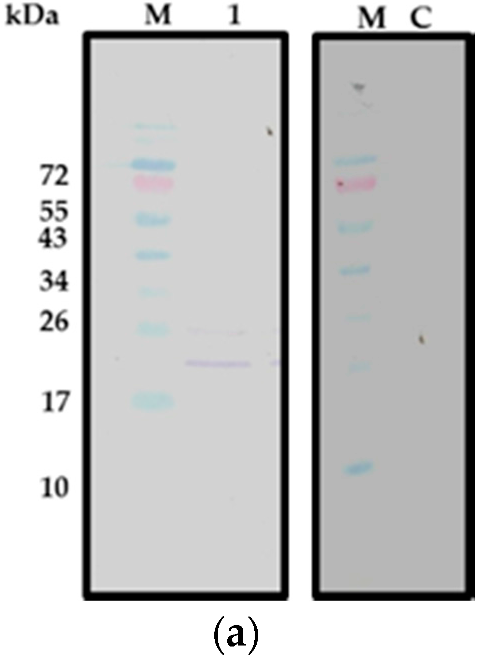
2.1. CecropinB2 Production in E. coli

To investigate the expression of cecropinB2 in E. coli, five constructs (pET26b-cecB2, pET28a-cecB2, pET28a-INT-cecB2, pET26b-INT-cecB2, pET26b-10K-INT-cecB2 and pET26b-10R-INT-cecB2, where 10K = 10 lysines, 10R = 10 arginines) were transformed into E. coli ER2566. The individual strain was cultivated and induced with 1 mM IPTG at 25 °C for 6 h. The results showed that three constructs, pET26b-cecB2, pET28a-cecB2 and pET-28a-INT-cecB2, did not express the target peptide, whereas the construct of pET26b-INT-cecB2/E. coli ER2566 did successfully express the target peptide as confirmed by Western blot (Figure 1a). The upper band of Figure 1a, lane 1 with the molecular weight of 23.5 kDa might be the incomplete cleavage segment of the signal peptide pelB (MW: 2.5 kDa) of the pET26b plus the cecropinB2 and INT (about 21 kDa). The results indicated that the target protein of INT-cecB2 might possess lower toxicity than cecropinB2. In addition, the plasmids affected the production of antibacterial peptides according to the result of pET26b (which can be produced with the construct pET26b-INT-cecB2) and pET28a (no production with the construct pET28a-INT-cecB2) systems, suggesting that the target peptide might give lower toxcity to the host when the peptide can be transferred to periplasm (pET26b). Moreover, to increase the solubility of the target protein, the 10K and 10R segments [17] were added to the N-terminal of INT-cecB2 and the results were observed by Western blot (data no shown). The addition of 10K and 10R segments did not improve the solubility of INT-cecB2, suggesting that the toxicity of target peptides (INT-cecB2) still had some effect on limiting the protein solubility. The antibacterial peptide cecropinB2 did not express well in E. coli ER2566. This might be due to the peptide’s toxicity against its host. The fusion protein approach by adding an INT seemed to slightly improve the expression; however, the production of INT-cecB2 was at a low level (<10 mg/L).
Figure 1. Expression of cecropinB2 in E. coli ER2566 and B. subtilis WB800. (a) Western blot of pET26b-INT-cecB2/E. coli ER2566; where Lane M, molecular weight markers (kDa); Lane 1, supernatant; Lane C: control (supernatant of ET26b/E. coli ER2566); The cell debris was centrifuged at 7000 g at 4 °C for 10 min to obtain the supernatant (Lane 1); (b) Western blot of PRPA-cecB2/B. subtilis WB800; where Lane M, molecular weight markers (kDa); Lane 1, cell crude extract; Lane 2, 100× concentrated extracellular fluid; Lane C: control (cell crude extract of PRPA-/B. subtilis WB800). The culture broth was centrifuged at 6000× g at 4 °C for 10 min to obtain the cell and extracellular fluid.
Molecules 21 00103 g001aMolecules 21 00103 g001b

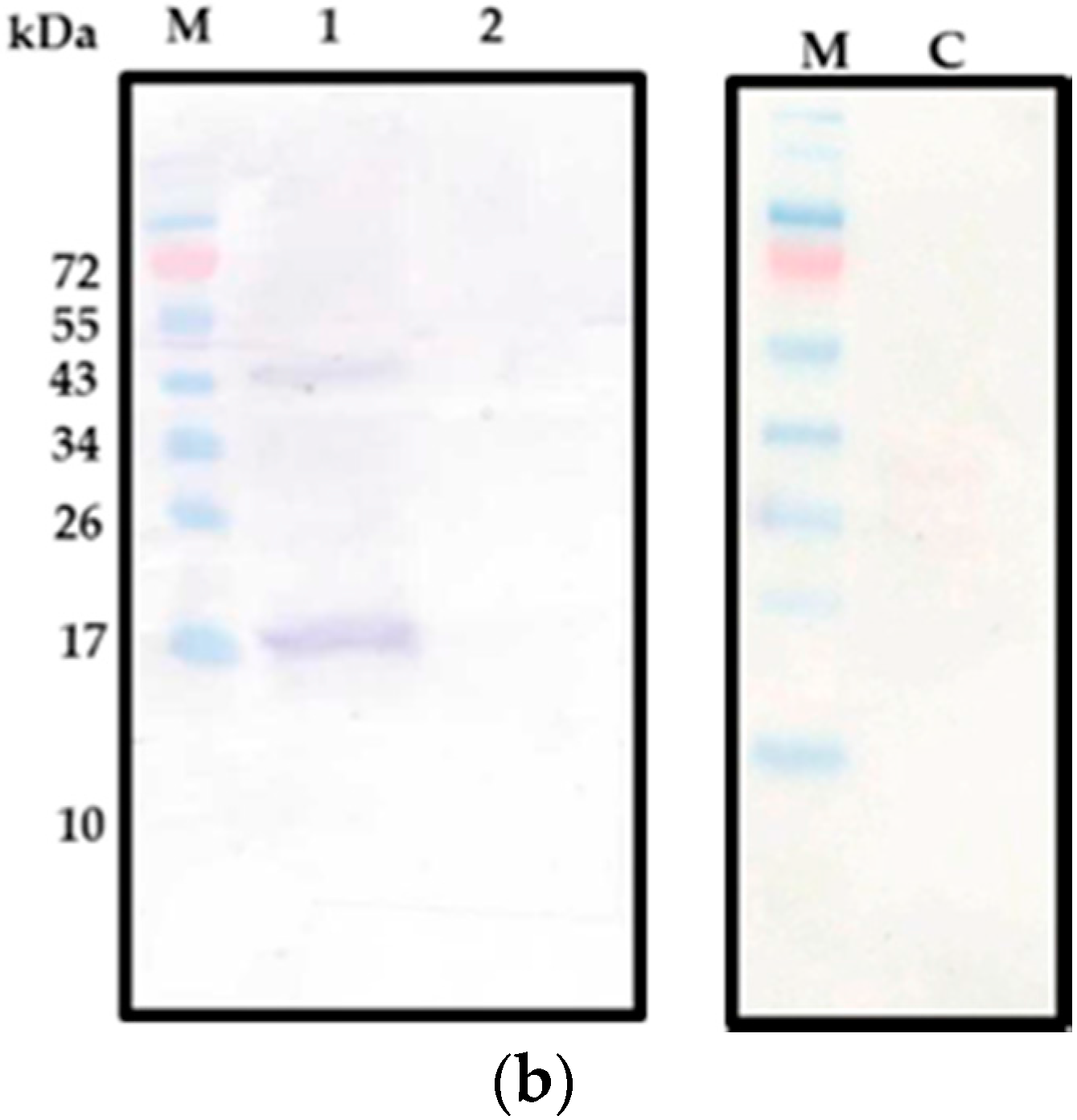
2.2. CecropinB2 Production in B. subtilis

To study the expression of cecB2 in B. subtilis, two constructs, PRPA-cecB2 and PRPA-INT-cecB2, were transformed into the B. subtilis WB800. The expression results were confirmed by Western blot. The construct of PRPA-INT-cecB2 in B. subtilis WB800 did not produce any target protein (INT-cecB2), indicating that the host B. subtilis could not express the INT protein well. The construct of PRPA-cecB2 in B. subtilis WB800 was found capable of expressing the target peptide; however, the expression was intracellular with very low yield (<10 mg/L), suggesting the highly-positive-charge property of the target peptide might still affect host’s secretion and expression. The larger molecular weight (17 kDa) shown in Western blot (Figure 1b) was likely the molecular size of the signal peptide (13 kDa) plus cecropinB2 (4 kDa). However, because of the low content of target protein, a longer staining time was needed, which resulted in the unspecific band observed in 43 kDa.

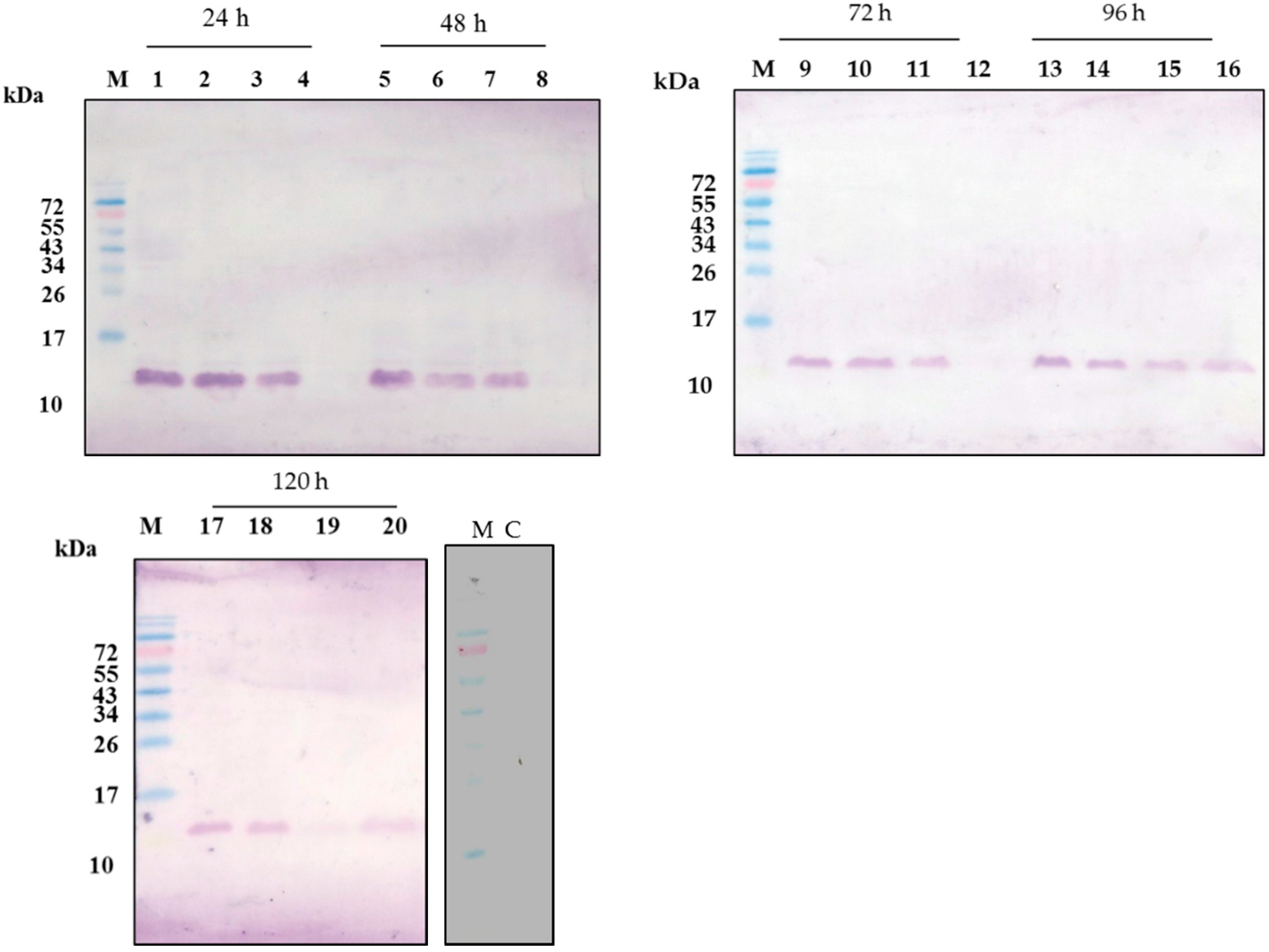
2.3. CecropinB2 Production in P. pastoris

To study the cecropinB2 expression in P. pastoris, three constructs, pGAPZαC-cecB2, pGAPZαC-INT-cecB2 and pGAPZαC-prepro-cecB2, were transformed into the host P. pastoris SMD1168. The construct of pGAPZαC-INT-cecB2 in host P. pastoris SMD1168 could not translate the target protein (INT-cecB2) well (data not shown). The protein INT originally discovered from bacteria might prohibit the expression in P. pastoris. In contrast, the constructs of pGAPZαC-cecB2 and pGAPZαC-prepro-cecB2 in host P. pastoris SMD1168 could properly express the target peptide. Among them, the construct pGAPZαC-prepro-cecB2 could express the target peptide with its signal peptide intracellularly to a high soluble level within 24 h (>80 mg/L) (Figure 2a). Meanwhile, the target peptides could be secreted out to the culture broth for a 96 h cultivation (Figure 2b). The target peptides with their highly positive charge might exhibit a large affinity to the host’s inner membrane (nagative-charge), which might cause the delay of the cecropinB2 secretion as shown in the time course of the production. For a longer cultivation time of 120 h, both the intracellular and extracellular target protein contents were significantly decreased, suggesting that the cells might gradually decompose the target protein (Figure 2c).
Figure 2. Western blot of pGAPZaC-prepro-cecB2/P. pastoris SMD1168. Lane M, molecular weight markers (kDa); Lane C: control (cell crude extract of pGAPZaC/P. pastoris SMD1168) lane 1–4, crude extract, pellet, supernatant, 100× concentrated extracellular fluid, at 24; lane 5–8, the same items at 48 h; lane 9–12, at 72 h; lane 13–16, at 96 h; lane 16–20, at 120 h cultivation time.
Molecules 21 00103 g002

2.4. Comparison of the Host Systems

The cecropinB2 production by applying various host strains of E. coli, B. subtilis and P. pastoris gave different results in the expression. The fusion protein approach could reduce the host directed toxicity of cecropinB2 and was successfully expressed in host E. coli. In contrast, it had a negative effect on hosts B. subtilis and P. pastoris. Comparison of cecropinB2 production by three expression systems showed that the highest yield occurred when using P. pastoris SMD1168 harboring the pGAPZαC-prepro-cecB2 (Table 1c). The highest yield was about 90 mg/L for a cultivation time of 24 h. This was better than that of the same construct pGAPZαC-cecB2 without prepro peptide (41 mg/L). Other constructs in this study gave yields lower than 40 mg/L. Based on the aforementioned results, it might be concluded that the secretion systems for B. subtilis and P. pastoris were greatly affected by the highly-positive-charge antibacterial peptide cecropinB2. The affinity between cecropinB2 and the cell membrane might build a barrier to limit the secretion process. This study also illustrated that, by properly constructing the host and plasmid system, the antibacterial peptide cecropinB2 production could be achieved.
Table 1. Yield of cecropinB2 under various expression systems.
ConstructTotal Protein Content (mg/L) 1Purity (%) 2Yield of CecropinB2 3
(a) E. coli
pET26b-cecB2/E. coli ER2566---
pET-28a-cecB2/E. coli ER2566---
pET28a-INT-cecB2/E. coli ER2566---
pET26b-INT-cecB2/E. coli ER256610220.8+
pET26b-10K-INT-cecB2/E. coli ER25669870.8+
pET26b-10R-INT-cecB2/E. coli ER256610550.8+
(b) B. subtilis
PRPA-cecB2/B. subtilis WB80011281.8+
PRPA-INT-cecB2/B. subtilis WB800---
(c) P. pastoris
pGAPZαC-cecB2/P. pastoris SMD116819802.4++
pGAPZαC-INT-cecB/P. pastoris SMD1168---
pGAPZαC-prepro-cecB2/P. pastoris SMD116820484.3+++
1 Total protein content was determined by the Bradford protein assay; symbol -: no detection; 2 Purity was determined by the image scanning; symbol -: no detection; 3 Definition of yield: purity of target protein (qualified via image scanning) × total protein content (quantified via Bradford); symbols -: no production; +: yield > 1 mg/L; ++: yield > 40 mg/L; +++: yield > 80 mg/L.

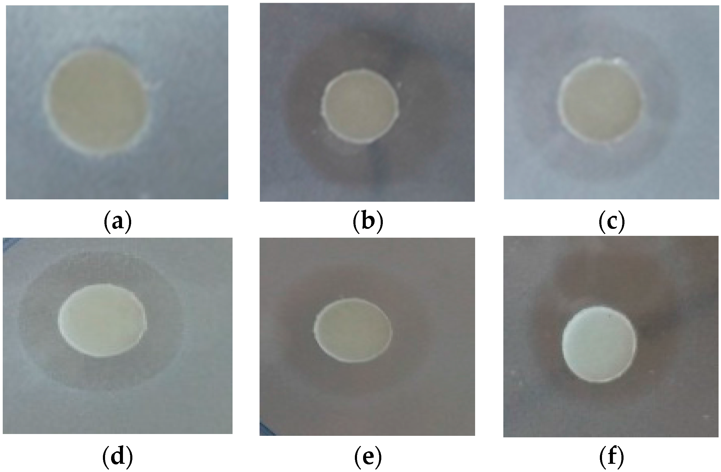
2.5. Antibacterial Tests

To check the antimicrobial activity effect, the purified cecropinB2 and the synthetic cecropinB2 were used to test the antibacterial activity, respectively. In the primary tests, the whole construct without gene CecB2 was expressed in the same host which didn’t show any activity against sensitive bacterial cells, indicating the fragments or chimeras did not respond to the antibacterial activity. In addition, the elution buffer and the extraction of host P. pastoris also displayed none of the inhibitory effect (data not shown). All these results gave evidence to the correct expression of cecropinB2 in the purified product. Figure 3 shows the antibactrial test results for E. coli strains. Various strains, i.e., E. coli strains (in Table 2), B. subtilis WB800, P. pastoris SMD1168, A. baumannii BCRC 15884 and A. baumannii E1359 (MDR) were used (Table 2). The results showed the purified cecropinB2 possessing the same activity as the synthetic peptides, which demonstrated strong antibacterial activity against the Gram-negative strains even in the case of A. baumannii; however, it did not exhibit toxicity toward the Gram-positive strains and eukaryotic cells such as P. pastoris (Table 2).
Figure 3. Antibacterial activity with agar diffusion test; where (a) elution buffer as blank; (bf) inhibition zone against E. coli ER2566, BL21, Rosetta, JM109 and DH1, respectively. The testing solution contains purified cecropinB2 (10 μg/mL) in the elution buffer.
Molecules 21 00103 g003
Table 2. Antibacterial spectrum of purified cecropinB2 1.
Bacteria StrainAntibacterial Activity 2
Purified CecropinB2 (10 μg/mL)Synthetic CecropinB2 (4 μg/mL)
E. coli ER25666.56.8
E. coli BL216.87.2
E. coli Rosetta7.17.2
E. coli JM1096.56.6
E. coli DH16.56.7
B. subtilis WB800--
P. pastoris SMD1168--
A. baumannii BCRC 158847.26.8
A. baumannii E13597.36.9
1 The cecropinB2 was purified from the host P. pastoris SMD1168 with the construct of pGAPZαC-prepro-cecB2; 2 Antibacterial activity was determined by the diameter (mm) of inhibition zone; -: no clear zone observed. Concentration of the purified cecropinB2 was determined via the Bradford assay and image scanning result.

3. Materials and Methods

3.1. Bacterial Strains, Plasmids and Synthetic CecropinB2

Four strains, i.e., E. coli strains DH5α, ER2566, B. subtilis strain WB800 and P. pastoris strain SMD1168, were used as the hosts for recombinant DNA manipulation and recombinant protein expression. The gene templates of antibacterial peptide pre-pro-cecropinB2 and plasmid PRPA were obtained, respectively, from Prof. Kuang-Hui Lu and Prof. Yeh Chuan-Mei [18] at the National Chung Hsing University, Taiwan. B. subtilis WB800 and P. pastoris SMD1168 were obtained from Prof. Chieh-Chen Huang and Prof. Huang Chien-jin at National Chung Hsing University, Taiwan, respectively. The synthetic cecropinB2 was purchased from Neogene Biomedicals Corp (Taiwan) as the standard. The number of amino acid and theoretical molecular mass of protein or peptide used in this study were summarized and are shown in Table 3.
Table 3. Number of amino acid and theoretical molecular mass.
ProteinNumber of Amino AcidTheoretical Molecular Mass (Da)
CecropinB2363749
INT-cecB219317,569
signal peptide (SP)182085
Propeptide5468

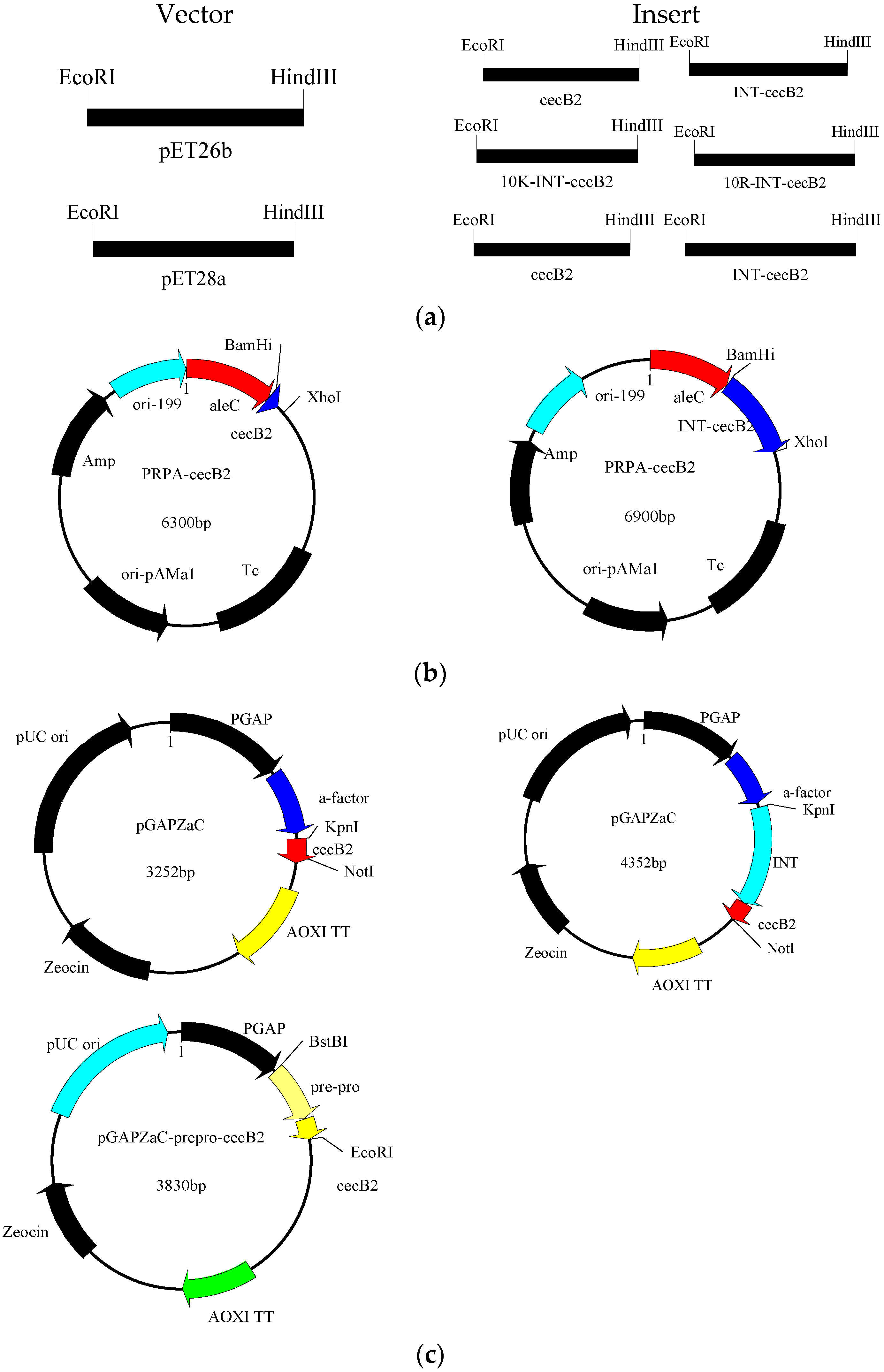
3.2. Construction of Expression Systems

The templates of prepero-cecropinB2 and plasmid pTWIN (New England Biolabs, Ipswich, MA, USA) were used to conduct an overlap extension PCR to obtain cecB2, pre-pro-cecropinB2 and INT-cecB2 which were further ligated to the vector pET26b, pET28a, PRPA and pGAPZaC, respectively. To increase the solubility, the amino acid segments of 10K and 10R were also used to fuse in the N-terminal of INT-cecB2 and ligated to the vector pET26b. The major plasmid constructs used in the study are showed in Figure 4. All recombinant DNA manipulations were performed following the standard procedures [19].
Figure 4. Plasmid constructs in this study. (a) Plasmid constructs in E. coli; (b) Plasmid constructs in B. subtilis; (c) Plasmid constructs in P. pastoris.
Molecules 21 00103 g004

3.3. Production of the Antibacterial Peptide

3.3.1. Culture of E. coli

The plasmid-transformed E. coli was grown overnight in 5 mL of Luria-Bertani (LB) medium, which was used to inoculate the main culture containing 100 mL of LB medium with 50 μg/mL kanamycin. The main culture was grown at 37 °C until OD600 reached 0.6. The cultivation was induced with 1 mM isopropyl-β-d-thiogalactopyranoside (IPTG) at 25 °C for another 6 h. Cells were then collected by centrifugation (6000× g, 5 min) and stored at 4 °C for further analysis.

3.3.2. Culture of B. subtilis

The plasmid-transformed B. subtilis was grown overnight in 5 mL of LB medium. The harvested broth was used to inoculate the main culture containing 100 mL of LB medium containing 10 μg/mL Tetracycline. The culture was grown at 30 °C for 24 h. Cells were then collected by centrifugation (6000× g, 5 min) and stored at 4 °C for further analysis.

3.3.3. Culture of P. pastoris

The plasmid-transformed P. pastoris was grown overnight in 5 mL of LB medium as the seed to inoculate 100 mL of YPD medium containing 50 μg/mL zeocin. The culture was grown at 30 °C for 120 h. The culture at different time intervals was collected and separated into cells and medium by centrifugation (6000× g, 5 min) and stored at 4 °C for further analysis.

3.4. Purification of Target Protein

The harvested cell pellet, with the the construct of pGAPZαC-prepro-cecB2/P. pastoris SMD1168, was re-suspended in Tris-HCl buffer (20 mM tris-HCl, pH 8.0, 50 mM NaCl) and then lysed by sonication using an ultrasonic processor at 20 W for 10 cycles (30 s working, 30 s free) to obtain the cell crude extract. The cell debris was pelleted at 6500 g at 4 °C for 20 min to obtain the supernatant and pellet. The supernatant was collected and applied to an immobilized-metal affinity chromatography resin (Ni2+-chelating) column. After being washed with the washing buffer (20 mM tris-HCl, pH 8.0, 50 mM NaCl, 20 mM imidazole), the product cecropinB2 was eluted with the elution buffer (20 mM tris-HCl, pH 8.0, 50 mM NaCl, 200 mM imidazole) and stored at 4 °C for futher use.

3.5. Analytical Methods

Protein analysis was carried out using 15% sodium dodecyl sulfate-polyacrylamide gel electrophoresis (SDS-PAGE) and coomassie blue staining according to the method of Laemmli [20]. For Western blot, the protein samples run on SDS-PAGE were transferred onto PVDF membrane. After being blocked with 1% BSA/TTBS (20 mM tris-HCl, pH 7.5, 0.9% NaCl, 0.1% tween-20) buffer, the membrane was washed three times with TTBS buffer and incubated with anti-His tag primary antibody (1:10,000) in 0.5% BSA/TTBS buffer at room temperature overnight. Then, the membrane was washed with TTBS buffer three times and incubated with anti-goat tag secondary antibody (1:10,000) in 0.5% BSA/TTBS buffer at room temperature for 2 h. After the three washings, staining was performed using the BCIP/NBT chromogenic system. The Bradford protein assay was used for total protein content measurement [21] and imageJ software (version 1.49s, NIH, Bethesda, MD, USA) was used for purity determination [22]. The yield of cecropinB2 was determined by multiple of the protein content with its purity.

3.6. Antibacterial Assay

Antibacterial activity was analyzed using the agar diffusion test [23]. Some 100 mL of LB broth containing 0.8% agar (LB agar) was autoclaved and then cooled to a temperature of 45 °C, followed by instantly adding an aliquot of 166 μL tested bacteria dilution solution (OD600 = 0.5, where OD600 represents the cell weight concentration). The 6 mL LB agar was poured over a 9.0 cm petri dish, giving an agar depth of 1 mm. After the agar solidified, 20 μL of the purified cecropinB2 was added to a paper disc (6 mm in diameter.) on the surface of the agar. The disc was incubated at 37 °C for 12 h. Antibacterial activity was indicated by the clear zone around the testing point. The antibacterial activity against MDR A. baumannii was tested in the experimental lab of Prof. Wu-Chun Tu at National Chung Hsing University, Taiwan.

4. Conclusions

In this study, three host strains were used to produce the antibacterial peptides. It was found that the relative yield of the cecropin B2 production could be rescribed in the order: P. pastoris > B. subtillis > E. coli. The best construct pGAPZαC-prepro-cecB2/P. pastoris SMD1168 could produce the antibacterial peptides within 24 h intracellularly, and the purified cecropinB2 showed strong antibacterial activity against the Gram-negative strains and even against that of MDR A. baumannii.

Acknowledgments

This work was supported by research grants provided by the National Science Council of Taiwan, R.O.C. (Grant No. NSC 103-2221-E-005-071-MY3).

Author Contributions

The author Wei-Shiang Lai is the major worker in the preparation of P. pastoris due to the frequent practice in this host. Shu-Chen Kan is the worker for the preparation of pasmid construct in host B. subtilis. Chia-Chi Lin is the worker for the preparation of pasmid construct in host E. coli. Chwen-Jen Shieh is the worker for the antibacterial test especially in the muti-drug resistant strain test. The author Yung-Chuan Liu is the major coindicator in this study and corresponding author for the whole work.

Conflicts of Interest

The authors declare no conflict of interest.

References

  1. Fleming, A. In-vitro test of pencillin potency. Lancet 1942, 239, 732–733. [Google Scholar] [CrossRef]
  2. Nikaido, H. Multidrug Resistance in Bacteria. Annu. Rev. Biochem. 2009, 78, 119–146. [Google Scholar] [CrossRef] [PubMed]
  3. Wu, J.M.; Jan, P.S.; Yu, H.C.; Haung, H.Y.; Fang, H.J.; Chang, Y.I.; Cheng, J.W.; Chen, H.M. Structure and function of a custom anticancer peptide, CB1a. Peptides 2009, 30, 839–848. [Google Scholar] [CrossRef] [PubMed]
  4. Hoskin, D.W.; Ramamoorthy, A. Studies on anticancer activities of antimicrobial peptides. Biochim. Biophys. Acta 2008, 1778, 357–375. [Google Scholar] [CrossRef] [PubMed]
  5. Bahar, A.; Ren, D. Antimicrobial Peptides. Pharmaceuticals 2013, 6, 1543. [Google Scholar] [CrossRef] [PubMed]
  6. Chen, H.M.; Wang, W.; Smith, D.; Chan, S.C. Effects of the anti-bacterial peptide cecropin B and its analogs, cecropins B-1 and B-2, on liposomes, bacteria, and cancer cells. Biochim. Biophys. Acta Gen. Subj. 1997, 1336, 171–179. [Google Scholar] [CrossRef]
  7. Steiner, H.; Hultmark, D.; Engstrom, A.; Bennich, H.; Boman, H.G. Sequence and specificity of two antibacterial proteins involved in insect immunity. Nature 1981, 292, 246–248. [Google Scholar] [CrossRef] [PubMed]
  8. Schweizer, F. Cationic amphiphilic peptides with cancer-selective toxicity. Eur. J. Pharmacol. 2009, 625, 190–194. [Google Scholar] [CrossRef] [PubMed]
  9. Xu, X.; Jin, F.; Yu, X.; Ji, S.; Wang, J.; Cheng, H.; Wang, C.; Zhang, W. Expression and purification of a recombinant antibacterial peptide, cecropin, from Escherichia coli. Protein Expr. Purif. 2007, 53, 293–301. [Google Scholar] [CrossRef] [PubMed]
  10. Xu, Z.; Zhong, Z.; Huang, L.; Peng, L.; Wang, F.; Cen, P. High-level production of bioactive human beta-defensin-4 in Escherichia coli by soluble fusion expression. Appl. Microbiol. Biotechnol. 2006, 72, 471–479. [Google Scholar] [CrossRef] [PubMed]
  11. Schumann, W. Production of Recombinant Proteins in Bacillus subtilis. Adv. Appl. Microbiol. 2007, 62, 137–189. [Google Scholar] [PubMed]
  12. Rosano, G.L.; Ceccarelli, E.A. Recombinant protein expression in Escherichia coli: Advances and challenges. Front. Microbiol. 2014, 5, 172. [Google Scholar] [CrossRef] [PubMed]
  13. Cregg, J.; Cereghino, J.; Shi, J.; Higgins, D. Recombinant protein expression in Pichia pastoris. Mol. Biotechnol. 2000, 16, 23–52. [Google Scholar] [CrossRef]
  14. Skosyrev, V.S.; Rudenko, N.V.; Yakhnin, A.V.; Zagranichny, V.E.; Popova, L.I.; Zakharov, M.V.; Gorokhovatsky, A.Y.; Vinokurov, L.M. EGFP as a fusion partner for the expression and organic extraction of small polypeptides. Protein Expr. Purif. 2003, 27, 55–62. [Google Scholar] [CrossRef]
  15. Zhang, J.; Movahedi, A.; Xu, J.; Wang, M.; Wu, X.; Xu, C.; Yin, T.; Zhuge, Q. In vitro production and antifungal activity of peptide ABP-dHC-cecropin A. J. Biotechnol. 2015, 199, 47–54. [Google Scholar] [CrossRef] [PubMed]
  16. Yang, K.; Su, Y.; Li, J.; Sun, J.; Yang, Y. Expression and purification of the antimicrobial peptide cecropin AD by fusion with cationic elastin-like polypeptides. Protein Expr. Purif. 2012, 85, 200–203. [Google Scholar] [CrossRef] [PubMed]
  17. Jung, H.-J.; Kim, S.-K.; Min, W.-K.; Lee, S.-S.; Park, K.; Park, Y.-C.; Seo, J.-H. Polycationic amino acid tags enhance soluble expression of Candida antarctica lipase B in recombinant Escherichia coli. Bioprocess Biosyst. Eng. 2011, 34, 833–839. [Google Scholar] [CrossRef] [PubMed]
  18. Yeh, C.-M.; Wang, J.-P.; Su, F.-S. Extracellular Production of a Novel Ice Structuring Protein byBacillus subtilis—A Case of Recombinant Food Peptide Additive Production. Food Biotechnol. 2007, 21, 119–128. [Google Scholar] [CrossRef]
  19. Sambrook, J.R. Molecular Cloning: A Laboratory Manual, 3rd ed.; Cold Spring Harbor Laboratory Press: New York, NY, USA, 2001. [Google Scholar]
  20. Laemmli, U.K. Cleavage of Structural Proteins during the Assembly of the Head of Bacteriophage T4. Nature 1970, 227, 680–685. [Google Scholar] [CrossRef] [PubMed]
  21. Bradford, M.M. A rapid and sensitive method for the quantitation of microgram quantities of protein utilizing the principle of protein-dye binding. Anal. Biochem. 1976, 72, 248–254. [Google Scholar] [CrossRef]
  22. Schneider, C.A.; Rasband, W.S.; Eliceiri, K.W. NIH Image to ImageJ: 25 years of image analysis. Nat. Meth. 2012, 9, 671–675. [Google Scholar] [CrossRef]
  23. Ekengren, S.; Hultmark, D. Drosophila cecropin as an antifungal agent. Insect Biochem. Mol. Biol. 1999, 29, 965–972. [Google Scholar] [CrossRef]
  • Sample Availability: Not available.

