Isolation and Crystal Structure of Marcanine A from Polyalthia plagioneura

Abstract
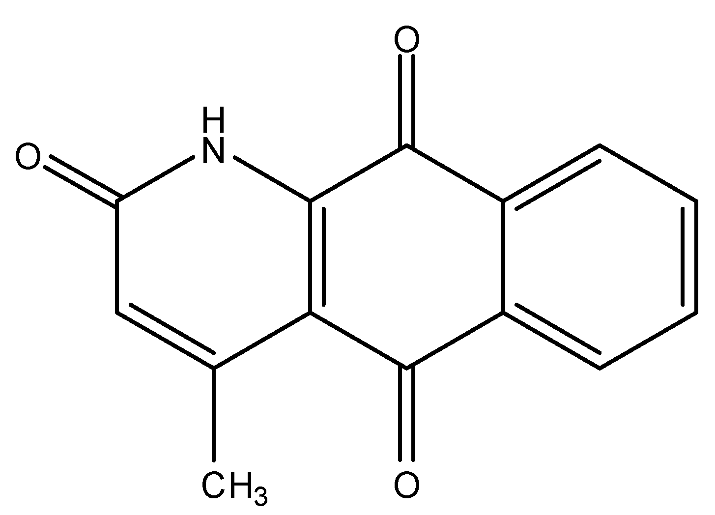
:1. Introduction

2. Results and Discussion
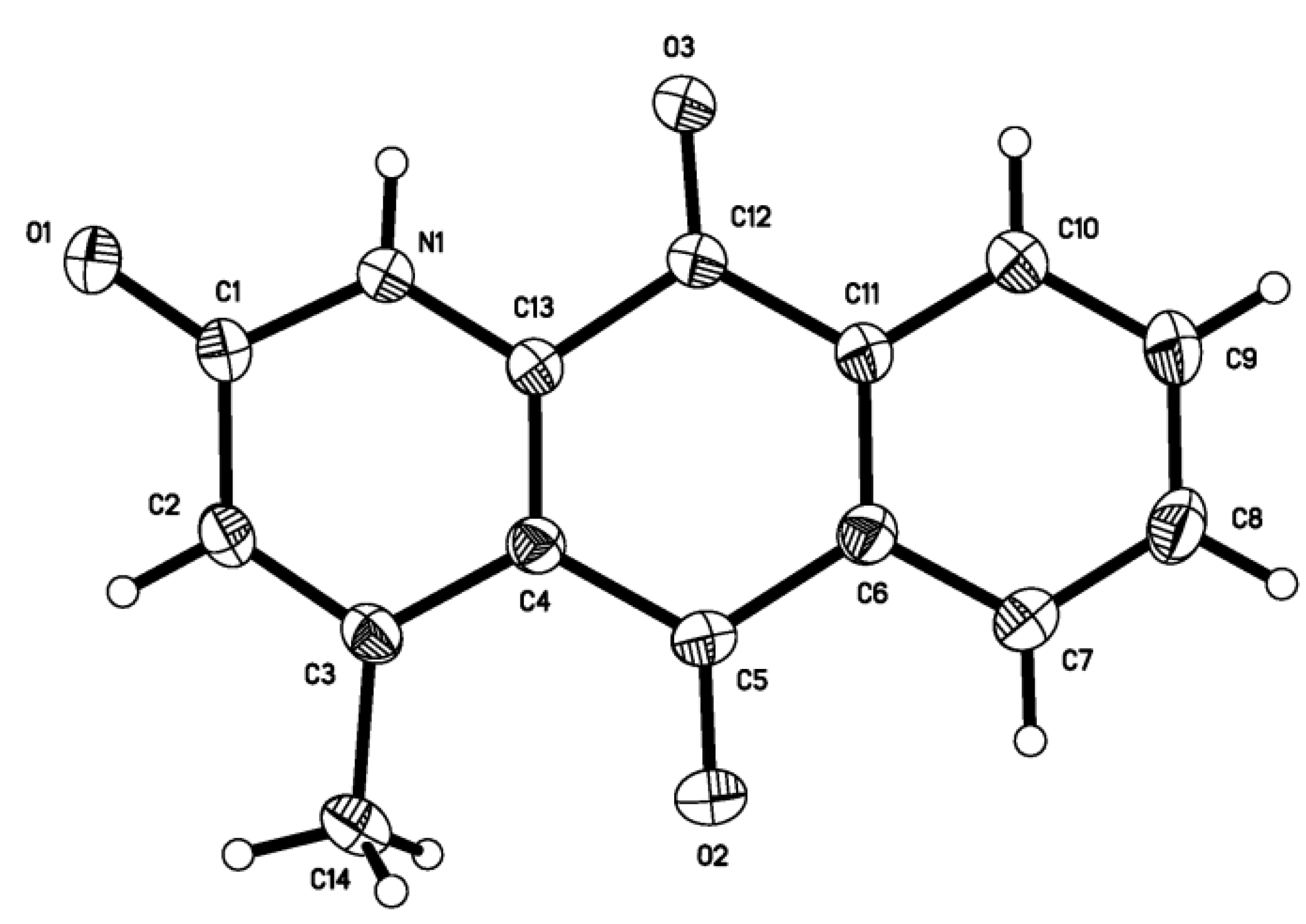
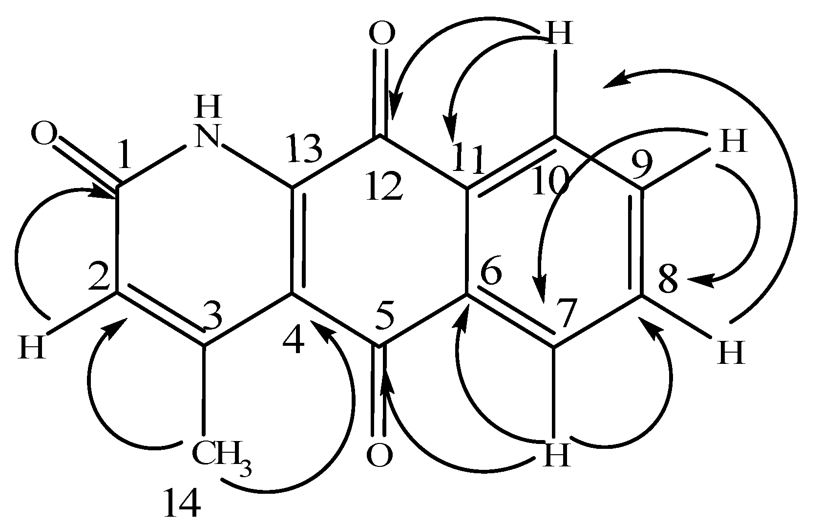
2.1. Crystal structure determination

| Empirical formula | C14H9NO3 | Volume (Å3) | 533.74(10) |
| Formula weight | Mr =239.22 | Absorption coefficient (mm-1) | 0.106 |
| Z | 2 | F(000) | 248 |
| Dc (Mg/m3) | 1.489 | Crystal size mm) | 0.50× 0.42 × 0.41 |
| Color, shape | light yellow, block | θ range for data collection | 2.03-25.01 |
| Temperature (K) | 293K | Index ranges | h = −6→6 |
| Wavelength (Å) | 0.71073 | k = −8→12 | |
| Crystal system | Triclinic | l = −13→9 | |
| Space group | P-1 | Reflections collected | 2776 |
| Cell dimensions | Independent reflections | 1849 | |
| a(Å) | 5.2140(5) | Rint | 0.0148 |
| b(Å) | 10.1871(11) | F2 | 1.032 |
| c(Å) | 11.0709(13) | Max. and min. transmission | 0.9577 and 0.9488 |
| α(º) | 110.452(2) | Data/restraints/parameters | 1849/0/164 |
| β(º) | 103.376(2) | Final R indices (I > 2σ(I)) | R=0.041 |
| γ(º) | 90.1870(10) | wR=0.1104 |
| Bond | Dist. | Bond | Dist. | Bond | Dist. |
|---|---|---|---|---|---|
| N(1)-C(13) | 1.352(2) | N(1)-C(1) | 1.383(2) | O(3)-C(12) | 1.2156(18) |
| O(1)-C(1) | 1.237(2) | O(2)-C(5) | 1.214(2) | C(4)-C(13) | 1.372(2) |
| C(2)-C(3) | 1.358(2) | C(6)-C(11) | 1.398(2) | C(3)-C(4) | 1.449(2) |
| C(1)-C(2) | 1.431(3) | C(3)-C(14) | 1.499(2) | C(6)-C(7) | 1.392(3) |
| C(4)-C(5) | 1.481(2) | C(5)-C(6) | 1.493(3) | C(9)-C(10) | 1.383(3) |
| C(7)-C(8) | 1.379(3) | C(8)-C(9) | 1.379(3) | C(12)-C(13) | 1.499(2) |
| C(10)-C(11) | 1.393(2) | C(11)-C(12) | 1.476(2) | ||
| Angle | (°) | Angle | (°) | ||
| N(1)-C(1)-C(2) | 114.28(15) | O(3)-C(12)-C(11) | 123.66(15) | ||
| O(1)-C(1)-N(1) | 120.79(16) | N(1)-C(13)-C(12) | 114.72(14) | ||
| C(13)-N(1)-C(1) | 123.43(14) | O(1)-C(1)-C(2) | 124.93(16) | ||
| O(2)-C(5)-C(6) | 120.10(16) | C(6)-C(11)-C(12) | 119.95(15) | ||
| O(3)-C(12)-C(13) | 118.93(15) | C(3)-C(2)-C(1) | 124.54(16) | ||
| C(2)-C(3)-C(14) | 118.98(15) | C(2)-C(3)-C(4) | 117.64(16) | ||
| C(13)-C(4)-C(3) | 117.98(15) | C(4)-C(3)-C(14) | 123.37(16) | ||
| C(3)-C(4)-C(5) | 122.70(15) | C(13)-C(4)-C(5) | 119.32(15) | ||
| C(4)-C(5)-C(6) | 118.36(15) | O(2)-C(5)-C(4) | 121.53(16) | ||
| C(7)-C(6)-C(5) | 119.17(16) | C(7)-C(6)-C(11) | 119.16(16) | ||
| C(8)-C(7)-C(6) | 120.08(18) | C(11)-C(6)-C(5) | 121.67(15) | ||
| C(8)-C(9)-C(10) | 119.82(17) | C(9)-C(10)-C(11) | 119.97(17) | ||
| C(10)-C(11)-C(6) | 120.10(16) | C(11)-C(12)-C(13) | 117.39(14) | ||
| C(9)-C(8)-C(7) | 120.87(18) | C(10)-C(11)-C(12) | 119.95(15) | ||
| N(1)-C(13)-C(4) | 122.09(14) | C(4)-C(13)-C(12) | 123.15(15) | ||
| C(13)-N(1)-C(1)-O(1) | -178.72(16) | C(13)-N(1)-C(1)-C(2) | 0.5(3) | ||
| O(1)-C(1)-C(2)-C(3) | -179.75(18) | N(1)-C(1)-C(2)-C(3) | 1.0(3) | ||
| C(1)-C(2)-C(3)-C(4) | -0.8(3) | C(1)-C(2)-C(3)-C(14) | -179.30(18) | ||
| C(2)-C(3)-C(4)-C(13) | -1.1(3) | C(14)-C(3)-C(4)-C(13) | 177.43(17) | ||
| C(2)-C(3)-C(4)-C(5) | 179.82(17) | C(14)-C(3)-C(4)-C(5) | -1.7(3) | ||
| C(13)-C(4)-C(5)-O(2) | 175.98(18) | C(3)-C(4)-C(5)-O(2) | -4.9(3) | ||
| C(13)-C(4)-C(5)-C(6) | -4.4(3) | C(3)-C(4)-C(5)-C(6) | 174.76(15) | ||
| O(2)-C(5)-C(6)-C(7) | 2.3(3) | C(4)-C(5)-C(6)-C(7) | -177.41(16) | ||
| O(2)-C(5)-C(6)-C(11) | -178.83(18) | C(4)-C(5)-C(6)-C(11) | 1.5(3) | ||
| C(11)-C(6)-C(7)-C(8) | -0.3(3) | C(5)-C(6)-C(7)-C(8) | 178.65(17) | ||
| C(6)-C(7)-C(8)-C(9) | 0.0(3) | C(7)-C(8)-C(9)-C(10) | 0.4(3) | ||
| C(8)-C(9)-C(10)-C(11) | -0.5(3) | C(9)-C(10)-C(11)-C(6) | 0.2(3) | ||
| C(9)-C(10)-C(11)-C(12) | -179.87(17) | C(7)-C(6)-C(11)-C(10) | 0.2(3) | ||
| C(5)-C(6)-C(11)-C(10) | -178.72(17) | C(7)-C(6)-C(11)-C(12) | -179.71(17) | ||
| C(5)-C(6)-C(11)-C(12) | 1.4(3) | C(10)-C(11)-C(12)-O(3) | 0.0(3) | ||
| C(6)-C(11)-C(12)-O(3) | 179.91(17) | C(10)-C(11)-C(12)-C(13) | 178.61(16) | ||
| C(6)-C(11)-C(12)-C(13) | -1.5(3) | C(1)-N(1)-C(13)-C(4) | -2.4(3) | ||
| C(1)-N(1)-C(13)-C(12) | 175.17(15) | C(3)-C(4)-C(13)-N(1) | 2.6(3) | ||
| C(5)-C(4)-C(13)-N(1) | -178.24(16) | C(3)-C(4)-C(13)-C(12) | -174.76(15) | ||
| C(5)-C(4)-C(13)-C(12) | 4.4(3) | O(3)-C(12)-C(13)-N(1) | -0.4(2) | ||
| C(11)-C(12)-C(13)-N(1) | -179.02(15) | O(3)-C(12)-C(13)-C(4) | 177.18(17) | ||
| C(11)-C(12)-C(13)-C(4) | -1.5(3) | ||||
| D-H···A | D-H | H‑A | D···A | D-H···A |
|---|---|---|---|---|
| N (1)-H (1)···O(1)a | 0.860 | 2.05 | 2.880 | 162 |
| C (10)-H (10)···O(3)b | 0.93 | 2.45 | 3.245(2) | 143 |
| C (7)-H (7)···O(2)c | 0.93 | 2.38 | 3.279(2) | 162 |


2.2. Cytotoxicity
| Tumor cell species | Inhibition(%) | IC50/μM | |||||
|---|---|---|---|---|---|---|---|
| 0.1μM | 1μM | 5μM | 10μM | 50μM | 100μM | ||
| BEL-7402 | -28.55 | -30.16 | 2.42 | 30.07 | 100.12 | 100.46 | 9.54 |
| K562 | -46.03 | -0.25 | 19.93 | 33.67 | 92.15 | 102.25 | 11.78 |
| SPCA-1 | -19.45 | 8.43 | 6.20 | 44.37 | 99.50 | 97.61 | 8.69 |
| SGC-7409 | 13.58 | 14.16 | 42.12 | 71.87 | 119.73 | 122.45 | 1.53 |
3. Experimental
3.1. Plant material
3.2. Extraction and separation
3.3. Sructure determination

3.4. Crystal structure determination
3.5. Biological activity
3.5.1. Cell culture
3.5.2. Cytotoxicity assay
4. Conclusions
Acknowledgements
- Samples Availability: Samples of the compounds are available from the authors.
References and Notes
- Jiang, Y.; Li, B.T. Flora of China; Science Press: Beijing, China, 1979; Volume 30, p. 87. [Google Scholar]
- Zheng, X.C.; Yang, R.Z.; Xu, R.S.; Qin, G.W. PlagionicinA with C5, a new monotetrahydrofuran acetogenin. Acta Bot. Sin. 1994, 36, 557–560. [Google Scholar]
- Padmaa, M.P.; Khosa, R.L. Phytoconstituents from the genus Polyalthia–a review. J. Pharm. Res. 2009, 2, 594–605. [Google Scholar]
- Soonthornchareonnon, N.; Suwanborirux, K.; Bavovada, R.; Patarapanich, C.; Cassady, J.M. Synthesis of Kalasinamide, a Putative Plant Defense Phototoxin. J. Nat. Prod. 1999, 62, 1390–1394. [Google Scholar] [CrossRef]
- Ichino, C.; Soonthornchareonnon, N.; Chuakul, W.; Kiyohara, H.; Ishiyama, A.; Sekiguchi, H.; Namatame, M.; Otoguro, K.; Omura, S.; Yamada, H. Screening of Thai medicinal plant extracts and their active constituents for in vitro antimalarial activity. Phytother. Res. 2006, 20, 307–309. [Google Scholar] [CrossRef]
- Marcos, A.; Pedregal, C.; Avendano, C. Annulations of 2-aminonaphthoquinone with aldehydes and acetals. Tetrahedron 1994, 50, 12941. [Google Scholar] [CrossRef]
- Michael, N.G.; Matthew, J.P. Synthesis of Kalasinamide, a Putative Plant Defense Phototoxin. J. Nat. Prod. 2008, 71, 866–868. [Google Scholar]
- Sheldrick, G.M. SHELXTL 5.10 for Windows NT: Structure Determination Software Programs. Bruker Analytical X-ray Systems: Madison, WI, USA, 1997. [Google Scholar]
- Tim, M. Rapid colorimetric assay for cellular growth and survival: application to proliferation and cytotoxicity assays. J. Immunol. Methods 1983, 65, 55–63. [Google Scholar] [CrossRef]
© 2010 by the authors; licensee MDPI, Basel, Switzerland. This article is an open access article distributed under the terms and conditions of the Creative Commons Attribution license (http://creativecommons.org/licenses/by/3.0/).
Share and Cite
Liu, B.; Wang, L.; Chen, G.; Han, C.; Wang, J. Isolation and Crystal Structure of Marcanine A from Polyalthia plagioneura. Molecules 2010, 15, 6349-6356. https://doi.org/10.3390/molecules15096349
Liu B, Wang L, Chen G, Han C, Wang J. Isolation and Crystal Structure of Marcanine A from Polyalthia plagioneura. Molecules. 2010; 15(9):6349-6356. https://doi.org/10.3390/molecules15096349
Chicago/Turabian StyleLiu, Bingjing, Lin Wang, Guangying Chen, Changri Han, and Jing Wang. 2010. "Isolation and Crystal Structure of Marcanine A from Polyalthia plagioneura" Molecules 15, no. 9: 6349-6356. https://doi.org/10.3390/molecules15096349