Topic Editors

Department of Orthodontics and Dentofacial Orthopedics, University of Bern, Freiburgstrasse 7, CH-3010 Bern, Switzerland
Prof. Dr. Carlalberta Verna
Department of Pediatric Oral Health and Orthodontics, University Center for Dental Medicine Basel UZB, University of Basel, Hebelstrasse 3, CH-4056 Basel, Switzerland

Diagnosis of Craniofacial Changes: Conventional Approaches and Novel Methodologies

Abstract submission deadline
closed (20 May 2023)
Manuscript submission deadline
closed (20 October 2023)
Viewed by
43890

Topic Information

Dear Colleagues,

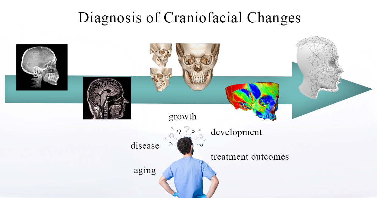
The assessment of changes in craniofacial structures is crucial for several disciplines for proper diagnosis, treatment planning, and outcome assessment. Such changes can be physiological, due to growth, development, and aging, pathological due to a disease, or can be induced by medical interventions. Apart from correct diagnosis and documentation, which are both needed for medical and legal reasons, the only way to control changes is to have accurate patient representations at different time points. However, it is expected that even consecutive craniofacial images, obtained within a short time span and under the same setting, will differ to each other due to factors related either to the imaging technique or to the object itself. Valid assessment of craniofacial changes over larger time spans is even more challenging.

The craniofacial area is highly important for human life, both in terms of functionality and esthetic appearance. Therefore, valid detection of associated changes is fundamental, and several approaches have been developed for this over the years. Traditional methods primarily concern two-dimensional images, but, lately, various 3D imaging techniques have been incorporated in daily research and practice. The forthcoming article collection will include literature reviews and original studies focusing on imaging modalities and methodologies used to assess craniofacial changes over time.

Dr. Nikolaos Gkantidis
Prof. Dr. Carlalberta Verna
Topic Editors

Keywords

  • craniofacial imaging
  • radiography
  • facial photography
  • anthropometry
  • cephalometrics
  • computed tomography
  • stereophotogrammetry
  • superimposition
  • geometric morphometrics
  • finite element analysis
  • machine learning
  • artificial intelligence

Participating Journals

Journal Name Impact Factor CiteScore Launched Year First Decision (median) APC
International Journal of Environmental Research and Public Health
ijerph
- 5.4 2004 29.6 Days CHF 2500
Journal of Clinical Medicine
jcm
3.9 5.4 2012 17.9 Days CHF 2600
Biology
biology
4.2 4.0 2012 18.7 Days CHF 2700
Diagnostics
diagnostics
3.6 3.6 2011 20.7 Days CHF 2600
Dentistry Journal
dentistry
2.6 4.0 2013 27.8 Days CHF 2000

Preprints.org is a multidiscipline platform providing preprint service that is dedicated to sharing your research from the start and empowering your research journey.

MDPI Topics is cooperating with Preprints.org and has built a direct connection between MDPI journals and Preprints.org. Authors are encouraged to enjoy the benefits by posting a preprint at Preprints.org prior to publication:

  1. Immediately share your ideas ahead of publication and establish your research priority;
  2. Protect your idea from being stolen with this time-stamped preprint article;
  3. Enhance the exposure and impact of your research;
  4. Receive feedback from your peers in advance;
  5. Have it indexed in Web of Science (Preprint Citation Index), Google Scholar, Crossref, SHARE, PrePubMed, Scilit and Europe PMC.

Published Papers (20 papers)

Order results
Result details
Journals
Select all
Export citation of selected articles as:
8 pages, 547 KiB  
Communication
Recognizability of Demographically Altered Computerized Facial Approximations in an Automated Facial Recognition Context for Potential Application in Unidentified Persons Data Repositories
by Connie L. Parks and Keith L. Monson
Biology 2023, 12(5), 682; https://doi.org/10.3390/biology12050682 - 04 May 2023
Viewed by 1021
Abstract
This study examined the recognizability of demographically altered facial approximations for potential utility in unidentified persons tracking systems. Five computer-generated approximations were generated for each of 26 African male participants using the following demographic parameters: (i) African male (true demographics), (ii) African female, [...] Read more.
This study examined the recognizability of demographically altered facial approximations for potential utility in unidentified persons tracking systems. Five computer-generated approximations were generated for each of 26 African male participants using the following demographic parameters: (i) African male (true demographics), (ii) African female, (iii) Caucasian male, (iv) Asian male, and (v) Hispanic male. Overall, 62% of the true demographic facial approximations for the 26 African male participants examined were matched to a corresponding life photo within the top 50 images of a candidate list generated from an automated blind search of an optimally standardized gallery of 6159 photographs. When the African male participants were processed as African females, the identification rate was 50%. In contrast, less congruent identification rates were observed when the African male participants were processed as Caucasian (42%), Asian (35%), and Hispanic (27%) males. The observed results suggest that approximations generated using the opposite sex may be operationally informative if sex is unknown. The performance of approximations generated using alternative ancestry assignments, however, was less congruent with the performance of the true demographic approximation (African male) and may not yield as operationally constructive data as sex-altered approximations. Full article
Show Figures

Figure 1

11 pages, 2703 KiB  
Article
A Cephalometric Analysis Assessing the Validity of Camper’s Plane to Establishing the Occlusal Plane in Edentulous Patients
by Lina Sharab, David Jensen, Gregory Hawk and Ahmad Kutkut
Dent. J. 2023, 11(3), 81; https://doi.org/10.3390/dj11030081 - 15 Mar 2023
Viewed by 2165
Abstract
Complete denture fabrication requires multiple clinical and laboratory steps. One of the most critical clinical steps is establishing an anatomical occlusal plane based on hard and soft tissue references. The aim of this study was to determine whether age or gender affects the [...] Read more.
Complete denture fabrication requires multiple clinical and laboratory steps. One of the most critical clinical steps is establishing an anatomical occlusal plane based on hard and soft tissue references. The aim of this study was to determine whether age or gender affects the level of the Ala-Tragus plane to establish which reference point on the Tragus should be used when fabricating the occlusal plane in edentulous patients. Clinical photographs and lateral cephalometric radiographs with complete dentitions were taken from 58 volunteers at the DMD clinic at the University of Kentucky. Each photograph was superimposed over its corresponding cephalometric image. An analysis was conducted to establish the angle of the occlusal plane relative to the Ala-Tragus landmarks; this data was then grouped according to age and gender. The analysis shows that age and gender did not significantly affect where the Camper’s plane should be approximated for complete denture treatment. However, it was found that the most parallel line to the occlusal plane was Ala’s inferior border to the ‘Tragus’s inferior border. It should be noted that the volunteers’ skeletal classification was significantly related to a Cl III malocclusion tendency. Still, with this new information, functionality and esthetics can be more adequately addressed for patients undergoing complete denture treatment. Given our results, we suggest redefining the ‘Camper’s plane with a line extending from ‘Ala’s inferior border to the ‘Tragus’s inferior border instead of the superior border. Further consideration should be taken if the patient is a skeletal CL III malocclusion. Full article
Show Figures

Figure 1

14 pages, 3579 KiB  
Article
Intraoral Scanners for In Vivo 3D Imaging of the Gingiva and the Alveolar Process
by Jonas Winkler, Anton Sculean and Nikolaos Gkantidis
J. Clin. Med. 2022, 11(21), 6389; https://doi.org/10.3390/jcm11216389 - 28 Oct 2022
Cited by 2 | Viewed by 1411
Abstract
This study aimed to assess the reliability of two intraoral surface scanners for the representation of the alveolar process in vivo. Complete maxillary scans (CS 3600, Carestream and TRIOS 3, 3Shape) were repeatedly obtained from 13 fully dentate individuals. Scanner precision and agreement [...] Read more.
This study aimed to assess the reliability of two intraoral surface scanners for the representation of the alveolar process in vivo. Complete maxillary scans (CS 3600, Carestream and TRIOS 3, 3Shape) were repeatedly obtained from 13 fully dentate individuals. Scanner precision and agreement were tested using 3D surface superimpositions on the following reference areas: the buccal front teeth area, the entire dental arch, the entire alveolar process, or single teeth by applying an iterative closest point algorithm. Following each superimposition, the mean absolute distance (MAD) between predefined 3D model surfaces was calculated. Outcomes were analyzed through non-parametric statistics and the visualization of color-coded distance maps. When superimpositions were performed on the alveolar process, the median scanner precision was below 0.05 mm, with statistically significant but negligible differences between scanners. The agreement between the scanners was approximately 0.06 mm. When single-tooth superimpositions were used to assess the precision of adjacent alveolar soft-tissue surfaces, the median error was 0.028 mm, and there was higher agreement between the scanners. The in vivo reliability of the intraoral scanners in the alveolar surface area was high overall. Single-tooth superimpositions should be preferred for the optimal assessment of neighboring alveolar surface areas relative to the dentition. Full article
Show Figures

Figure 1

19 pages, 1741 KiB  
Article
Novel Candidate Genes for Non-Syndromic Tooth Agenesis Identified Using Targeted Next-Generation Sequencing
by Barbara Biedziak, Ewa Firlej, Justyna Dąbrowska, Agnieszka Bogdanowicz, Małgorzata Zadurska and Adrianna Mostowska
J. Clin. Med. 2022, 11(20), 6089; https://doi.org/10.3390/jcm11206089 - 15 Oct 2022
Cited by 6 | Viewed by 1791
Abstract
Non-syndromic tooth agenesis (ns-TA) is one of the most common dental anomalies characterized by the congenital absence of at least one permanent tooth (excluding third molars). Regarding the essential role of genetic factors in ns-TA aetiology, the present study aimed to identify novel [...] Read more.
Non-syndromic tooth agenesis (ns-TA) is one of the most common dental anomalies characterized by the congenital absence of at least one permanent tooth (excluding third molars). Regarding the essential role of genetic factors in ns-TA aetiology, the present study aimed to identify novel pathogenic variants underlying hypodontia and oligodontia. In a group of 65 ns-TA patients and 127 healthy individuals from the genetically homogenous Polish population, the coding sequences of 423 candidate genes were screened using targeted next-generation sequencing. Pathogenic and likely pathogenic variants were identified in 37 (56.92%) patients, including eight nucleotide alternations of genes not previously implicated in ns-TA (CHD7, CREBBP, EVC, LEF1, ROR2, TBX22 and TP63). However, since only single variants were detected, future research is required to confirm and fully understand their role in the aetiology of ns-TA. Additionally, our results support the importance of already known ns-TA candidate genes (AXIN2, EDA, EDAR, IRF6, LAMA3, LRP6, MSX1, PAX9 and WNT10A) and provide additional evidence that ns-TA might be an oligogenic condition involving the cumulative effect of rare variants in two or more distinct genes. Full article
Show Figures

Figure 1

12 pages, 3967 KiB  
Article
Precision of a Hand-Held 3D Surface Scanner in Dry and Wet Skeletal Surfaces: An Ex Vivo Study
by Jannis Probst, Konstantinos Dritsas, Demetrios Halazonetis, Yijin Ren, Christos Katsaros and Nikolaos Gkantidis
Diagnostics 2022, 12(9), 2251; https://doi.org/10.3390/diagnostics12092251 - 18 Sep 2022
Cited by 8 | Viewed by 1585
Abstract
Three-dimensional surface scans of skeletal structures have various clinical and research applications in medicine, anthropology, and other relevant fields. The aim of this study was to test the precision of a widely used hand-held surface scanner and the associated software’s 3D model generation-error [...] Read more.
Three-dimensional surface scans of skeletal structures have various clinical and research applications in medicine, anthropology, and other relevant fields. The aim of this study was to test the precision of a widely used hand-held surface scanner and the associated software’s 3D model generation-error in both dry and wet skeletal surfaces. Ten human dry skulls and ten mandibles (dry and wet conditions) were scanned twice with an industrial scanner (Artec Space Spider) by one operator. Following a best-fit superimposition of corresponding surface model pairs, the mean absolute distance (MAD) between them was calculated on ten anatomical regions on the skulls and six on the mandibles. The software’s 3D model generation process was repeated for the same scan of four dry skulls and four mandibles (wet and dry conditions), and the results were compared in a similar manner. The median scanner precision was 31 μm for the skulls and 25 μm for the mandibles in dry conditions, whereas in wet conditions it was slightly lower at 40 μm for the mandibles. The 3D model generation-error was negligible (range: 5–10 μm). The Artec Space Spider scanner exhibits very high precision in the scanning of dry and wet skeletal surfaces. Full article
Show Figures

Figure 1

18 pages, 6794 KiB  
Article
Semantic Segmentation of Maxillary Teeth and Palatal Rugae in Two-Dimensional Images
by Abdul Rehman El Bsat, Elie Shammas, Daniel Asmar, George E. Sakr, Kinan G. Zeno, Anthony T. Macari and Joseph G. Ghafari
Diagnostics 2022, 12(9), 2176; https://doi.org/10.3390/diagnostics12092176 - 08 Sep 2022
Cited by 4 | Viewed by 3191
Abstract
The superimposition of sequential radiographs of the head is commonly used to determine the amount and direction of orthodontic tooth movement. A harmless method includes the timely unlimited superimposition on the relatively stable palatal rugae, but the method is performed manually and, if [...] Read more.
The superimposition of sequential radiographs of the head is commonly used to determine the amount and direction of orthodontic tooth movement. A harmless method includes the timely unlimited superimposition on the relatively stable palatal rugae, but the method is performed manually and, if automated, relies on the best fit of surfaces, not only rugal structures. In the first step, motion estimation requires segmenting and detecting the location of teeth and rugae at any time during the orthodontic intervention. Aim: to develop a process of tooth segmentation that eliminates all manual steps to achieve an autonomous system of assessment of the dentition. Methods: A dataset of 797 occlusal views from photographs of teeth was created. The photographs were manually semantically segmented and labeled. Machine learning methods were applied to identify a robust deep network architecture able to semantically segment teeth in unseen photographs. Using well-defined metrics such as accuracy, precision, and the average mean intersection over union (mIoU), four network architectures were tested: MobileUnet, AdapNet, DenseNet, and SegNet. The robustness of the trained network was additionally tested on a set of 47 image pairs of patients before and after orthodontic treatment. Results: SegNet was the most accurate network, producing 95.19% accuracy and an average mIoU value of 86.66% for the main sample and 86.2% for pre- and post-treatment images. Conclusions: Four architectural tests were developed for automated individual teeth segmentation and detection in two-dimensional photos that required no post-processing. Accuracy and robustness were best achieved with SegNet. Further research should focus on clinical applications and 3D system development. Full article
Show Figures

Figure 1

10 pages, 2034 KiB  
Article
Improving the Treatment Outcome of Naso-Orbito-Ethmoido-Maxillary Fractures Using Virtual Three-Dimensional Anthropometric Data
by Andrei-Mihail Roșu, Daniela Șulea, Geanina Bandol, Bogdan Mihail Cobzeanu, Liliana Moisii, Florentina Severin, Luiza-Maria Cobzeanu, Dragoș Negru, Oana Cristina Roșu, Dragoș Octavian Palade, Victor Vlad Costan and Mihail Dan Cobzeanu
Int. J. Environ. Res. Public Health 2022, 19(16), 10412; https://doi.org/10.3390/ijerph191610412 - 21 Aug 2022
Cited by 1 | Viewed by 1873
Abstract
Background: Naso-orbito-ethmoido-maxillary (NOEM) fractures are usually the result of a high or moderate intensity impact to the upper midface. These types of fractures are difficult to treat and are frequently misdiagnosed. Craniometric analysis can be of real aid in the treatment of NOEM [...] Read more.
Background: Naso-orbito-ethmoido-maxillary (NOEM) fractures are usually the result of a high or moderate intensity impact to the upper midface. These types of fractures are difficult to treat and are frequently misdiagnosed. Craniometric analysis can be of real aid in the treatment of NOEM complex fractures by establishing midfacial proportions. Aim: This study aims to establish the distances between selected anthropometric points and midfacial proportions found in the adult Caucasian population and to determine if any differences exist between genders. Methodology: Measurements between anthropometric points, nasion (N), dacryon (D), infraorbital foramen (IOF), frontomalare orbitale (FMO), rhinion (Rhi) and porion (Po), were made on 3D models obtained using patients’ CT exams. Results: Significant differences were found between genders for the orbital dimensions represented by N–FMO (p = 0.000), N–IOF (p = 0.000), Rhi–FMO (p = 0.000), Rhi–IOF (p = 0.000), nose bridge width N–D (p = 0.001), Rhi–D (p = 0.016), D–D (p = 0.038) and the projection of the nose evaluated by Rhi–Po (p = 0.000), N–Po (p = 0.000), while a t-test showed that there are no significant differences between males and females for the N–Rhi (p = 0.254). Conclusions: The values of these measurements can be utilized during skeletal reconstruction after NOEM fractures, especially for bilateral comminuted fractures where no points of comparison are available. Full article
Show Figures

Figure 1

12 pages, 3517 KiB  
Article
Signs and Symptoms of Temporomandibular Dysfunction and Radiographic Condylar Morphology in Patients with Idiopathic Condylar Resorption
by Yanfang Yu, Sijie Wang, Mengjie Wu, Xiaoyan Chen and Fuming He
J. Clin. Med. 2022, 11(15), 4289; https://doi.org/10.3390/jcm11154289 - 23 Jul 2022
Cited by 1 | Viewed by 1718
Abstract
Background: Little is known about the clinical characteristics of idiopathic condylar resorption (ICR). The aim of this study was to examine the signs and symptoms of temporomandibular dysfunction (TMD) and evaluate the morphological characteristics of the condyles in patients with ICR. Methods: Sixty [...] Read more.
Background: Little is known about the clinical characteristics of idiopathic condylar resorption (ICR). The aim of this study was to examine the signs and symptoms of temporomandibular dysfunction (TMD) and evaluate the morphological characteristics of the condyles in patients with ICR. Methods: Sixty patients with ICR (41 in the bilateral ICR group and 19 in the unilateral ICR group) and forty-one healthy controls were examined. Signs and symptoms of TMD were described, and three-dimensional models of the condyles were measured and analyzed. Results: In total, 81.7% of ICR patients had self-reported symptoms and 78.3% of ICR patients had objective-found signs. The anteroposterior diameter, transverse diameter, height, maximal sectional area, volume of the condyles, axial angle, and the distance from the posterior point of the condyle to the Saggittal standard line were significantly smaller in the ICR condyles compared with the controls (p < 0.05). The condylar neck angle was significantly larger in the ICR condyles compared with the controls (p < 0.05). Conclusions: Most patients with ICR had signs and symptoms of TMD. The prevalence of clicking and opening–closing deviation was significantly different between the bilateral and the unilateral ICR groups. In patients with ICR, the size of the condyles decreased significantly; the condyles also rotated inward, moved forward, and inclined posteriorly. Full article
Show Figures

Figure 1

8 pages, 915 KiB  
Article
Combined Effects of Botulinum Toxin Injection and Oral Appliance Therapy on Lower Facial Contouring: A Randomized Controlled Trial
by YounJung Park, Sang Kyun Ku, Debora H. Lee and Seong Taek Kim
J. Clin. Med. 2022, 11(14), 4092; https://doi.org/10.3390/jcm11144092 - 14 Jul 2022
Viewed by 1300
Abstract
(1) Background: Botulinum toxin (BoNT) injection is an esthetically effective and safe treatment for contouring the lower face. This study aimed to evaluate the combined effects of BoNT and supplementary oral appliance (OA) therapy on lower facial contouring. (2) Methods: We conducted a [...] Read more.
(1) Background: Botulinum toxin (BoNT) injection is an esthetically effective and safe treatment for contouring the lower face. This study aimed to evaluate the combined effects of BoNT and supplementary oral appliance (OA) therapy on lower facial contouring. (2) Methods: We conducted a prospective randomized controlled trial from January 2015 to June 2016 at the Yonsei University Dental Hospital. Volunteers aged 20–45 years with masseter hypertrophy were randomly assigned to one of two groups: the non-OA group and the OA group. The non-OA group received BoNT injections alone, whereas the OA group received an OA in addition to BoNT injections. Changes in the bulkiest height of the lower face were evaluated by three-dimensional laser scanning before and 4, 8, 12, and 24 weeks after injections in both groups. (3) Results: In both groups, the bulkiest height reductions decreased, with a significant interaction between group (p = 0.046) and time (p < 0.001), although the overall reduction was at a similar level at 24 weeks. (4) Conclusions: The pattern of the bulkiest height reduction of the lower face after BoNT injection differed between standalone treatment and OA therapy, implying a normalizing effect of OA on masseter muscle activity. Full article
Show Figures

Figure 1

14 pages, 4416 KiB  
Article
Novel Anterior Cranial Base Area for Voxel-Based Superimposition of Craniofacial CBCTs
by Georgios Kanavakis, Mohammed Ghamri and Nikolaos Gkantidis
J. Clin. Med. 2022, 11(12), 3536; https://doi.org/10.3390/jcm11123536 - 20 Jun 2022
Cited by 5 | Viewed by 1518
Abstract
A standard method to assess changes in craniofacial morphology over time is through the superimposition of serial patient images. This study evaluated the reliability of a novel anterior cranial base reference area, principally including stable midline structures (EMACB) after an early age, and [...] Read more.
A standard method to assess changes in craniofacial morphology over time is through the superimposition of serial patient images. This study evaluated the reliability of a novel anterior cranial base reference area, principally including stable midline structures (EMACB) after an early age, and compared it to the total anterior cranial base (TACB) and an area including only midline structures (MACB). Fifteen pairs of pre-existing serial CBCT images acquired from growing patients were superimposed with all techniques by applying a best-fit registration algorithm of corresponding voxel intensities (Dolphin 3D software). The research outcomes were the reproducibility of each technique and the agreement between them in skeletal change detection, as well as their validity. The TACB and EMACB methods were valid, since the superimposed midline ACB structures consistently showed adequate overlap. They also presented perfect overall reproducibility (median error < 0.01 mm) and agreement (median difference < 0.01 mm). MACB showed reduced validity, higher errors, and a moderate agreement to the TACB. Thus, the EMACB method performed efficiently and mainly included the stable midline ACB structures during growth. Based on the technical, anatomical, and biological principles applied when superimposing serial 3D data to assess craniofacial changes, we recommend the EMACB method as the method of choice to fulfil this purpose. Full article
Show Figures

Figure 1

10 pages, 1883 KiB  
Article
Correlation between the Photographic Cranial Angles and Radiographic Cervical Spine Alignment
by Tomoko Kawasaki, Shunsuke Ohji, Junya Aizawa, Tomoko Sakai, Kenji Hirohata, Hironobu Kuruma, Hirohisa Koseki, Atsushi Okawa and Tetsuya Jinno
Int. J. Environ. Res. Public Health 2022, 19(10), 6278; https://doi.org/10.3390/ijerph19106278 - 22 May 2022
Cited by 4 | Viewed by 3323
Abstract
The cranial vertical angle (CVA) and cranial rotation angle (CRA) are used in clinical settings because they can be measured on lateral photographs of the head and neck. We aimed to clarify the relationship between CVA and CRA photographic measurements and radiographic cervical [...] Read more.
The cranial vertical angle (CVA) and cranial rotation angle (CRA) are used in clinical settings because they can be measured on lateral photographs of the head and neck. We aimed to clarify the relationship between CVA and CRA photographic measurements and radiographic cervical spine alignment. Twenty-six healthy volunteers were recruited for this study. Lateral photographs and cervical spine radiographs were obtained in the sitting position. The CVA and CRA were measured using lateral photographs of the head and neck. The C2-7 sagittal vertical axis (SVA), cervical lordosis (C2-7), and occipito-C2 lordosis (O-C2) were measured using radiographic imaging as a standard method of evaluating cervical spine alignment. Correlations between the CVA and CRA on photographs and cervical spine alignment on radiographs were analyzed. The CVA and SVA were significantly negatively correlated (ρ = −0.51; p < 0.05). Significant positive correlations were found between CVA and C2-7 (ρ = 0.59; p < 0.01) and between CRA and O-C2 (ρ = 0.65; p < 0.01). Evaluating the CVA and CRA on photographs may be useful for ascertaining head and neck alignment in the mid-lower and upper parts of the sagittal plane. Full article
Show Figures

Figure 1

14 pages, 2816 KiB  
Article
Smile Reproducibility and Its Relationship to Self-Perceived Smile Attractiveness
by Denitsa Dobreva, Nikolaos Gkantidis, Demetrios Halazonetis, Carlalberta Verna and Georgios Kanavakis
Biology 2022, 11(5), 719; https://doi.org/10.3390/biology11050719 - 07 May 2022
Cited by 5 | Viewed by 2216
Abstract
The reproducibility of facial expressions has been previously explored, however, there is no detailed information regarding the reproducibility of lip morphology forming a social smile. In this study, we recruited 93 young adults, aged 21–35 years old, who agreed to participate in two [...] Read more.
The reproducibility of facial expressions has been previously explored, however, there is no detailed information regarding the reproducibility of lip morphology forming a social smile. In this study, we recruited 93 young adults, aged 21–35 years old, who agreed to participate in two consecutive study visits four weeks apart. On each visit, they were asked to perform a social smile, which was captured on a 3D facial image acquired using the 3dMD camera system. Assessments of self-perceived smile attractiveness were also performed using a VAS scale. Lip morphology, including smile shape, was described using 62 landmarks and semi-landmarks. A Procrustes superimposition of each set of smiling configurations (first and second visit) was performed and the Euclidean distance between each landmark set was calculated. A linear regression model was used to test the association between smile consistency and self-perceived smile attractiveness. The results show that the average landmark distance between sessions did not exceed 1.5 mm, indicating high repeatability, and that females presented approximately 15% higher smile consistecy than males (p < 0.05). There was no statistically significant association between smile consistency and self-perceived smile attractiveness (η2 = 0.015; p = 0.252), when controlling for the effect of sex and age. Full article
Show Figures

Figure 1

17 pages, 5287 KiB  
Article
Development and Validation of Rapid In-House Diagnostic ELISA Kits for Detection of Human Orthopneumovirus in Clinical Samples
by Ibrahim M. Aziz, Mohamed A. Farrag, Rauf Bhat, Anwar Ahmed, Noorah A. Alkubaisi, Rasha M. Alzayed, Gani Asa Dudin and Fahad N. Almajhdi
Diagnostics 2022, 12(4), 912; https://doi.org/10.3390/diagnostics12040912 - 06 Apr 2022
Cited by 1 | Viewed by 2140
Abstract
Currently, the standard assay employed to diagnose human orthopneumovirus infection is real-time reverse transcriptase PCR assay (rRT-PCR), a costly and time-consuming procedure that requires the manipulation of infectious viruses. In addition to RT-PCR, serological tests can complement the molecular diagnostic methods and have [...] Read more.
Currently, the standard assay employed to diagnose human orthopneumovirus infection is real-time reverse transcriptase PCR assay (rRT-PCR), a costly and time-consuming procedure that requires the manipulation of infectious viruses. In addition to RT-PCR, serological tests can complement the molecular diagnostic methods and have proven to be important tools in sero-surveillance. In this study, we report the development, optimization, and validation of a novel and rapid in-house diagnostic ELISA kit to detect human orthopneumovirus in clinical samples. We developed three sensitive ELISA formats through the immunization of rats with novel recombinant pPOE-F or pPOE-TF vectors. The two vectors expressed either the full-length (pPOE-F) or the truncated form (pPOE-TF) of the fusion (F) protein. The developed ELISA kits were optimized for coating buffer, capture antibody, blocking buffer, sample antigen, detection antibodies, and peroxidase-conjugated antibody, and validated using 75 rRT-PCR-confirmed nasopharyngeal aspirate (NPA) human orthopneumovirus samples and 25 negative samples collected from hospitalized children during different epidemic seasons between 2014 and 2017. Our results indicate that rats immunized with pPOE-F or pPOE-TF showed significant induction of high levels of MPAs. Validation of the ELISA method was compared to the rRT-PCR and the sensitivity hierarchy of these developed ELISA assays was considered from highest to lowest: indirect competitive inhibition ELISA (93.3%) > indirect antigen-capture ELISA (90.6%) > direct antigen-capture ELISA (86.6%). The development of the rapid in-house diagnostic ELISA kits described in this study demonstrates that a specific, rapid and sensitive test for human orthopneumovirus antigens could be successfully applied to samples collected from hospitalized children during different epidemics and can help in the efficient diagnosis of respiratory syncytial viral infections. Full article
Show Figures

Figure 1

16 pages, 1775 KiB  
Article
Number of Teeth Is Related to Craniofacial Morphology in Humans
by Elias S. Oeschger, Georgios Kanavakis, Alina Cocos, Demetrios J. Halazonetis and Nikolaos Gkantidis
Biology 2022, 11(4), 544; https://doi.org/10.3390/biology11040544 - 01 Apr 2022
Cited by 9 | Viewed by 2364
Abstract
One of the most common dental anomalies in humans is the congenital absence of teeth, referred to as tooth agenesis. The association of tooth agenesis to craniofacial morphology has been previously investigated but remains unclear. We investigated this association by applying geometric morphometric [...] Read more.
One of the most common dental anomalies in humans is the congenital absence of teeth, referred to as tooth agenesis. The association of tooth agenesis to craniofacial morphology has been previously investigated but remains unclear. We investigated this association by applying geometric morphometric methods in a large sample of modern humans. In line with previous studies, we report here that a reduced teeth number is linked to a less convex profile, as well as to a shorter face. The effects were similar for males and females; they increased as the severity of the tooth agenesis increased and remained unaltered by the inclusion of third molars and of allometry in the analysis. Furthermore, in cases with tooth agenesis only in the maxilla, there was no detectable effect in mandibular shape, whereas maxillary shape was affected independently of the location of missing teeth. The robustness of the present sample along with the shape analysis and the statistical approach applied, allowed for thorough testing of various contributing factors regarding the presence but also the magnitude of effects. The present findings suggest a relationship between number of teeth and overall craniofacial development and have evolutionary implications. Full article
Show Figures

Figure 1

13 pages, 5959 KiB  
Article
Fluorescence-Aided Identification Technique (FIT) Improves Tooth Surface Clean-Up after Debonding of Buccal and Lingual Orthodontic Appliances
by Olivia Engeler, Oliver Stadler, Simone Horn, Christian Dettwiler, Thomas Connert, Carlalberta Verna and Georgios Kanavakis
J. Clin. Med. 2022, 11(1), 213; https://doi.org/10.3390/jcm11010213 - 31 Dec 2021
Cited by 4 | Viewed by 1628
Abstract
The aim of this study was to evaluate the use of fluorescence inducing light to aid the clean-up of tooth surfaces after bracket removal when using buccal or lingual orthodontic appliances. Two full sets of dental arches using extracted human teeth were assembled, [...] Read more.
The aim of this study was to evaluate the use of fluorescence inducing light to aid the clean-up of tooth surfaces after bracket removal when using buccal or lingual orthodontic appliances. Two full sets of dental arches using extracted human teeth were assembled, with 14 teeth per arch. All teeth were bonded on their buccal and lingual surfaces. After debonding, a single blinded operator performed the tooth surface clean-up, as commonly performed in clinical practice; without the use of fluorescent light (non-FIT) and with two methods using fluorescent light to identify composite remnants on the tooth surfaces (FIT; OPAL and BRACE). Tooth surfaces were scanned before bonding and after clean-up, and the two scans were superimposed using the best-fit method. The results showed that the debonding method, type of tooth and type of tooth surface had a significant effect on the presence of composite remnants, enamel defects, and on debonding time. Contrary to the non-FIT method, there were no composite remnants after clean-up with the use of fluorescence inducing light. Clean-up time was significantly reduced on the buccal surfaces when using the FIT methods. On the lingual surfaces, the FIT methods resulted in larger enamel defects. Full article
Show Figures

Figure 1

17 pages, 1384 KiB  
Article
Differentiation Model for Insomnia Disorder and the Respiratory Arousal Threshold Phenotype in Obstructive Sleep Apnea in the Taiwanese Population Based on Oximetry and Anthropometric Features
by Cheng-Yu Tsai, Yi-Chun Kuan, Wei-Han Hsu, Yin-Tzu Lin, Chia-Rung Hsu, Kang Lo, Wen-Hua Hsu, Arnab Majumdar, Yi-Shin Liu, Shin-Mei Hsu, Shu-Chuan Ho, Wun-Hao Cheng, Shang-Yang Lin, Kang-Yun Lee, Dean Wu, Hsin-Chien Lee, Cheng-Jung Wu and Wen-Te Liu
Diagnostics 2022, 12(1), 50; https://doi.org/10.3390/diagnostics12010050 - 27 Dec 2021
Cited by 6 | Viewed by 2896
Abstract
Insomnia disorder (ID) and obstructive sleep apnea (OSA) with respiratory arousal threshold (ArTH) phenotypes often coexist in patients, presenting similar symptoms. However, the typical diagnosis examinations (in-laboratory polysomnography (lab-PSG) and other alternatives methods may therefore have limited differentiation capacities. Hence, this study established [...] Read more.
Insomnia disorder (ID) and obstructive sleep apnea (OSA) with respiratory arousal threshold (ArTH) phenotypes often coexist in patients, presenting similar symptoms. However, the typical diagnosis examinations (in-laboratory polysomnography (lab-PSG) and other alternatives methods may therefore have limited differentiation capacities. Hence, this study established novel models to assist in the classification of ID and low- and high-ArTH OSA. Participants reporting insomnia as their chief complaint were enrolled. Their sleep parameters and body profile were accessed from the lab-PSG database. Based on the definition of low-ArTH OSA and ID, patients were divided into three groups, namely, the ID, low- and high-ArTH OSA groups. Various machine learning approaches, including logistic regression, k-nearest neighbors, naive Bayes, random forest (RF), and support vector machine, were trained using two types of features (Oximetry model, trained with oximetry parameters only; Combined model, trained with oximetry and anthropometric parameters). In the training stage, RF presented the highest cross-validation accuracy in both models compared with the other approaches. In the testing stage, the RF accuracy was 77.53% and 80.06% for the oximetry and combined models, respectively. The established models can be used to differentiate ID, low- and high-ArTH OSA in the population of Taiwan and those with similar craniofacial features. Full article
Show Figures

Figure 1

11 pages, 1013 KiB  
Article
Geometric Reproducibility of Three-Dimensional Oral Implant Planning Based on Magnetic Resonance Imaging and Cone-Beam Computed Tomography
by Franz Sebastian Schwindling, Sophia Boehm, Christopher Herpel, Dorothea Kronsteiner, Lorenz Vogel, Alexander Juerchott, Sabine Heiland, Martin Bendszus, Peter Rammelsberg and Tim Hilgenfeld
J. Clin. Med. 2021, 10(23), 5546; https://doi.org/10.3390/jcm10235546 - 26 Nov 2021
Cited by 2 | Viewed by 2075
Abstract
This study aimed to investigate the geometric reproducibility of three-dimensional (3D) implant planning based on magnetic resonance imaging (MRI) and cone-beam computed tomography (CBCT). Four raters used a backward-planning approach based on CBCT imaging and standard software to position 41 implants in 27 [...] Read more.
This study aimed to investigate the geometric reproducibility of three-dimensional (3D) implant planning based on magnetic resonance imaging (MRI) and cone-beam computed tomography (CBCT). Four raters used a backward-planning approach based on CBCT imaging and standard software to position 41 implants in 27 patients. Implant planning was repeated, and the first and second plans were analyzed for geometric differences regarding implant tip, entry-level, and axis. The procedure was then repeated for MRI data of the same patients. Thus, 656 implant plans were available for analysis of intra-rater reproducibility. For both imaging modalities, the second-round 3D implant plans were re-evaluated regarding inter-rater reproducibility. Differences between the modalities were analyzed using paired t-tests. Intra- and inter-rater reproducibility were higher for CBCT than for MRI. Regarding intra-rater deviations, mean values for MRI were 1.7 ± 1.1 mm/1.5 ± 1.1 mm/5.5 ± 4.2° at implant tip/entry-level/axis. For CBCT, corresponding values were 1.3 ± 0.8 mm/1 ± 0.6 mm/4.5 ± 3.1°. Inter-rater comparisons revealed mean values of 2.2 ± 1.3 mm/1.7 ± 1 mm/7.5 ± 4.9° for MRI, and 1.7 ± 1 mm/1.2 ± 0.7 mm/6 ± 3.7° for CBCT. CBCT-based implant planning was more reproducible than MRI. Nevertheless, more research is needed to increase planning reproducibility—for both modalities—thereby standardizing 3D implant planning. Full article
Show Figures

Figure 1

12 pages, 1895 KiB  
Article
Biplanar Low-Dose Radiograph Is Suitable for Cephalometric Analysis in Patients Requiring 3D Evaluation of the Whole Skeleton
by Adeline Kerbrat, Isabelle Rivals, Pauline Dupuy, Gauthier Dot, Britt-Isabelle Berg, Valérie Attali and Thomas Schouman
J. Clin. Med. 2021, 10(23), 5477; https://doi.org/10.3390/jcm10235477 - 23 Nov 2021
Cited by 2 | Viewed by 1961
Abstract
Background: The biplanar 2D/3D X-ray technology (BPXR) is a 2D/3D imaging system allowing simultaneous stereo-corresponding posteroanterior (PA) and lateral 2D views of the whole body. The aim of our study was to assess the feasibility of cephalometric analysis based on the BPXR lateral [...] Read more.
Background: The biplanar 2D/3D X-ray technology (BPXR) is a 2D/3D imaging system allowing simultaneous stereo-corresponding posteroanterior (PA) and lateral 2D views of the whole body. The aim of our study was to assess the feasibility of cephalometric analysis based on the BPXR lateral skull view to accurately characterize facial morphology. Method: A total of 17 landmarks and 11 angles were placed and/or calculated on lateral BPXR and lateral cephalograms of 13 patients by three investigators. Five methods of angle identification were performed: the direct construction of straight lines on lateral cephalograms (LC-A) and on BPXR (BPXR-A), as well as the calculation of angles based on landmark identification on lateral cephalograms (LA-L) and on BPXR with the PA image (BPXR-LPA) or without (BPXR-L). Intra- and interoperator reliability of landmark identification and angle measurement of each method were calculated. To determine the most reliable method among the BPXR-based methods, their concordance with the reference method, LC-A, was evaluated. Results: Both imaging techniques had excellent intra- and interoperator reliability for landmark identification. On lateral BPXR, BPXR-A presented the best concordance with the reference method and a good intra- and interoperator reliability. Conclusion: BPXR provides a lateral view of the skull suitable for cephalometric analysis with good reliability. Full article
Show Figures

Figure 1

12 pages, 2454 KiB  
Article
Reliability of Different Anterior Cranial Base Reference Areas for Voxel-Based Superimposition
by Mohammed Ghamri, Georgios Kanavakis and Nikolaos Gkantidis
J. Clin. Med. 2021, 10(22), 5429; https://doi.org/10.3390/jcm10225429 - 20 Nov 2021
Cited by 4 | Viewed by 1714
Abstract
The study aimed to evaluate the reliability and reproducibility and compare the outcomes of two 3D voxel-based superimposition techniques for craniofacial CBCT images, using anterior cranial base areas of different extent as references. Fifteen preexisting pairs of serial CBCTs (initial age: 11.7 ± [...] Read more.
The study aimed to evaluate the reliability and reproducibility and compare the outcomes of two 3D voxel-based superimposition techniques for craniofacial CBCT images, using anterior cranial base areas of different extent as references. Fifteen preexisting pairs of serial CBCTs (initial age: 11.7 ± 0.6 years; interval: 1.7 ± 0.4 years) were superimposed on total anterior cranial base (TACB) or middle anterior cranial base (MACB) structures through the Dolphin 3D software. The overlap of the reference structures was assessed visually to indicate reliability. All superimpositions were repeated by the same investigator. Outcomes were compared to assess the agreement between the two methods. Reliability was perfect for the TACB and moderate for the MACB method (p = 0.044). Both areas showed good overall reproducibility, though in individual cases there were notable differences for MACB superimpositions, ranging from −1.84 to 1.64 mm (TACB range: −0.48 to 0.31 mm). The overall agreement in the detected T0/T1 changes was also good, though it was significantly reduced for individual measurements (median < 0.01 mm, IQR: 0.46 mm, range: −2.81 to 0.73 mm). In conclusion, the voxel-based superimposition on TACB was more reliable and showed higher reproducibility than the superimposition on MACB. Thus, the extended anterior cranial base area is recommended for the assessment of craniofacial changes. Full article
Show Figures

Figure 1

12 pages, 2911 KiB  
Article
Safe Debonding of Fixed Appliances: A Comparison of Traditional Techniques and LODI Devices on Different Bracket Types in Terms of Enamel Cracks, Site of Bond Failure, and Bracket Reusability
by Marta Gibas-Stanek and Małgorzata Pihut
Int. J. Environ. Res. Public Health 2021, 18(19), 10267; https://doi.org/10.3390/ijerph181910267 - 29 Sep 2021
Cited by 4 | Viewed by 2240
Abstract
The objective of this study was to compare the effects of the debonding of three different bracket types by means of three popular debonding methods. A total of 180 human third molars was divided into six groups, consisting of 20 teeth each. Three [...] Read more.
The objective of this study was to compare the effects of the debonding of three different bracket types by means of three popular debonding methods. A total of 180 human third molars was divided into six groups, consisting of 20 teeth each. Three bracket types were bonded to the enamel (metal brackets with an integral base and a foil mesh base, and ceramic brackets) and three methods of bracket debonding were employed (bracket removal pliers, Weingart pliers, and Lift-Off Debonding Instrument). The samples were examined with scanning electron microscopy to assess the number of enamel cracks, measure the area of adhesive remaining on the enamel, and calculate the adhesive remnant index (ARI). There were no statistically significant differences between the groups in terms of the number of enamel cracks after bracket debonding. The amount of adhesive remaining on the teeth after the brackets were removed was significantly different between the groups. LODI and Weingart pliers are considered to be the safest methods of debonding brackets with an integral base, while LODI is the best tool for brackets with foil mesh. Bracket removal pliers are considered to be the preferred method for ceramic bracket debonding. Full article
Show Figures

Figure 1

Back to TopTop