Biomedical Imaging and Sensing
Share This Topical Collection
Editors
 Prof. Dr. Alessandro Bevilacqua
Prof. Dr. Alessandro Bevilacqua
E-Mail
Website
Collection Editor
Department of Computer Science and Engineering (DISI), University of Bologna,c/o ARCES - Via Toffano, 2/2, 40125 Bologna, BO, Italy
Interests: machine learning; medical and biomedical image processing and analysis
Dr. Margherita Mottola
Dr. Margherita Mottola
E-Mail
Website
Collection Editor
Department of Experimental, Diagnostic and Specialty Medicine (DIMES), University of Bologna, c/o ARCES - Via Toffano, 2/2, 40125 Bologna BO, Italy
Interests: machine learning; medical and biomedical image processing and analysis
Topical Collection Information
Dear Colleagues,
Human health monitoring technologies have recently seen a rapid and exciting growth. Advances in hardware and software systems have enabled new ways of detecting, monitoring, and tracking health-related biomarkers in clinical, preclinical, and home environments. This technological shift has led to improved healthcare efficacy, including earlier detection of diseases, novel treatment procedures, and quantitative self-monitoring.
Recent growth in computational power and sensors has enabled both new sensing technologies and new analysis techniques with well-established modalities. For example, advances in machine learning and image processing techniques have become widespread across different facets of biomedical imaging and sensing for identifying complex patterns. Likewise, hardware advances have yielded health monitoring technologies in difficult environments, such as ambulatory monitoring. To bring different facets of health monitoring together, papers reporting novel imaging and/or sensing methods with preclinical/clinical applications are invited for submission to this Topical Collection. The core themes of this topic include but are not limited to:
- Classical machine learning techniques for image analysis
- Machine learning and health outcomes;
- Machine learning for biomedical sensing;
- The generation of synthetic patient data;
- Quantitative image analysis;
- Deep learning and diagnosis;
- Biomedical image reconstruction;
- The generation of natural language descriptions of biomedical images;
- Computer-aided diagnosis.
Prof. Dr. Alessandro Bevilacqua
Dr. Margherita Mottola
Collection Editors
Manuscript Submission Information
Manuscripts should be submitted online at www.mdpi.com by registering and logging in to this website. Once you are registered, click here to go to the submission form. Manuscripts can be submitted until the deadline. All submissions that pass pre-check are peer-reviewed. Accepted papers will be published continuously in the journal (as soon as accepted) and will be listed together on the collection website. Research articles, review articles as well as short communications are invited. For planned papers, a title and short abstract (about 100 words) can be sent to the Editorial Office for announcement on this website.
Submitted manuscripts should not have been published previously, nor be under consideration for publication elsewhere (except conference proceedings papers). All manuscripts are thoroughly refereed through a single-blind peer-review process. A guide for authors and other relevant information for submission of manuscripts is available on the Instructions for Authors page. Sensors is an international peer-reviewed open access semimonthly journal published by MDPI.
Please visit the Instructions for Authors page before submitting a manuscript.
The Article Processing Charge (APC) for publication in this open access journal is 2600 CHF (Swiss Francs).
Submitted papers should be well formatted and use good English. Authors may use MDPI's
English editing service prior to publication or during author revisions.
Published Papers (20 papers)
Open AccessArticle
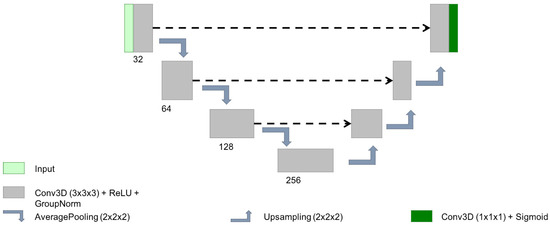
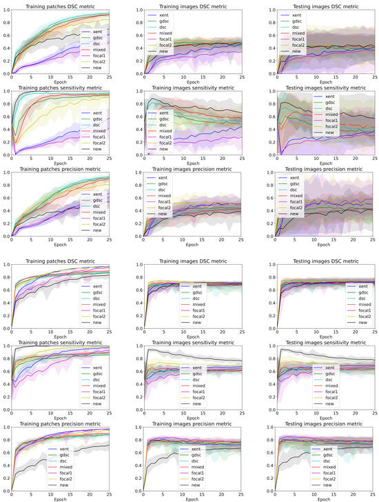
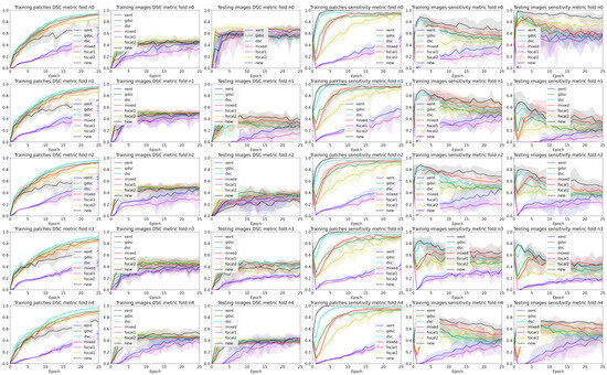
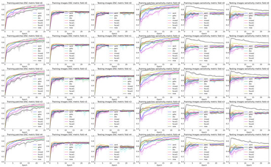
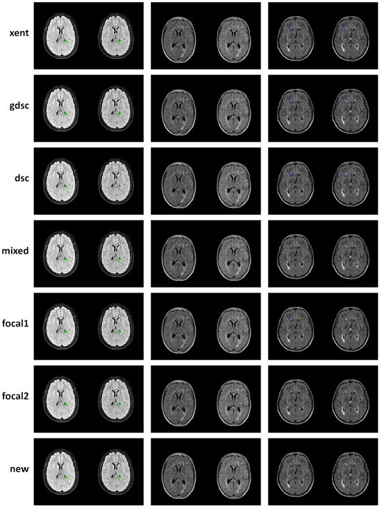
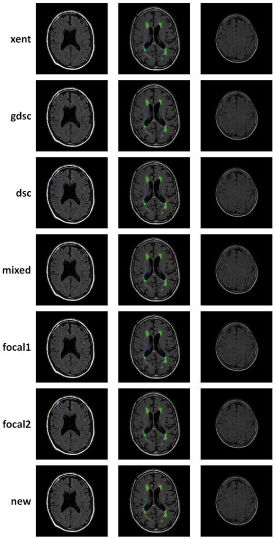
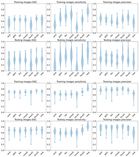
An Analysis of Loss Functions for Heavily Imbalanced Lesion Segmentation
by
Mariano Cabezas and Yago Diez
Viewed by 443
Abstract
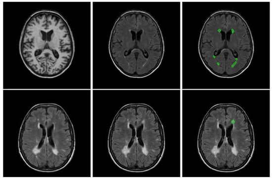
Heavily imbalanced datasets are common in lesion segmentation. Specifically, the lesions usually comprise less than 5% of the whole image volume when dealing with brain MRI. A common solution when training with a limited dataset is the use of specific loss functions that
[...] Read more.
Heavily imbalanced datasets are common in lesion segmentation. Specifically, the lesions usually comprise less than 5% of the whole image volume when dealing with brain MRI. A common solution when training with a limited dataset is the use of specific loss functions that rebalance the effect of background and foreground voxels. These approaches are usually evaluated running a single cross-validation split without taking into account other possible random aspects that might affect the true improvement of the final metric (i.e., random weight initialisation or random shuffling). Furthermore, the evolution of the effect of the loss on the heavily imbalanced class is usually not analysed during the training phase. In this work, we present an analysis of different common loss metrics during training on public datasets dealing with brain lesion segmentation in heavy imbalanced datasets. In order to limit the effect of hyperparameter tuning and architecture, we chose a 3D Unet architecture due to its ability to provide good performance on different segmentation applications. We evaluated this framework on two public datasets and we observed that weighted losses have a similar performance on average, even though heavily weighting the gradient of the foreground class gives better performance in terms of true positive segmentation.
Full article
►▼
Show Figures
Open AccessReview
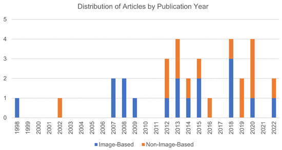
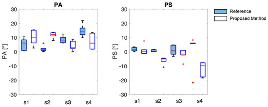
Intraoperative Angle Measurement of Anatomical Structures: A Systematic Review
by
João Cruz, Sérgio B. Gonçalves, Manuel Cassiano Neves, Hugo Plácido Silva and Miguel Tavares Silva
Viewed by 616
Abstract
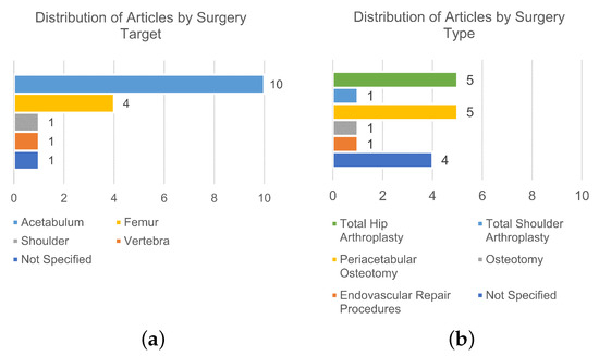
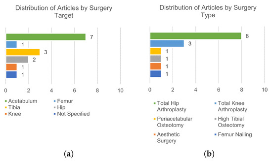
Ensuring precise angle measurement during surgical correction of orientation-related deformities is crucial for optimal postoperative outcomes, yet there is a lack of an ideal commercial solution. Current measurement sensors and instrumentation have limitations that make their use context-specific, demanding a methodical evaluation of
[...] Read more.
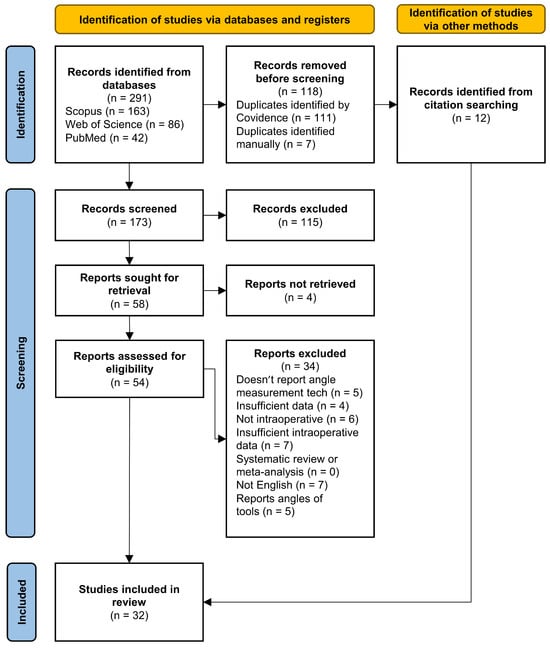
Ensuring precise angle measurement during surgical correction of orientation-related deformities is crucial for optimal postoperative outcomes, yet there is a lack of an ideal commercial solution. Current measurement sensors and instrumentation have limitations that make their use context-specific, demanding a methodical evaluation of the field. A systematic review was carried out in March 2023. Studies reporting technologies and validation methods for intraoperative angular measurement of anatomical structures were analyzed. A total of 32 studies were included, 17 focused on image-based technologies (6 fluoroscopy, 4 camera-based tracking, and 7 CT-based), while 15 explored non-image-based technologies (6 manual instruments and 9 inertial sensor-based instruments). Image-based technologies offer better accuracy and 3D capabilities but pose challenges like additional equipment, increased radiation exposure, time, and cost. Non-image-based technologies are cost-effective but may be influenced by the surgeon’s perception and require careful calibration. Nevertheless, the choice of the proper technology should take into consideration the influence of the expected error in the surgery, surgery type, and radiation dose limit. This comprehensive review serves as a valuable guide for surgeons seeking precise angle measurements intraoperatively. It not only explores the performance and application of existing technologies but also aids in the future development of innovative solutions.
Full article
►▼
Show Figures
Open AccessArticle
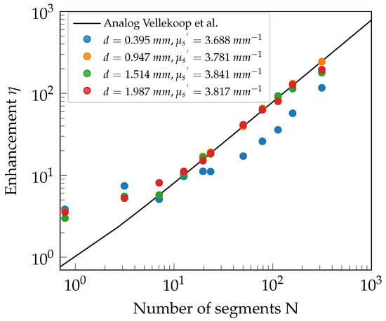
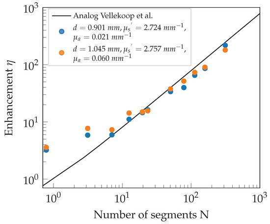
Focusing Coherent Light through Volume Scattering Phantoms via Wavefront Shaping
by
Niklas Fritzsche, Felix Ott, Karsten Pink and Alwin Kienle
Viewed by 1003
Abstract
Manipulating the wavefront of coherent light incident on scattering media to enhance the imaging depth, sensitivity, and resolution is a common technique in biomedical applications. Local phase variations cause changes in the interference and can be used to create a focus inside or
[...] Read more.
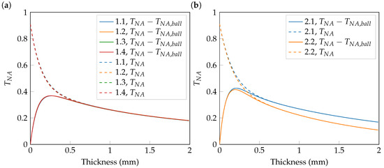
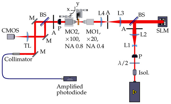
Manipulating the wavefront of coherent light incident on scattering media to enhance the imaging depth, sensitivity, and resolution is a common technique in biomedical applications. Local phase variations cause changes in the interference and can be used to create a focus inside or behind a scattering medium. We use wavefront shaping (WFS) to force constructive interference at an arbitrary location. The amount of light transmitted into a given region strongly depends on the scattering and absorption characteristics. These are described by their respective coefficients
and
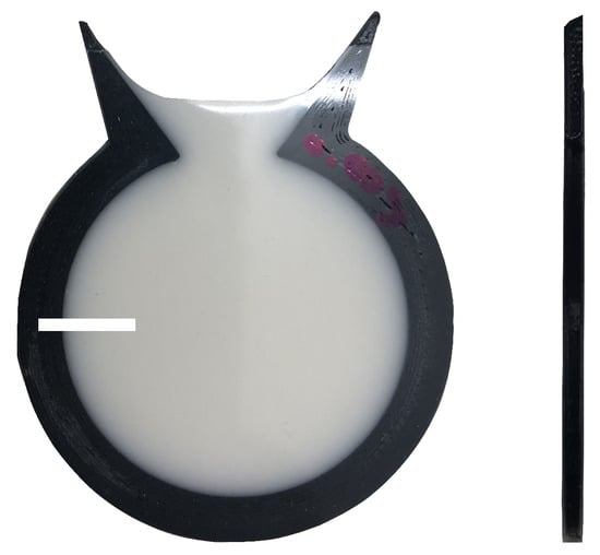
and the scattering phase function. Controlling the scattering and absorption coefficients, we study the behavior of wavefront shaping and the achievable intensity enhancement behind volume scattering media with well-defined optical properties. The phantoms designed in this publication are made of epoxy resin. Into these epoxy matrices, specific amounts of scattering and absorbing particles, such as titanium dioxide pigments and molecular dyes, are mixed. The mixture obtained is filled into 3D-printed frames of various thicknesses. After a precise fabrication procedure, an integrating sphere-based setup characterizes the phantoms experimentally. It detects the total hemispherical transmission and reflection. Further theoretical characterization is performed with a newly developed hybrid
method. This method senses the flux of light into a particular angular range at the lower boundary of a slab. The calculations are performed without suffering from ringing and fulfill the exact boundary conditions there. A decoupled two-path detection system allows for fast optimization as well as sensitive detection. The measurements yield results that agree well with the theoretically expected behavior.
Full article
►▼
Show Figures
Open AccessCommunication
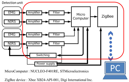
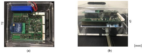
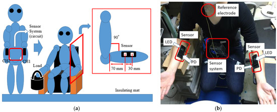
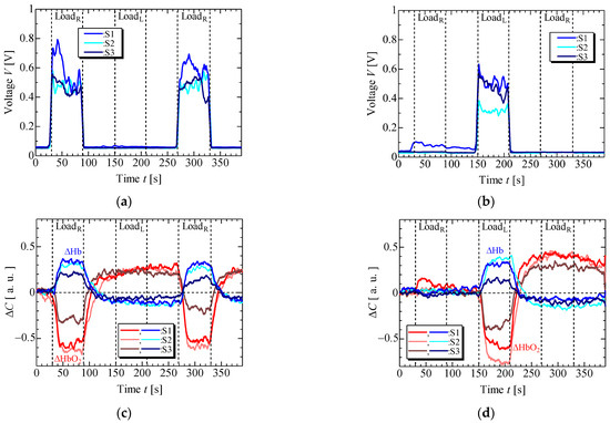
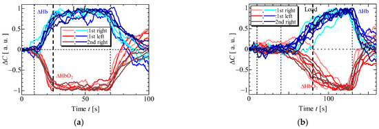
A Wireless 2-Channel Layered EMG/NIRS Sensor System for Local Muscular Activity Evaluation
by
Akira Kimoto, Yuya Oishi and Masanao Machida
Cited by 1 | Viewed by 739
Abstract
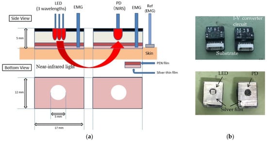
A wireless 2-channel layered sensor system that enables electromyography (EMG) and near-infrared spectroscopy (NIRS) measurements at two local positions was developed. The layered sensor consists of a thin silver electrode and a photosensor consisting of a photoemitting diode (LED) or photodiode (PD). The
[...] Read more.
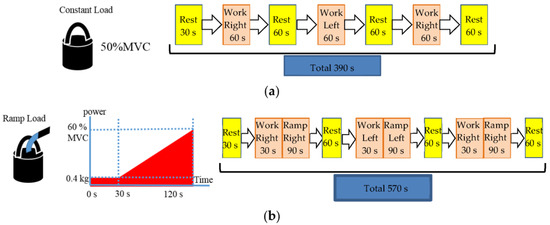
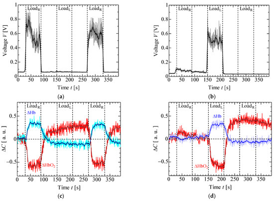
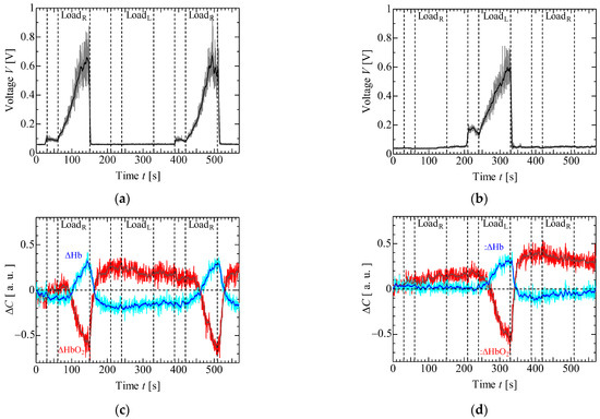
A wireless 2-channel layered sensor system that enables electromyography (EMG) and near-infrared spectroscopy (NIRS) measurements at two local positions was developed. The layered sensor consists of a thin silver electrode and a photosensor consisting of a photoemitting diode (LED) or photodiode (PD). The EMG and NIRS signals were simultaneously measured using a pair of electrodes and photosensors for the LED and PD, respectively. Two local muscular activities are presented in detail using layered sensors. In the experiments, EMG and NIRS signals were measured for isometric constant and ramp contractions at each forearm using layered sensors. The results showed that local muscle activity analysis is possible using simultaneous EMG and NIRS signals at each local position.
Full article
►▼
Show Figures
Open AccessArticle
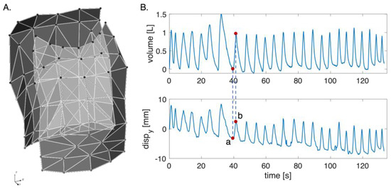
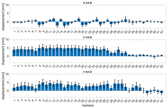
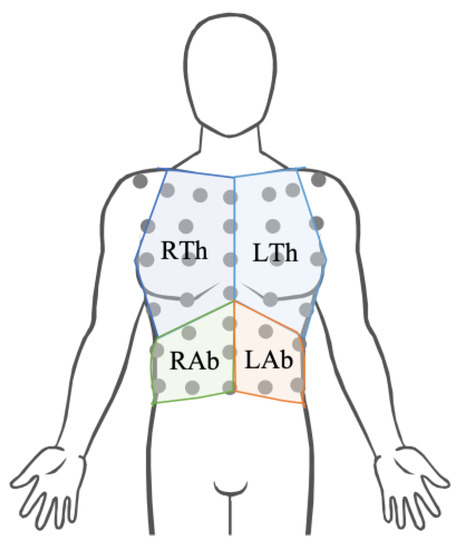
Breathing Chest Wall Kinematics Assessment through a Single Digital Camera: A Feasibility Study
by
Nunzia Molinaro, Emiliano Schena, Sergio Silvestri and Carlo Massaroni
Viewed by 899
Abstract
The identification of respiratory patterns based on the movement of the chest wall can assist in monitoring an individual’s health status, particularly those with neuromuscular disorders, such as hemiplegia and Duchenne muscular dystrophy. Thoraco-abdominal asynchrony (TAA) refers to the lack of coordination between
[...] Read more.
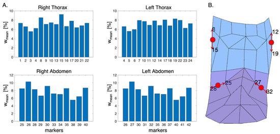
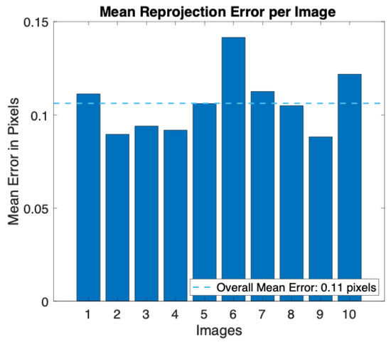
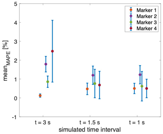
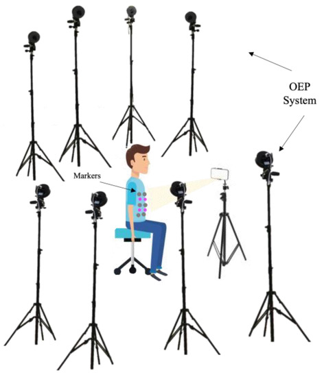
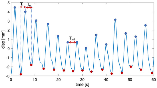
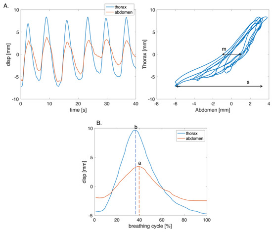
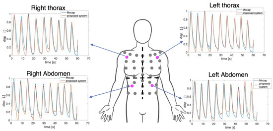
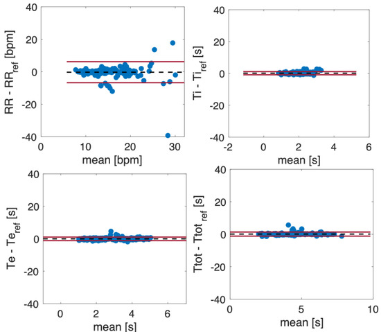
The identification of respiratory patterns based on the movement of the chest wall can assist in monitoring an individual’s health status, particularly those with neuromuscular disorders, such as hemiplegia and Duchenne muscular dystrophy. Thoraco-abdominal asynchrony (TAA) refers to the lack of coordination between the rib cage and abdominal movements, characterized by a time delay in their expansion. Motion capture systems, like optoelectronic plethysmography (OEP), are commonly employed to assess these asynchronous movements. However, alternative technologies able to capture chest wall movements without physical contact, such as RGB digital cameras and time-of-flight digital cameras, can also be utilized due to their accessibility, affordability, and non-invasive nature. This study explores the possibility of using a single RGB digital camera to record the kinematics of the thoracic and abdominal regions by placing four non-reflective markers on the torso. In order to choose the positions of these markers, we previously investigated the movements of 89 chest wall landmarks using OEP. Laboratory tests and volunteer experiments were conducted to assess the viability of the proposed system in capturing the kinematics of the chest wall and estimating various time-related respiratory parameters (i.e.,
fR,
Ti,
Te, and
Ttot) as well as TAA indexes. The results demonstrate a high level of agreement between the detected chest wall kinematics and the reference data. Furthermore, the system shows promising potential in estimating time-related respiratory parameters and identifying phase shifts indicative of TAA, thus suggesting its feasibility in detecting abnormal chest wall movements without physical contact with a single RGB camera.
Full article
►▼
Show Figures
Open AccessArticle
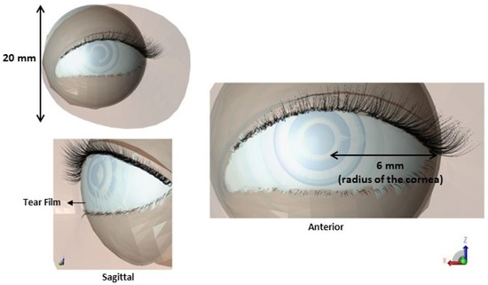
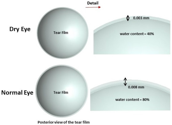
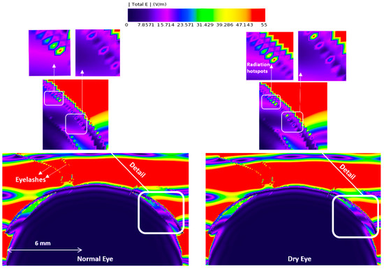
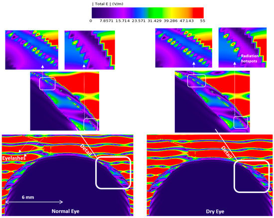
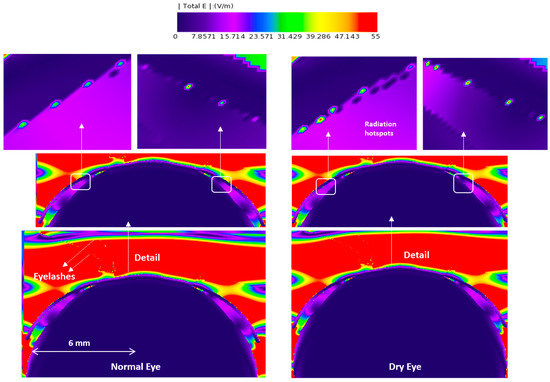
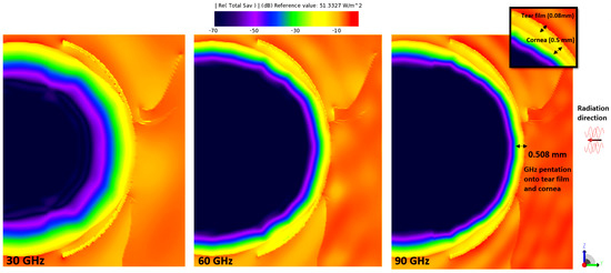
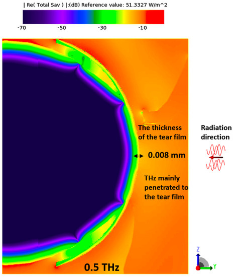
The Effects of mmW and THz Radiation on Dry Eyes: A Finite-Difference Time-Domain (FDTD) Computational Simulation Using XFdtd
by
Negin Foroughimehr, Zoltan Vilagosh, Ali Yavari and Andrew Wood
Cited by 2 | Viewed by 1371
Abstract
The importance of investigating the health effects of RF radiation on the cornea cannot be overstated. This study aimed to address this need by utilizing a mathematical simulation to examine the absorption of millimeter wave (mmW) and terahertz (THz) waves by the cornea,
[...] Read more.
The importance of investigating the health effects of RF radiation on the cornea cannot be overstated. This study aimed to address this need by utilizing a mathematical simulation to examine the absorption of millimeter wave (mmW) and terahertz (THz) waves by the cornea, considering both normal and pathological conditions. The simulation incorporated variations in tear film thickness and hydration levels, as these factors play a crucial role in corneal health. To assess the impact of RF radiation on the cornea, the study calculated temperature rises, which indicate heating effects for both dry and normal eyes. XFdtd, a widely used commercial software based on the Finite-Difference Time Domain (FDTD) method, was employed to evaluate the radiation absorption and resulting temperature changes. The outcomes of this study demonstrated a crucial finding, i.e., that changes in the water ratio and thickness of the tear film, which are associated with an increased risk of dry eye syndrome, directly impact the absorption of mmW and THz waves by the cornea. This insight provides valuable evidence supporting the interconnection between tear film properties and the vulnerability of the cornea to RF radiation.
Full article
►▼
Show Figures
Open AccessArticle
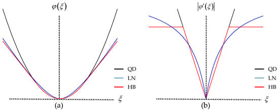
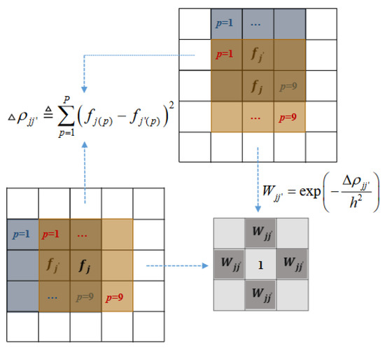
Similarity-Driven Fine-Tuning Methods for Regularization Parameter Optimization in PET Image Reconstruction
by
Wen Zhu and Soo-Jin Lee
Viewed by 832
Abstract
We present an adaptive method for fine-tuning hyperparameters in edge-preserving regularization for PET image reconstruction. For edge-preserving regularization, in addition to the smoothing parameter that balances data fidelity and regularization, one or more control parameters are typically incorporated to adjust the sensitivity of
[...] Read more.
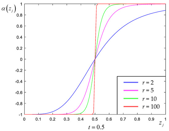
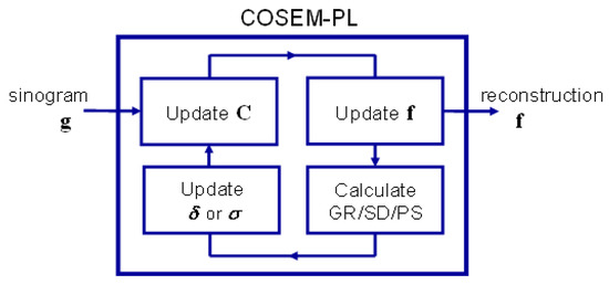
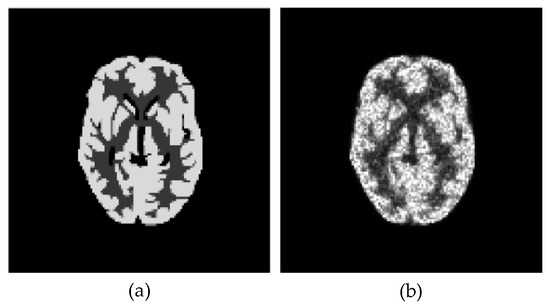
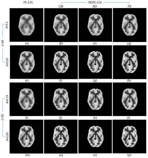
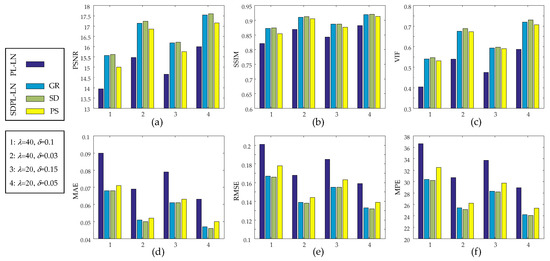
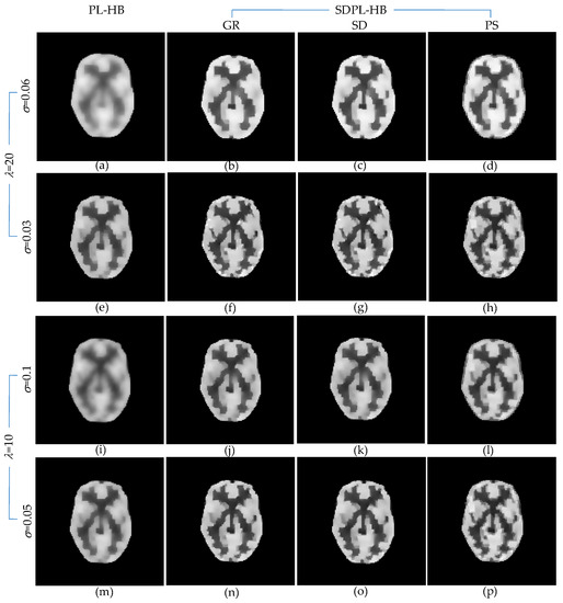
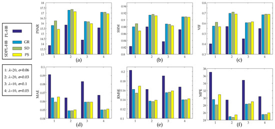
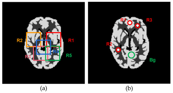
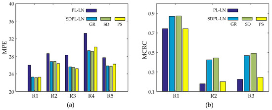
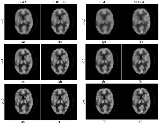
We present an adaptive method for fine-tuning hyperparameters in edge-preserving regularization for PET image reconstruction. For edge-preserving regularization, in addition to the smoothing parameter that balances data fidelity and regularization, one or more control parameters are typically incorporated to adjust the sensitivity of edge preservation by modifying the shape of the penalty function. Although there have been efforts to develop automated methods for tuning the hyperparameters in regularized PET reconstruction, the majority of these methods primarily focus on the smoothing parameter. However, it is challenging to obtain high-quality images without appropriately selecting the control parameters that adjust the edge preservation sensitivity. In this work, we propose a method to precisely tune the hyperparameters, which are initially set with a fixed value for the entire image, either manually or using an automated approach. Our core strategy involves adaptively adjusting the control parameter at each pixel, taking into account the degree of patch similarities calculated from the previous iteration within the pixel’s neighborhood that is being updated. This approach allows our new method to integrate with a wide range of existing parameter-tuning techniques for edge-preserving regularization. Experimental results demonstrate that our proposed method effectively enhances the overall reconstruction accuracy across multiple image quality metrics, including peak signal-to-noise ratio, structural similarity, visual information fidelity, mean absolute error, root-mean-square error, and mean percentage error.
Full article
►▼
Show Figures
Open AccessArticle
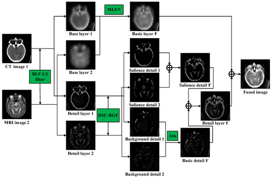
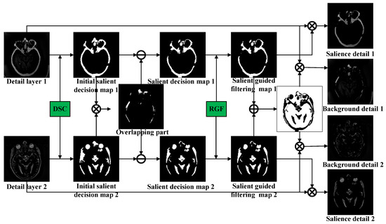
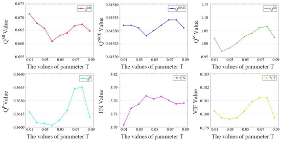
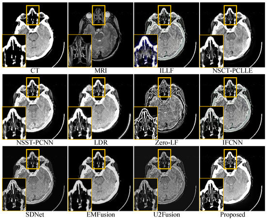
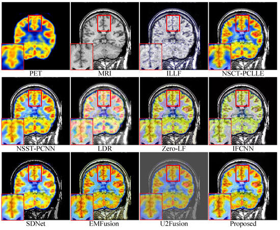
Multi-Sensor Medical-Image Fusion Technique Based on Embedding Bilateral Filter in Least Squares and Salient Detection
by
Jiangwei Li, Dingan Han, Xiaopan Wang, Peng Yi, Liang Yan and Xiaosong Li
Cited by 10 | Viewed by 1458
Abstract
A multi-sensor medical-image fusion technique, which integrates useful information from different single-modal images of the same tissue and provides a fused image that is more comprehensive and objective than a single-source image, is becoming an increasingly important technique in clinical diagnosis and treatment
[...] Read more.
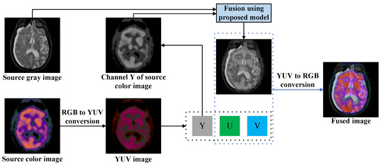
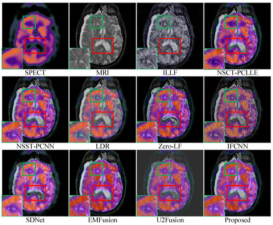
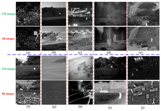
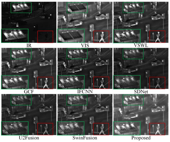
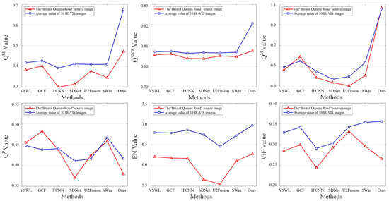
A multi-sensor medical-image fusion technique, which integrates useful information from different single-modal images of the same tissue and provides a fused image that is more comprehensive and objective than a single-source image, is becoming an increasingly important technique in clinical diagnosis and treatment planning. The salient information in medical images often visually describes the tissue. To effectively embed salient information in the fused image, a multi-sensor medical image fusion method is proposed based on an embedding bilateral filter in least squares and salient detection via a deformed smoothness constraint. First, source images are decomposed into base and detail layers using a bilateral filter in least squares. Then, the detail layers are treated as superpositions of salient regions and background information; a fusion rule for this layer based on the deformed smoothness constraint and guided filtering was designed to successfully conserve the salient structure and detail information of the source images. A base-layer fusion rule based on modified Laplace energy and local energy is proposed to preserve the energy information of these source images. The experimental results demonstrate that the proposed method outperformed nine state-of-the-art methods in both subjective and objective quality assessments on the Harvard Medical School dataset.
Full article
►▼
Show Figures
Open AccessArticle
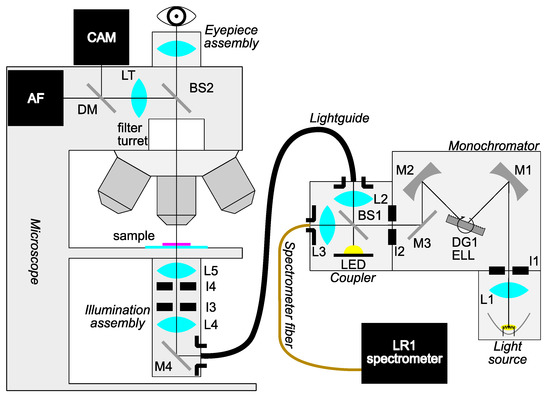
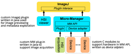
Design and Validation of a Custom-Made Hyperspectral Microscope Imaging System for Biomedical Applications
by
Jošt Stergar, Rok Hren and Matija Milanič
Cited by 2 | Viewed by 1465
Abstract
Hyperspectral microscope imaging (HMI) is an emerging modality that integrates spatial information collected by standard laboratory microscopy and the spectral-based contrast obtained by hyperspectral imaging and may be instrumental in establishing novel quantitative diagnostic methodologies, particularly in histopathology. Further expansion of HMI capabilities
[...] Read more.
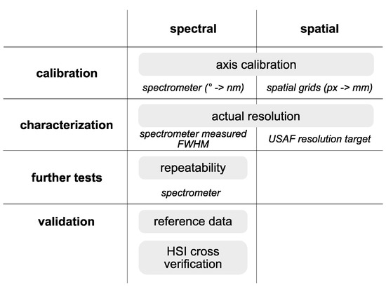
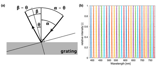
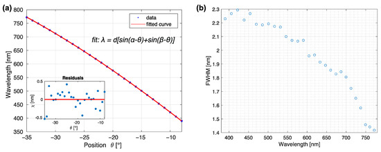
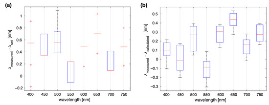
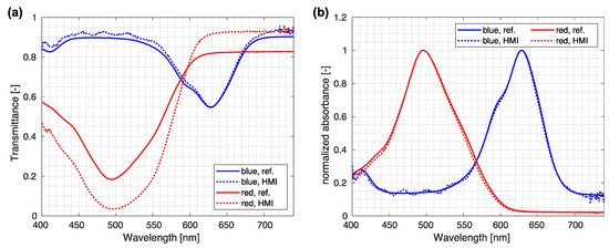
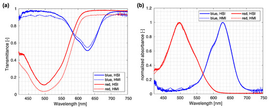
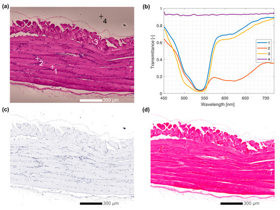
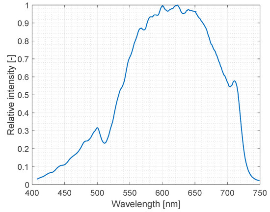
Hyperspectral microscope imaging (HMI) is an emerging modality that integrates spatial information collected by standard laboratory microscopy and the spectral-based contrast obtained by hyperspectral imaging and may be instrumental in establishing novel quantitative diagnostic methodologies, particularly in histopathology. Further expansion of HMI capabilities hinges upon the modularity and versatility of systems and their proper standardization. In this report, we describe the design, calibration, characterization, and validation of the custom-made laboratory HMI system based on a Zeiss Axiotron fully motorized microscope and a custom-developed Czerny-Turner-type monochromator. For these important steps, we rely on a previously designed calibration protocol. Validation of the system demonstrates a performance comparable to classic spectrometry laboratory systems. We further demonstrate validation against a laboratory hyperspectral imaging system for macroscopic samples, enabling future comparison of spectral imaging results across length scales. An example of the utility of our custom-made HMI system on a standard hematoxylin and eosin-stained histology slide is also shown.
Full article
►▼
Show Figures
Open AccessArticle
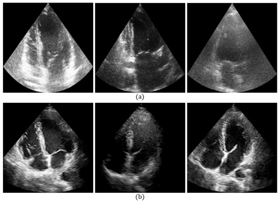
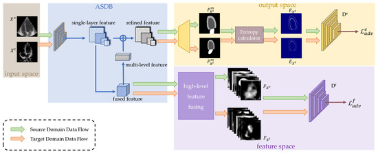
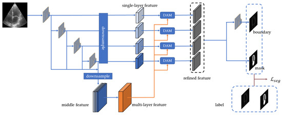
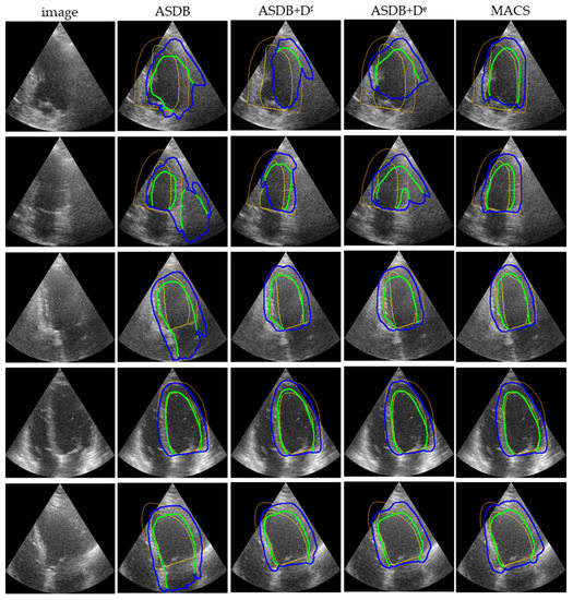
Cross-Domain Echocardiography Segmentation with Multi-Space Joint Adaptation
by
Tongwaner Chen, Menghua Xia, Yi Huang, Jing Jiao and Yuanyuan Wang
Cited by 1 | Viewed by 1624
Abstract
The segmentation of the left ventricle endocardium (LV
endo) and the left ventricle epicardium (LV
epi) in echocardiography plays an important role in clinical diagnosis. Recently, deep neural networks have been the most commonly used approach for echocardiography segmentation. However, the
[...] Read more.
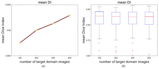
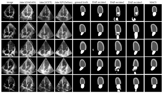
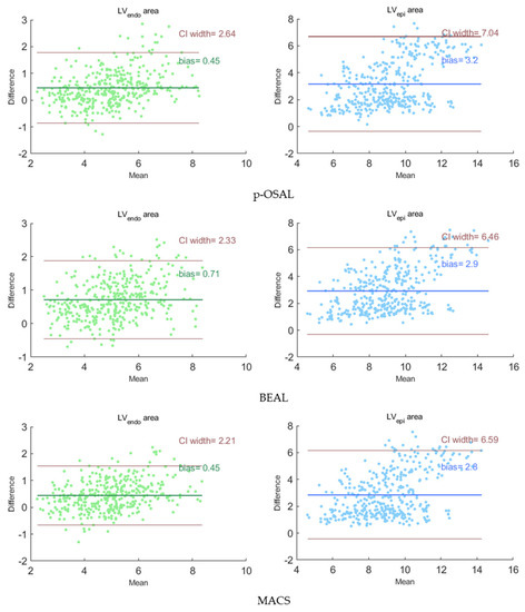
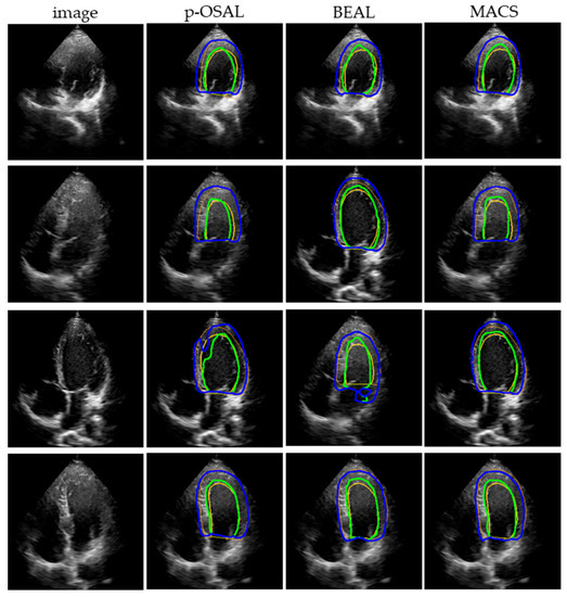
The segmentation of the left ventricle endocardium (LV
endo) and the left ventricle epicardium (LV
epi) in echocardiography plays an important role in clinical diagnosis. Recently, deep neural networks have been the most commonly used approach for echocardiography segmentation. However, the performance of a well-trained segmentation network may degrade in unseen domain datasets due to the distribution shift of the data. Adaptation algorithms can improve the generalization of deep neural networks to different domains. In this paper, we present a multi-space adaptation-segmentation-joint framework, named MACS, for cross-domain echocardiography segmentation. It adopts a generative adversarial architecture; the generator fulfills the segmentation task and the multi-space discriminators align the two domains on both the feature space and output space. We evaluated the MACS method on two echocardiography datasets from different medical centers and vendors, the publicly available CAMUS dataset and our self-acquired dataset. The experimental results indicated that the MACS could handle unseen domain datasets well, without requirements for manual annotations, and improve the generalization performance by 2.2% in the Dice metric.
Full article
►▼
Show Figures
Open AccessReview
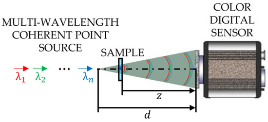
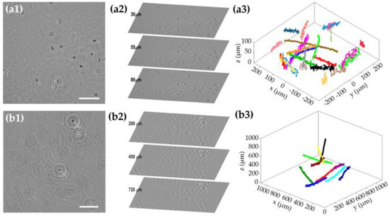
Multi-Illumination Single-Holographic-Exposure Lensless Fresnel (MISHELF) Microscopy: Principles and Biomedical Applications
by
José Ángel Picazo-Bueno, Martín Sanz, Luis Granero, Javier García and Vicente Micó
Cited by 1 | Viewed by 1693
Abstract
Lensless holographic microscopy (LHM) comes out as a promising label-free technique since it supplies high-quality imaging and adaptive magnification in a lens-free, compact and cost-effective way. Compact sizes and reduced prices of LHMs make them a perfect instrument for point-of-care diagnosis and increase
[...] Read more.
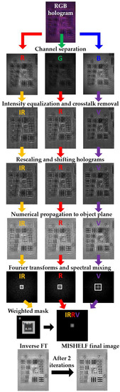
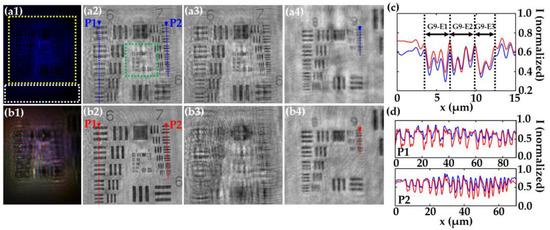
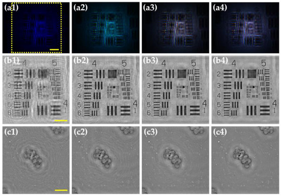
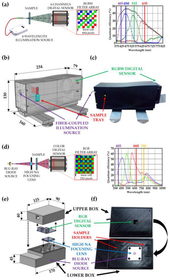
Lensless holographic microscopy (LHM) comes out as a promising label-free technique since it supplies high-quality imaging and adaptive magnification in a lens-free, compact and cost-effective way. Compact sizes and reduced prices of LHMs make them a perfect instrument for point-of-care diagnosis and increase their usability in limited-resource laboratories, remote areas, and poor countries. LHM can provide excellent intensity and phase imaging when the twin image is removed. In that sense, multi-illumination single-holographic-exposure lensless Fresnel (MISHELF) microscopy appears as a single-shot and phase-retrieved imaging technique employing multiple illumination/detection channels and a fast-iterative phase-retrieval algorithm. In this contribution, we review MISHELF microscopy through the description of the principles, the analysis of the performance, the presentation of the microscope prototypes and the inclusion of the main biomedical applications reported so far.
Full article
►▼
Show Figures
Open AccessArticle
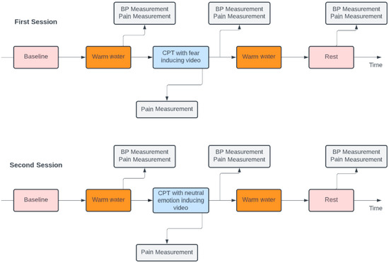
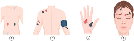
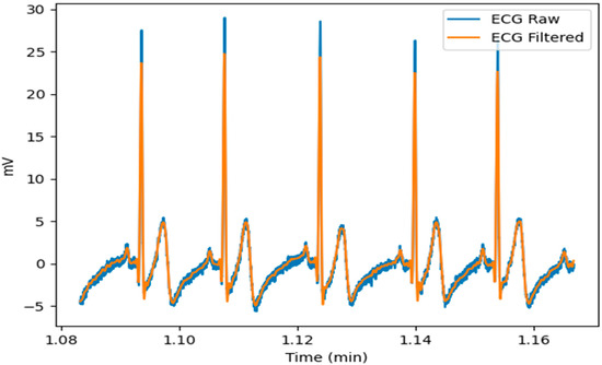
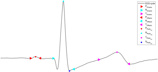
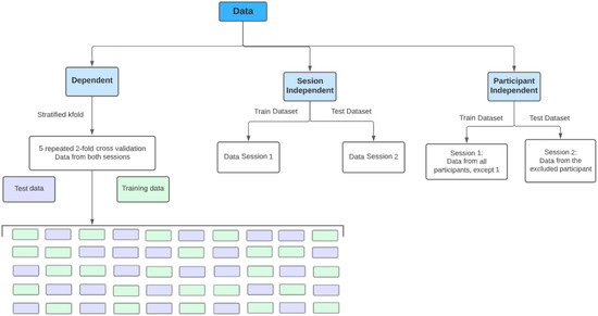
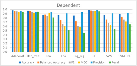
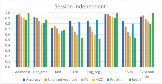
Using the Electrocardiogram for Pain Classification under Emotional Contexts
by
Pedro Silva and Raquel Sebastião
Cited by 2 | Viewed by 1320
Abstract
The adequate characterization of pain is critical in diagnosis and therapy selection, and currently is subjectively assessed by patient communication and self-evaluation. Thus, pain recognition and assessment have been a target of study in past years due to the importance of objective measurement.
[...] Read more.
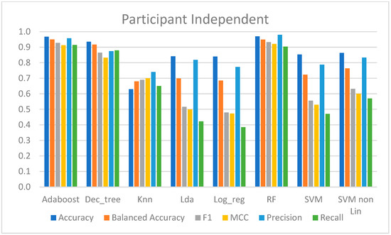
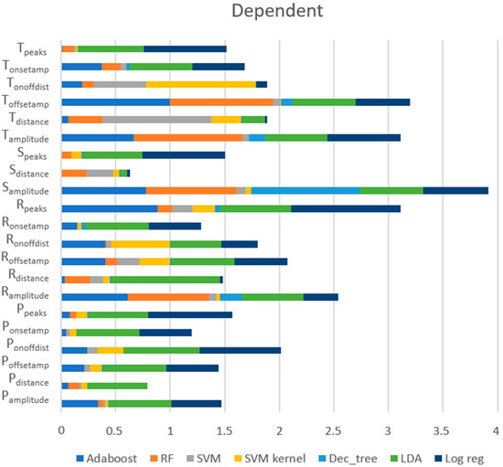
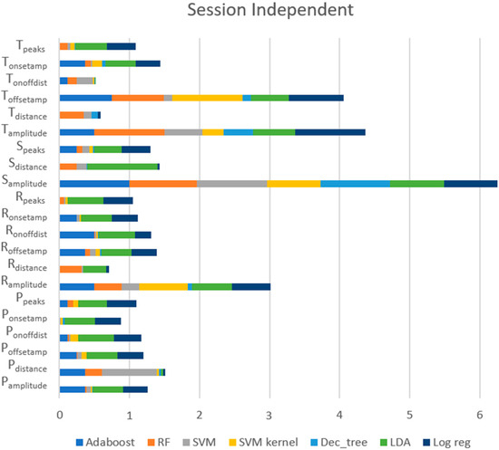
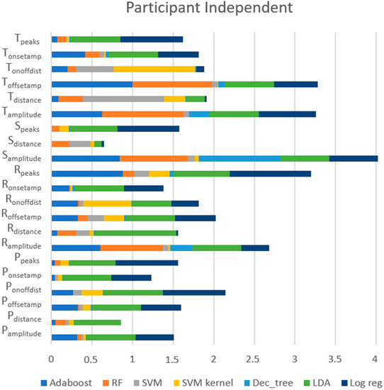
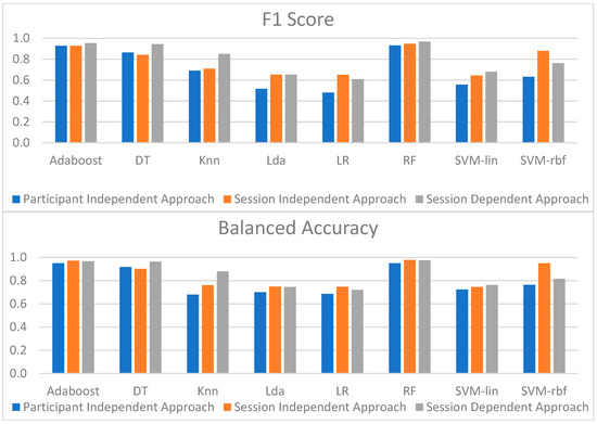
The adequate characterization of pain is critical in diagnosis and therapy selection, and currently is subjectively assessed by patient communication and self-evaluation. Thus, pain recognition and assessment have been a target of study in past years due to the importance of objective measurement. The goal of this work is the analysis of the electrocardiogram (ECG) under emotional contexts and reasoning on the physiological classification of pain under neutral and fear conditions. Using data from both contexts for pain classification, a balanced accuracy of up to 97.4% was obtained. Using an emotionally independent approach and using data from one emotional context to learn pain and data from the other to evaluate the models, a balanced accuracy of up to 97.7% was reached. These similar results seem to support that the physiological response to pain was maintained despite the different emotional contexts. Attempting a participant-independent approach for pain classification and using a leave-one-out cross-validation strategy, data from the fear context were used to train pain classification models, and data from the neutral context were used to evaluate the performance, achieving a balanced accuracy of up to 94.9%. Moreover, across the different learning strategies, Random Forest outperformed the remaining models. These results show the feasibility of identifying pain through physiological characteristics of the ECG response despite the presence of autonomic nervous system perturbations.
Full article
►▼
Show Figures
Open AccessArticle
Voting Ensemble Approach for Enhancing Alzheimer’s Disease Classification
by
Subhajit Chatterjee and Yung-Cheol Byun
Cited by 5 | Viewed by 1946
Abstract
Alzheimer’s disease is dementia that impairs one’s thinking, behavior, and memory. It starts as a moderate condition affecting areas of the brain that make it challenging to retain recently learned information, causes mood swings, and causes confusion regarding occasions, times, and locations. The
[...] Read more.
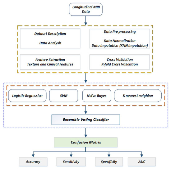
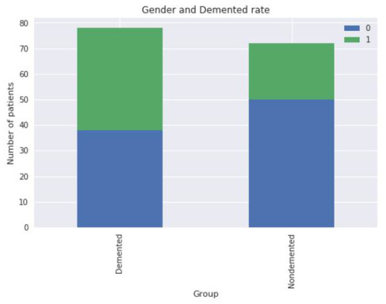
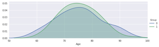
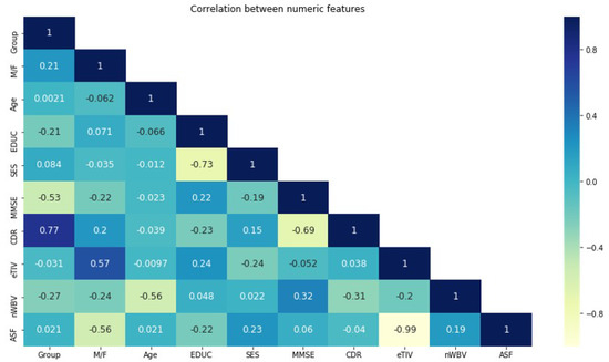
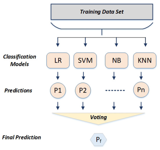
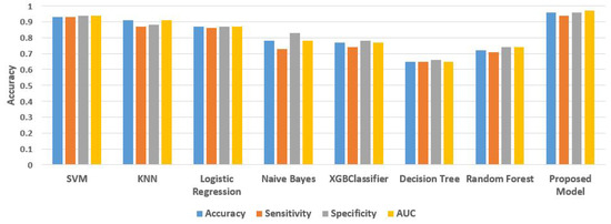
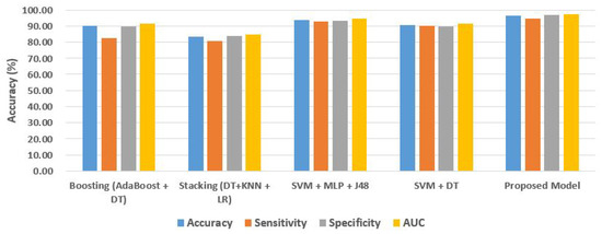
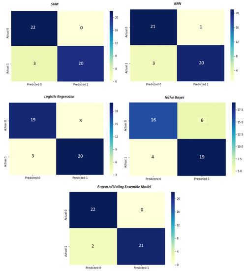
Alzheimer’s disease is dementia that impairs one’s thinking, behavior, and memory. It starts as a moderate condition affecting areas of the brain that make it challenging to retain recently learned information, causes mood swings, and causes confusion regarding occasions, times, and locations. The most prevalent type of dementia, called Alzheimer’s disease (AD), causes memory-related problems in patients. A precise medical diagnosis that correctly classifies AD patients results in better treatment. Currently, the most commonly used classification techniques extract features from longitudinal MRI data before creating a single classifier that performs classification. However, it is difficult to train a reliable classifier to achieve acceptable classification performance due to limited sample size and noise in longitudinal MRI data. Instead of creating a single classifier, we propose an ensemble voting method that generates multiple individual classifier predictions and then combines them to develop a more accurate and reliable classifier. The ensemble voting classifier model performs better in the Open Access Series of Imaging Studies (OASIS) dataset for older adults than existing methods in important assessment criteria such as accuracy, sensitivity, specificity, and AUC. For the binary classification of with dementia and no dementia, an accuracy of 96.4% and an AUC of 97.2% is attained.
Full article
►▼
Show Figures
Open AccessReview
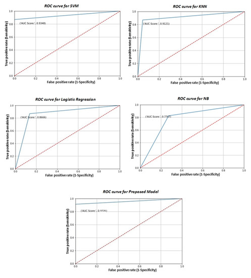
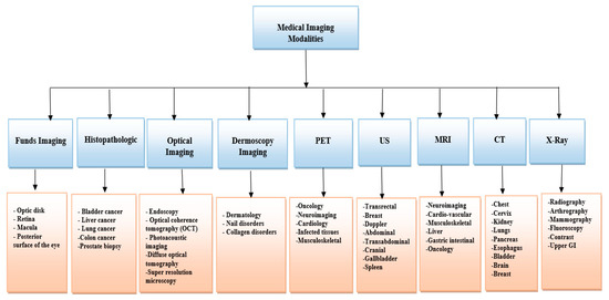
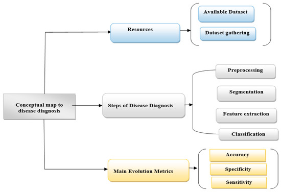
Critical Analysis of the Current Medical Image-Based Processing Techniques for Automatic Disease Evaluation: Systematic Literature Review
by
Baidaa Mutasher Rashed and Nirvana Popescu
Cited by 4 | Viewed by 5886
Abstract
Medical image processing and analysis techniques play a significant role in diagnosing diseases. Thus, during the last decade, several noteworthy improvements in medical diagnostics have been made based on medical image processing techniques. In this article, we reviewed articles published in the most
[...] Read more.
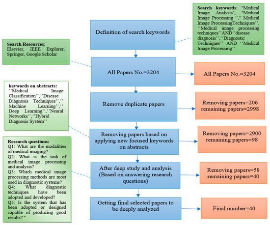
Medical image processing and analysis techniques play a significant role in diagnosing diseases. Thus, during the last decade, several noteworthy improvements in medical diagnostics have been made based on medical image processing techniques. In this article, we reviewed articles published in the most important journals and conferences that used or proposed medical image analysis techniques to diagnose diseases. Starting from four scientific databases, we applied the PRISMA technique to efficiently process and refine articles until we obtained forty research articles published in the last five years (2017–2021) aimed at answering our research questions. The medical image processing and analysis approaches were identified, examined, and discussed, including preprocessing, segmentation, feature extraction, classification, evaluation metrics, and diagnosis techniques. This article also sheds light on machine learning and deep learning approaches. We also focused on the most important medical image processing techniques used in these articles to establish the best methodologies for future approaches, discussing the most efficient ones and proposing in this way a comprehensive reference source of methods of medical image processing and analysis that can be very useful in future medical diagnosis systems.
Full article
►▼
Show Figures
Open AccessArticle
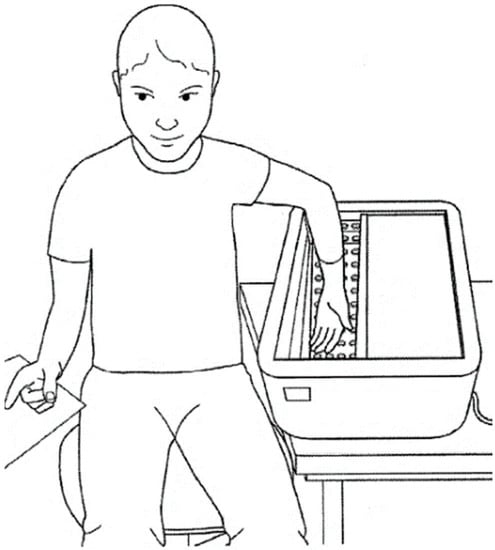
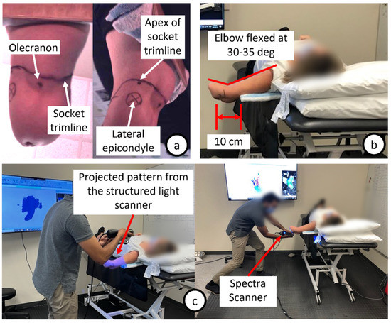
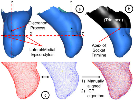
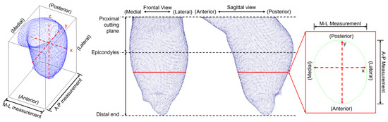
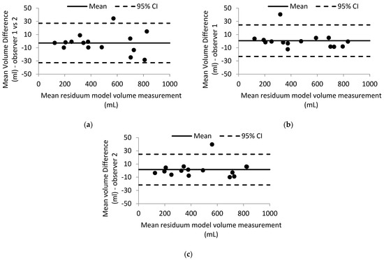
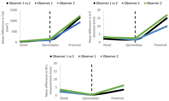
Evaluating the Reliability of a Shape Capturing Process for Transradial Residual Limb Using a Non-Contact Scanner
by
Calvin C. Ngan, Harry Sivasambu, Sandra Ramdial and Jan Andrysek
Cited by 2 | Viewed by 1366
Abstract
Advancements in digital imaging technologies hold the potential to transform prosthetic and orthotic practices. Non-contact optical scanners can capture the shape of the residual limb quickly, accurately, and reliably. However, their suitability in clinical practice, particularly for the transradial (below-elbow) residual limb, is
[...] Read more.
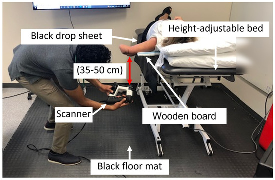
Advancements in digital imaging technologies hold the potential to transform prosthetic and orthotic practices. Non-contact optical scanners can capture the shape of the residual limb quickly, accurately, and reliably. However, their suitability in clinical practice, particularly for the transradial (below-elbow) residual limb, is unknown. This project aimed to evaluate the reliability of an optical scanner-based shape capture process for transradial residual limbs related to volumetric measurements and shape assessment in a clinical setting. A dedicated setup for digitally shape capturing transradial residual limbs was developed, addressing challenges with scanning of small residual limb size and aspects such as positioning and patient movement. Two observers performed three measurements each on 15 participants with transradial-level limb absence. Overall, the developed shape capture process was found to be highly repeatable, with excellent intra- and inter-rater reliability that was comparable to the scanning of residual limb cast models. Future work in this area should compare the differences between residual limb shapes captured through digital and manual methods.
Full article
►▼
Show Figures
Open AccessArticle
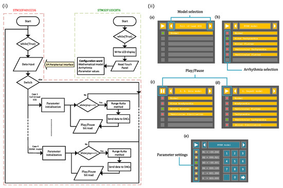
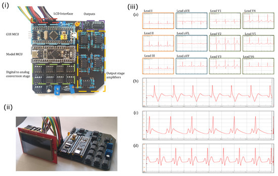
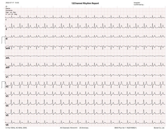
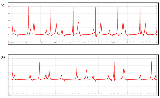
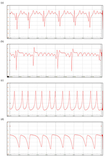
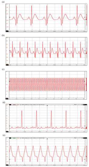
ECG Patient Simulator Based on Mathematical Models
by
Mario Alan Quiroz-Juárez, Juan Alberto Rosales-Juárez, Omar Jiménez-Ramírez, Rubén Vázquez-Medina and José Luis Aragón
Cited by 2 | Viewed by 4075
Abstract
In this work, we propose a versatile, low-cost, and tunable electronic device to generate realistic electrocardiogram (ECG) waveforms, capable of simulating ECG of patients within a wide range of possibilities. A visual analysis of the clinical ECG register provides the cardiologist with vital
[...] Read more.
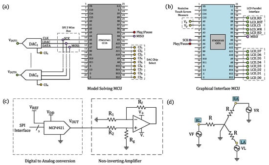
In this work, we propose a versatile, low-cost, and tunable electronic device to generate realistic electrocardiogram (ECG) waveforms, capable of simulating ECG of patients within a wide range of possibilities. A visual analysis of the clinical ECG register provides the cardiologist with vital physiological information to determine the patient’s heart condition. Because of its clinical significance, there is a strong interest in algorithms and medical ECG measuring devices that acquire, preserve, and process ECG recordings with high fidelity. Bearing this in mind, the proposed electronic device is based on four different mathematical models describing macroscopic heartbeat dynamics with ordinary differential equations. Firstly, we produce full 12-lead ECG profiles by implementing a model comprising a network of heterogeneous oscillators. Then, we implement a discretized reaction–diffusion model in our electronic device to reproduce ECG waveforms from various rhythm disorders. Finally, in order to show the versatility and capabilities of our system, we include two additional models, a ring of three coupled oscillators and a model based on a quasiperiodic motion, which can reproduce a wide range of pathological conditions. With this, the proposed device can reproduce around thirty-two cardiac rhythms with the possibility of exploring different parameter values to simulate new arrhythmias with the same hardware. Our system, which is a hybrid analog–digital circuit, generates realistic ECG signals through digital-to-analog converters whose amplitudes and waveforms are controlled through an interactive and friendly graphic interface. Our ECG patient simulator arises as a promising platform for assessing the performance of electrocardiograph equipment and ECG signal processing software in clinical trials. Additionally the produced 12-lead profiles can be tested in patient monitoring systems.
Full article
►▼
Show Figures
Open AccessArticle
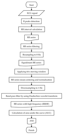
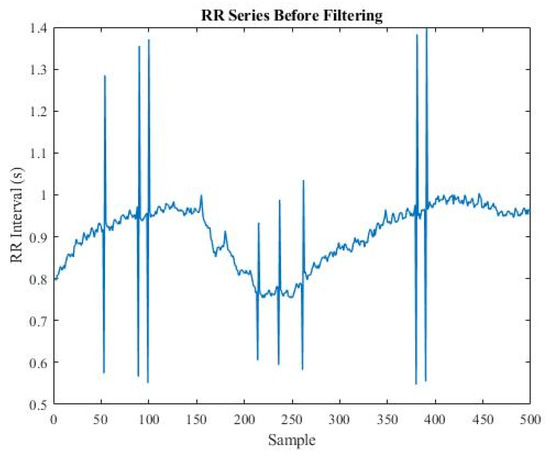
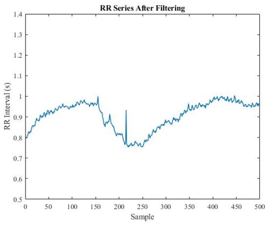
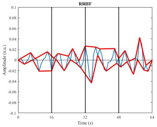
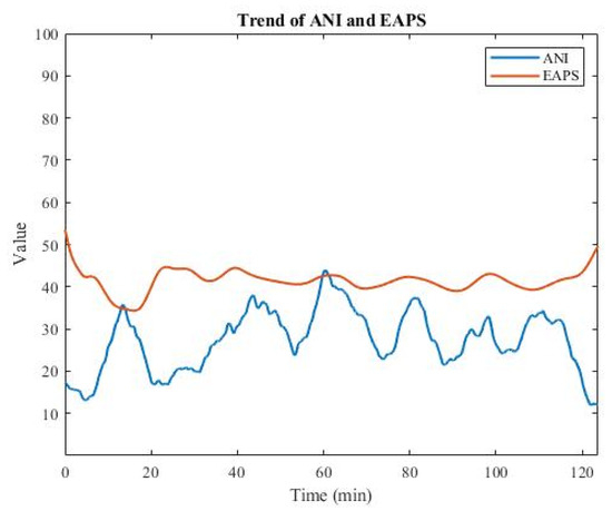
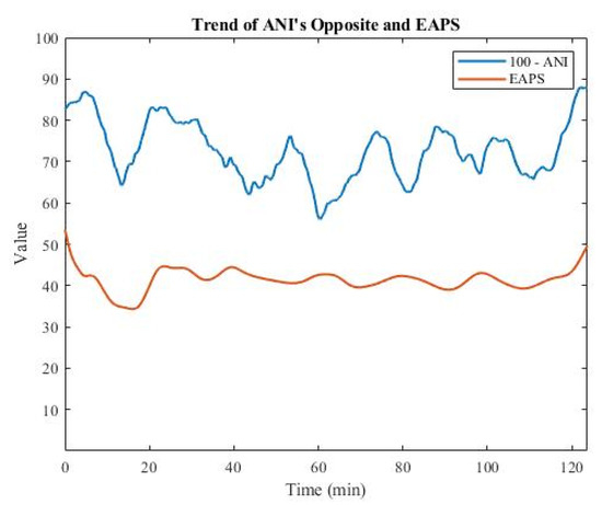
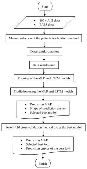
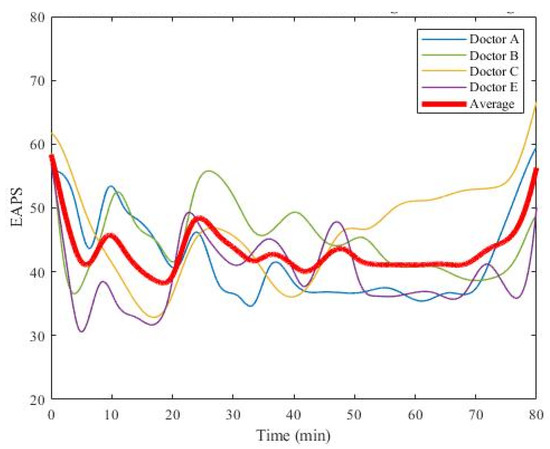
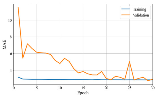
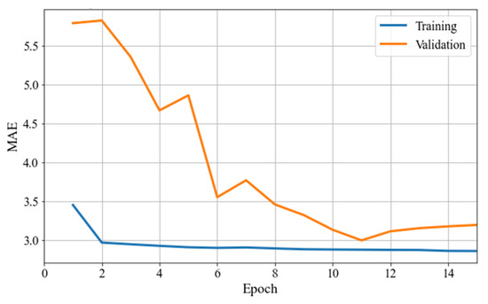
Comparison of Deep Learning Algorithms in Predicting Expert Assessments of Pain Scores during Surgical Operations Using Analgesia Nociception Index
by
Wei-Horng Jean, Peter Sutikno, Shou-Zen Fan, Maysam F. Abbod and Jiann-Shing Shieh
Cited by 5 | Viewed by 1497
Abstract
There are many surgical operations performed daily in operation rooms worldwide. Adequate anesthesia is needed during an operation. Besides hypnosis, adequate analgesia is critical to prevent autonomic reactions. Clinical experience and vital signs are usually used to adjust the dosage of analgesics. Analgesia
[...] Read more.
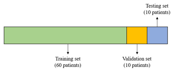
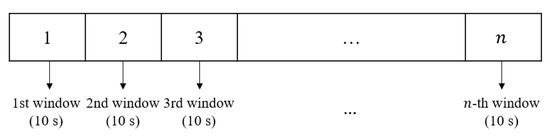
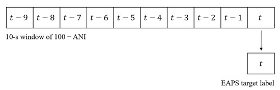
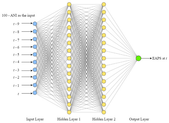
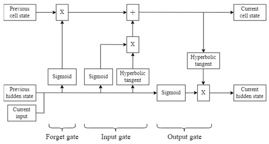
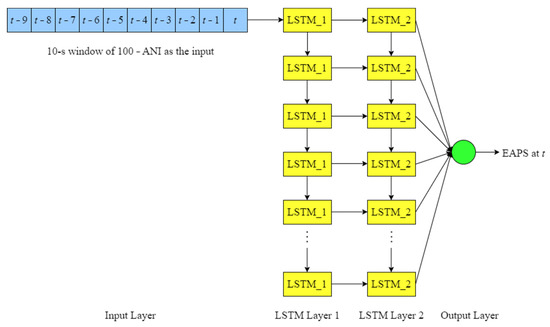
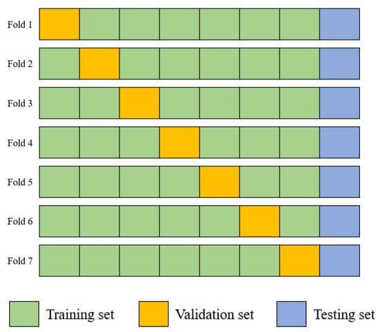
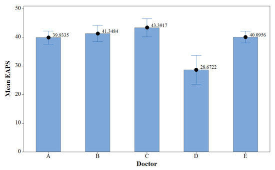
There are many surgical operations performed daily in operation rooms worldwide. Adequate anesthesia is needed during an operation. Besides hypnosis, adequate analgesia is critical to prevent autonomic reactions. Clinical experience and vital signs are usually used to adjust the dosage of analgesics. Analgesia nociception index (ANI), which ranges from 0 to 100, is derived from heart rate variability (HRV) via electrocardiogram (ECG) signals, for pain evaluation in a non-invasive manner. It represents parasympathetic activity. In this study, we compared the performance of multilayer perceptron (MLP) and long short-term memory (LSTM) algorithms in predicting expert assessment of pain score (EAPS) based on patient′s HRV during surgery. The objective of this study was to analyze how deep learning models differed from the medical doctors′ predictions of EAPS. As the input and output features of the deep learning models, the opposites of ANI and EAPS were used. This study included 80 patients who underwent operations at National Taiwan University Hospital. Using MLP and LSTM, a holdout method was first applied to 60 training patients, 10 validation patients, and 10 testing patients. As compared to the LSTM model, which had a testing mean absolute error (MAE) of 2.633 ± 0.542, the MLP model had a testing MAE of 2.490 ± 0.522, with a more appropriate shape of its prediction curves. The model based on MLP was selected as the best. Using MLP, a seven-fold cross validation method was then applied. The first fold had the lowest testing MAE of 2.460 ± 0.634, while the overall MAE for the seven-fold cross validation method was 2.848 ± 0.308. In conclusion, HRV analysis using MLP algorithm had a good correlation with EAPS; therefore, it can play role as a continuous monitor to predict intraoperative pain levels, to assist physicians in adjusting analgesic agent dosage. Further studies may consider obtaining more input features, such as photoplethysmography (PPG) and other kinds of continuous variable, to improve the prediction performance.
Full article
►▼
Show Figures
Open AccessArticle
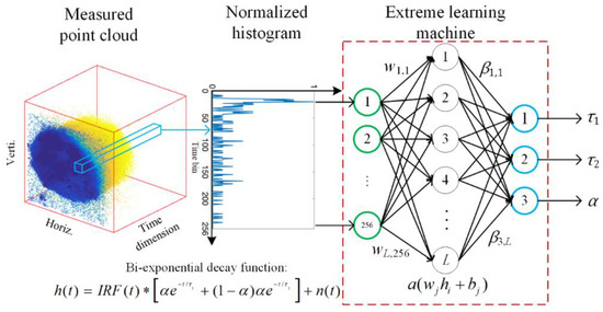
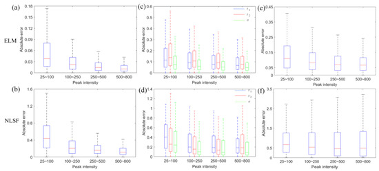
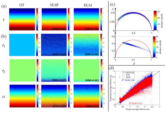
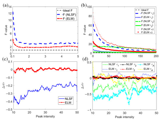
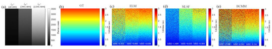
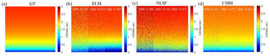
Fast Analysis of Time-Domain Fluorescence Lifetime Imaging via Extreme Learning Machine
by
Zhenya Zang, Dong Xiao, Quan Wang, Zinuo Li, Wujun Xie, Yu Chen and David Day Uei Li
Cited by 4 | Viewed by 2682
Abstract
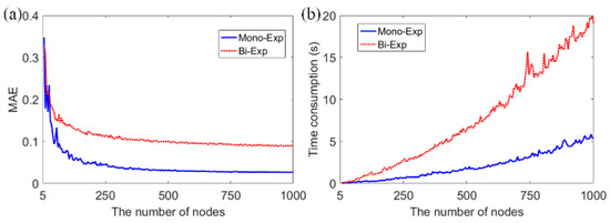
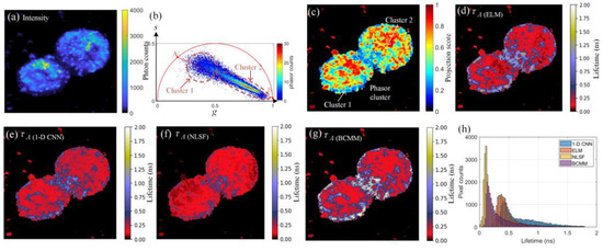
We present a fast and accurate analytical method for fluorescence lifetime imaging microscopy (FLIM), using the extreme learning machine (ELM). We used extensive metrics to evaluate ELM and existing algorithms. First, we compared these algorithms using synthetic datasets. The results indicate that ELM
[...] Read more.
We present a fast and accurate analytical method for fluorescence lifetime imaging microscopy (FLIM), using the extreme learning machine (ELM). We used extensive metrics to evaluate ELM and existing algorithms. First, we compared these algorithms using synthetic datasets. The results indicate that ELM can obtain higher fidelity, even in low-photon conditions. Afterwards, we used ELM to retrieve lifetime components from human prostate cancer cells loaded with gold nanosensors, showing that ELM also outperforms the iterative fitting and non-fitting algorithms. By comparing ELM with a computational efficient neural network, ELM achieves comparable accuracy with less training and inference time. As there is no back-propagation process for ELM during the training phase, the training speed is much higher than existing neural network approaches. The proposed strategy is promising for edge computing with online training.
Full article
►▼
Show Figures
Open AccessArticle
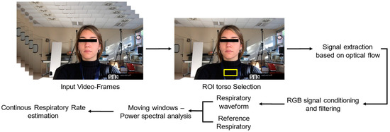
Multi-ROI Spectral Approach for the Continuous Remote Cardio-Respiratory Monitoring from Mobile Device Built-In Cameras
by
Nunzia Molinaro, Emiliano Schena, Sergio Silvestri and Carlo Massaroni
Cited by 14 | Viewed by 1878
Abstract
Heart rate (HR) and respiratory rate (
fR) can be estimated by processing videos framing the upper body and face regions without any physical contact with the subject. This paper proposed a technique for continuously monitoring HR and
fR via
[...] Read more.
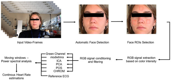
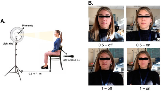
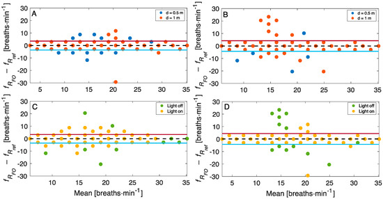
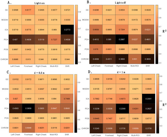
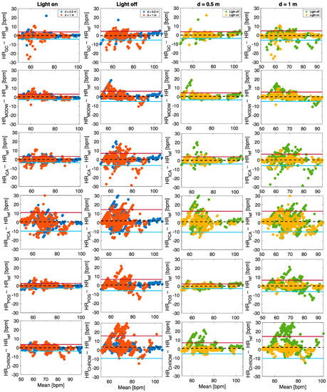
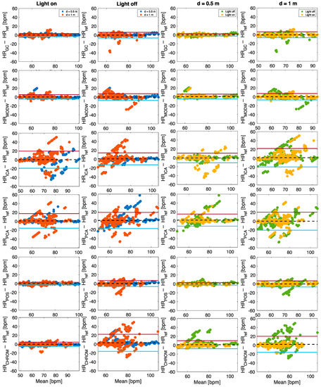
Heart rate (HR) and respiratory rate (
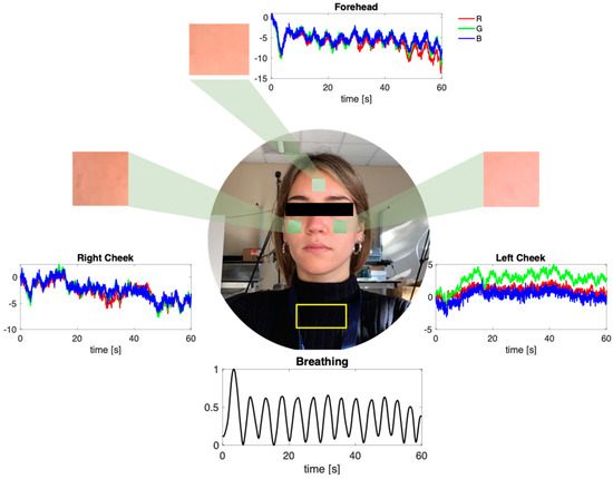
fR) can be estimated by processing videos framing the upper body and face regions without any physical contact with the subject. This paper proposed a technique for continuously monitoring HR and
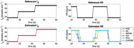
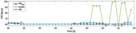
fR via a multi-ROI approach based on the spectral analysis of RGB video frames recorded with a mobile device (i.e., a smartphone’s camera). The respiratory signal was estimated by the motion of the chest, whereas the cardiac signal was retrieved from the pulsatile activity at the level of right and left cheeks and forehead. Videos were recorded from 18 healthy volunteers in four sessions with different user-camera distances (i.e., 0.5 m and 1.0 m) and illumination conditions (i.e., natural and artificial light). For HR estimation, three approaches were investigated based on single or multi-ROI approaches. A commercially available multiparametric device was used to record reference respiratory signals and electrocardiogram (ECG). The results demonstrated that the multi-ROI approach outperforms the single-ROI approach providing temporal trends of both the vital parameters comparable to those provided by the reference, with a mean absolute error (MAE) consistently below 1 breaths·min
−1 for
fR in all the scenarios, and a MAE between 0.7 bpm and 6 bpm for HR estimation, whose values increase at higher distances.
Full article
►▼
Show Figures
Open AccessArticle
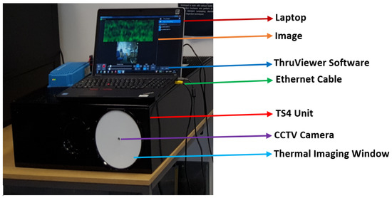
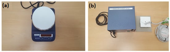
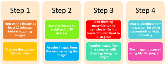
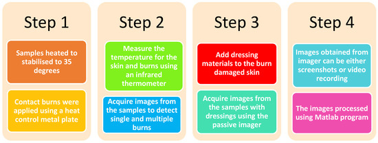
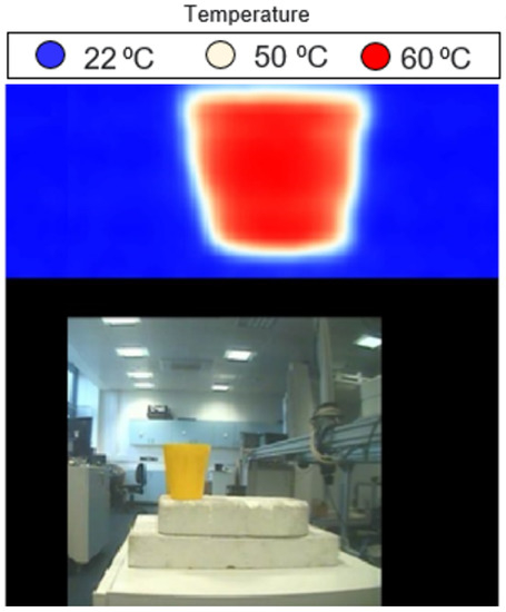
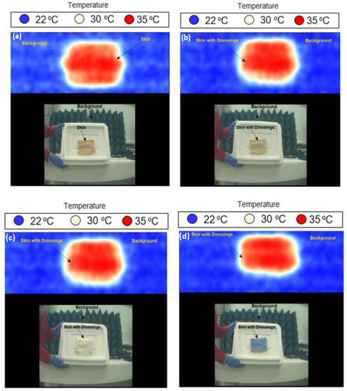
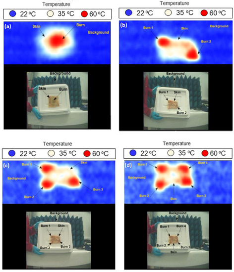
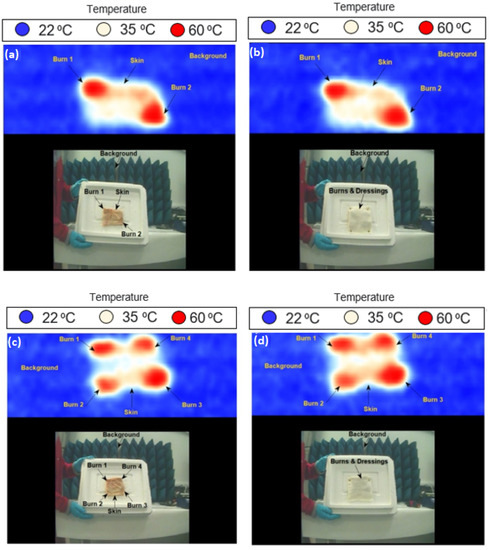
Passive Millimeter-Wave Imaging for Burns Diagnostics under Dressing Materials
by
Amani Yousef Owda
Cited by 4 | Viewed by 1689
Abstract
This paper presents a feasibility study of using a passive millimeter-wave imaging (PMMWI) system to assess burn wounds and the potential for monitoring the healing process under dressing materials, without their painful removal. Experimental images obtained from ex vivo porcine skin samples indicate
[...] Read more.
This paper presents a feasibility study of using a passive millimeter-wave imaging (PMMWI) system to assess burn wounds and the potential for monitoring the healing process under dressing materials, without their painful removal. Experimental images obtained from ex vivo porcine skin samples indicate that a ThruVision passive imager operating over the band 232–268 GHz can be used for diagnosing burns and for potentially monitoring the healing under dressing materials. Experimental images show that single and multiple burns are observed throughout dressing materials. As the interaction of millimeter-wave (MMW) radiation with the human body is almost exclusively with the skin, the major outcomes of the research are that PMMWI is capable of discriminating burn-damaged skin from unburned skin, and these measurements can be made through bandages without the imager making any physical contact with the skin or the bandage. This highlights the opportunity that the healing of burn wounds can be assessed and monitored without the removal of dressing materials. The key innovation in this work is in detecting single and multiple burns under dressing materials in noncontact with the skin and without exposing the skin to any type of manmade radiation (i.e., passive sensing technology). These images represent the first demonstration of burns wound under dressing materials using a passive sensing imager.
Full article
►▼
Show Figures


