


Molecules 2022, 27(16), 5175; https://doi.org/10.3390/molecules27165175 - 14 Aug 2022
Cited by 1 | Viewed by 2330
Abstract
►
Show Figures
Hydroxychloroquine (HCQ) is an autophagy inhibitor that has been used for the treatment of many diseases, such as malaria, rheumatoid arthritis, systemic lupus erythematosus, and cancer. Despite the therapeutic advances in these diseases, the underlying mechanisms have not been well determined and hinder
[...] Read more.
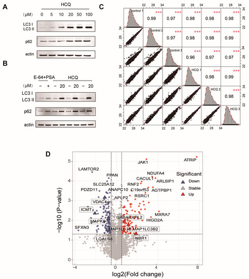
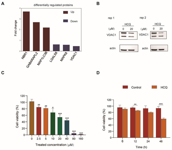
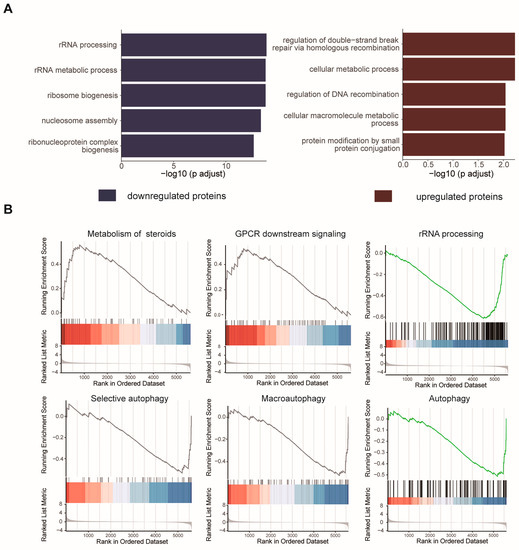
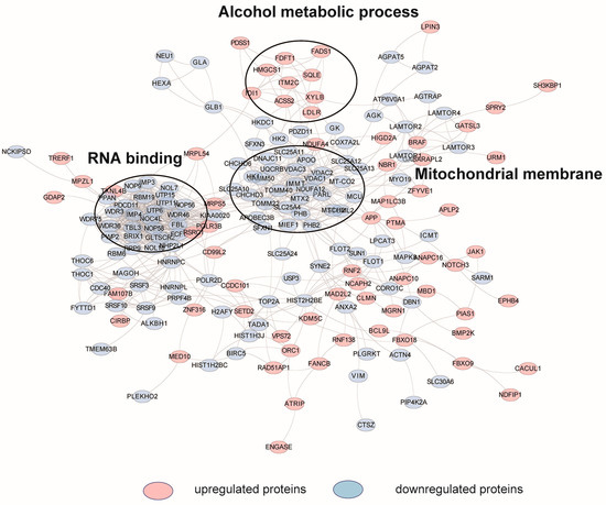
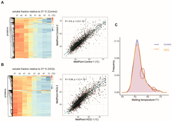
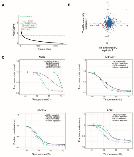
Hydroxychloroquine (HCQ) is an autophagy inhibitor that has been used for the treatment of many diseases, such as malaria, rheumatoid arthritis, systemic lupus erythematosus, and cancer. Despite the therapeutic advances in these diseases, the underlying mechanisms have not been well determined and hinder the rational use of this drug in the future. Here, we explored the possible mechanisms and identified the potential binding targets of HCQ by performing quantitative proteomics and thermal proteome profiling on MIA PaCa-2 cells. This study revealed that HCQ may exert its functions by targeting some autophagy-related proteins such as ribosyldihydronicotinamide dehydrogenase (NQO2) and transport protein Sec23A (SEC23A), or regulating the expression of galectin-8 (LGALS8), mitogen-activated protein kinase 8 (MAPK8), and so on. Furthermore, HCQ may prevent the progression of pancreatic cancer by regulating the expression of nesprin-2 (SYNE2), protein-S-isoprenylcysteine O-methyltransferase (ICMT), and cotranscriptional regulator FAM172A (FAM172A). Together, these findings not only identified potential binding targets for HCQ but also revealed the non-canonical mechanisms of HCQ that may contribute to pancreatic cancer treatment.
Full article
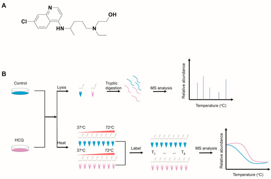
Figure 1