
Micro 2024, 4(1), 185-195; https://doi.org/10.3390/micro4010013 - 16 Mar 2024
Viewed by 553
Abstract
►
Show Figures
Acoustic focusing of particle flow in microfluidics has been shown to be an efficient tool for particle separation for various chemical and biomedical applications. The mechanism behind the method is the selective effect of the acoustic radiation force on distinct particles. In this
[...] Read more.
Acoustic focusing of particle flow in microfluidics has been shown to be an efficient tool for particle separation for various chemical and biomedical applications. The mechanism behind the method is the selective effect of the acoustic radiation force on distinct particles. In this way, they can be selectively focused and separated. The technique can also be applied under stationary conditions, i.e., in the absence of fluid flows. In this study, the manipulation of self-propelled particles, such as Janus particles, in an acoustofluidic setup was investigated. In experiments with self-propelled Janus particles and passive beads, we explored the interplay between self-propulsion and the acoustic radiation force. Our results demonstrated unusual and potentially useful effects such as selective trapping, escape, and assisted escape in binary mixtures of active and passive particles. We also analyzed various aspects related to the behavior of Janus particles in acoustic traps in the presence and absence of flows.
Full article
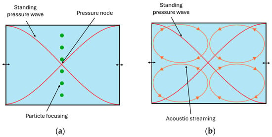
Figure 1