


Membranes 2023, 13(4), 370; https://doi.org/10.3390/membranes13040370 - 23 Mar 2023
Viewed by 1320
Abstract
►
Show Figures
The interaction of antimicrobial and amyloid peptides with cell membranes is a critical step in their activities. Peptides of the uperin family obtained from the skin secretion of Australian amphibians demonstrate antimicrobial and amyloidogenic properties. All-atomic molecular dynamics and an umbrella sampling approach
[...] Read more.
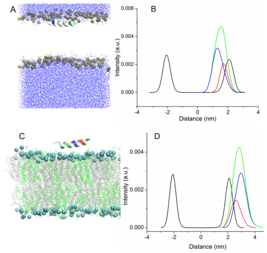
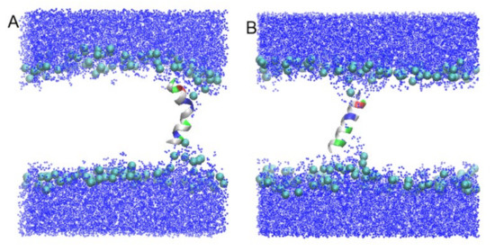
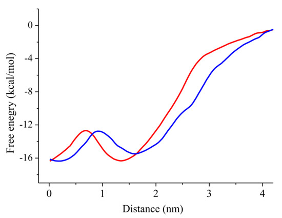
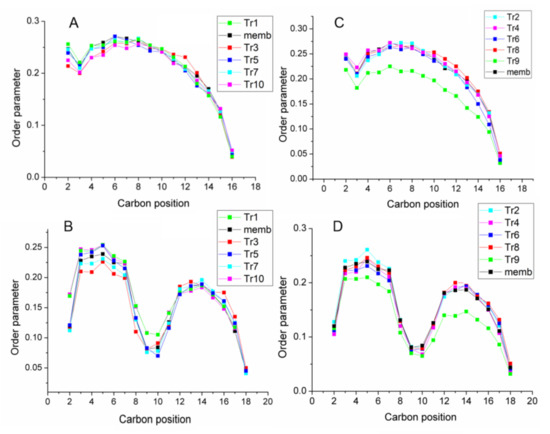
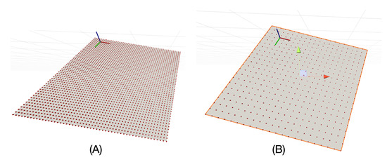
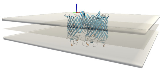
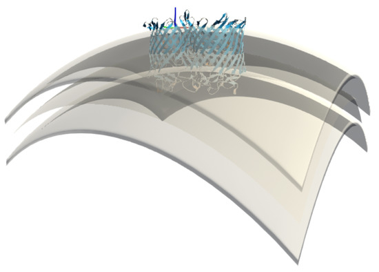
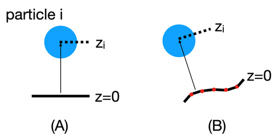
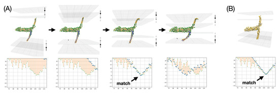
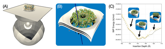
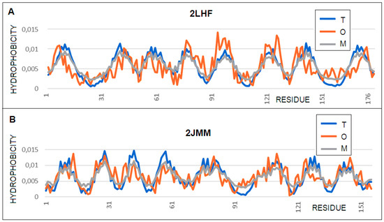
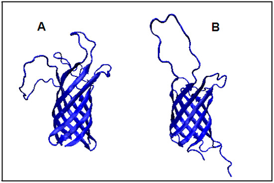
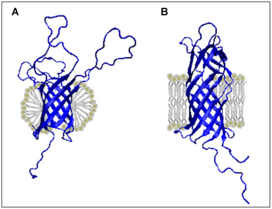
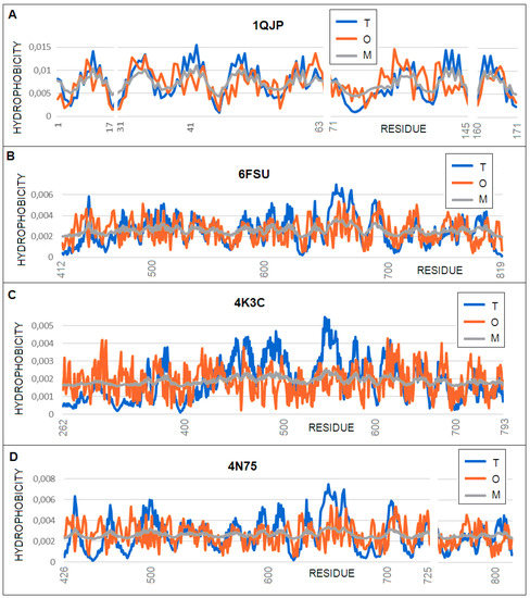
The interaction of antimicrobial and amyloid peptides with cell membranes is a critical step in their activities. Peptides of the uperin family obtained from the skin secretion of Australian amphibians demonstrate antimicrobial and amyloidogenic properties. All-atomic molecular dynamics and an umbrella sampling approach were used to study the interaction of uperins with model bacterial membrane. Two stable configurations of peptides were found. In the bound state, the peptides in helical form were located right under the head group region in parallel orientation with respect to the bilayer surface. Stable transmembrane configuration was observed for wild-type uperin and its alanine mutant in both alpha-helical and extended unstructured forms. The potential of mean force characterized the process of peptide binding from water to the lipid bilayer and its insertion into the membrane, and revealed that the transition of uperins from the bound state to the transmembrane position was accompanied by the rotation of peptides and passes through the energy barrier of 4–5 kcal/mol. Uperins have a weak effect on membrane properties.
Full article
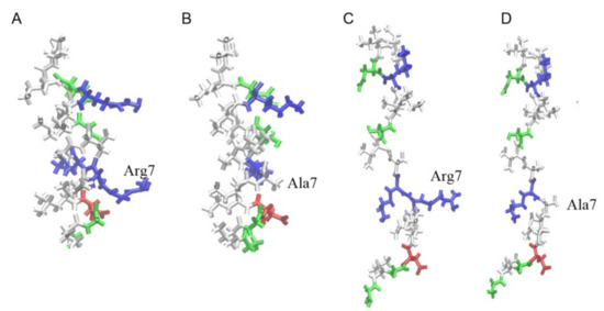
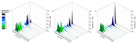
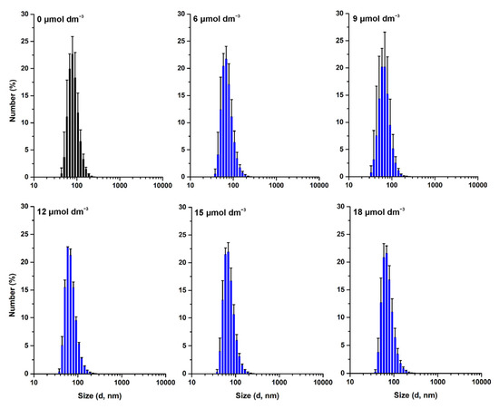
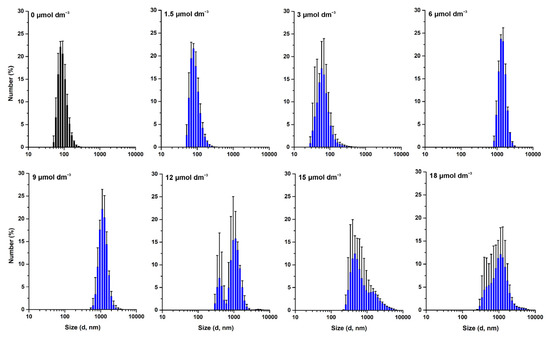
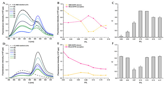
Figure 1


