Advances in the Pathogenesis of Neurodegenerative Diseases

A topical collection in Medical Sciences (ISSN 2076-3271). This collection belongs to the section "Neurosciences".

Viewed by 44217

Editor


E-Mail Website1 Website2
Collection Editor
National Research Council of Italy, Institute of Biomedical Technologies - Section of Bari, 70126 Bari, Italy
Interests: multiple sclerosis; neurodegenerative diseases; serum biomarkers; neuroimaging
Special Issues, Collections and Topics in MDPI journals

Topical Collection Information

Dear Colleagues,

The term “Neurodegenerative Diseases” (NDDs) identifies a heterogeneous group of disorders characterized by progressive degeneration of the structure and function of the central or peripheral nervous systems. The evidence of common features, represented, e.g., by intracellular accumulation and aggregation of misfolded proteins, abnormal cellular transport and mitochondrial deficits, suggest converging pathogenic pathways that lead to neurodegeneration. Quite recently, inflammation has been confirmed as another essential contributing factor, in which reactive astroglia and/or microglia seem to play key roles.

Despite the current knowledge, at present, the whole picture of neurodegeneration is still missing. Furthermore, besides well-known degenerative disorders like Alzheimer's, Parkinson's, Huntington's diseases and Amyotrophic Lateral Sclerosis, a crucial neurodegenerative component has been recognized, e.g., in inflammatory demyelinating diseases like Multiple Sclerosis, thus representing a significant piece of the puzzle in their characterization, which may narrow a more successful therapeutic window. Another important consideration concerns the wide heterogeneity of clinical or instrumental (e.g., neuroradiological) phenotypes in most of NDDs that may explain the sometimes-different response to treatment of each patient.

A fundamental support for the possible solution of these different issues is represented by the use of innovative extensive approaches in the molecular analysis like massive high-throughput sequencing (DNA, RNA) and proteomics that allowed to show the involvement of multiple cellular pathways in a given pathogenic process. As examples, transcriptomics studies showed that misregulated microRNAs (miRNAs, endogenous small RNAs binding the target sites of protein-coding genes, which lead to their degradation or the repression of translation) seem to justify several neurodegenerative processes, suggesting that alterations in miRNA regulatory pathways may contribute significantly to NDDs pathogenesis. Considering the growing interest in the potential of miRNAs, it is also plausible that these findings may lead to develop miRNAs therapeutics also for NDDs.

To our view, understanding the interplay of these (and others) key factors (i.e., extracellular vesicles and their cargos) may be of invaluable help in the effort of shedding lights on these still progressive and irreversible processes.

On these grounds, the big challenge of this Special Issue is to further explore the novel molecular hypotheses that underline the degenerative features of NDDs, in order: 1) to better define their pathogenic processes; 2) to picture peculiar molecular profiles of clinical/instrumental phenotypes; 3) to possibly identify early circulating biomarkers of clinical prognosis and treatment efficacy. The final goal of this approach will be to possibly offer suggestions for controlling the diseases and their progression, thus avoiding the irreversible and dramatic events of more advanced conditions. This will lead to more attention regarding the patient's quality of life, by promoting treatment plans based on the individual molecular signature (i.e.: the most effective dose with least side effects), a fundamental step towards the implementation of so-called Personalized Medicine.

Dr. Maria Liguori
Guest Editor

Manuscript Submission Information

Manuscripts should be submitted online at www.mdpi.com by registering and logging in to this website. Once you are registered, click here to go to the submission form. Manuscripts can be submitted until the deadline. All submissions that pass pre-check are peer-reviewed. Accepted papers will be published continuously in the journal (as soon as accepted) and will be listed together on the collection website. Research articles, review articles as well as short communications are invited. For planned papers, a title and short abstract (about 100 words) can be sent to the Editorial Office for announcement on this website.

Submitted manuscripts should not have been published previously, nor be under consideration for publication elsewhere (except conference proceedings papers). All manuscripts are thoroughly refereed through a single-blind peer-review process. A guide for authors and other relevant information for submission of manuscripts is available on the Instructions for Authors page. Medical Sciences is an international peer-reviewed open access quarterly journal published by MDPI.

Please visit the Instructions for Authors page before submitting a manuscript. The Article Processing Charge (APC) for publication in this open access journal is 1400 CHF (Swiss Francs). Submitted papers should be well formatted and use good English. Authors may use MDPI's English editing service prior to publication or during author revisions.


Keywords

Neurodegenerative diseases

  • Neurobiology
  • Extracellular vesicles
  • Massive high-throughput sequencing (DNA, RNA)
  • Non-coding RNAs, gene targets
  • Molecular pathways
  • Circulating biomarkers of diseases
  • Phenotype Biomarkers
  • Biomarkers of treatment efficacy
Published Papers (7 papers)

2020

Jump to: 2018, 2017

12 pages, 341 KiB  
Review
The Potential Role of Astrocytes in Parkinson’s Disease (PD)
by Hiluf Hindeya Gebreyesus and Teklu Gebrehiwot Gebremichael
Med. Sci. 2020, 8(1), 7; https://doi.org/10.3390/medsci8010007 - 27 Jan 2020
Cited by 16 | Viewed by 5772
Abstract
Astrocytes are multi-functional cells, now recognized as critical participants in many brain functions. They play a critical physiological role in the clearance of neurotransmitters, such as glutamate and gamma-aminobutyric acid (GABA), and in the regulation of K+ from the space of synaptic [...] Read more.
Astrocytes are multi-functional cells, now recognized as critical participants in many brain functions. They play a critical physiological role in the clearance of neurotransmitters, such as glutamate and gamma-aminobutyric acid (GABA), and in the regulation of K+ from the space of synaptic clefts. Astrocytes also express the excitatory amino acid transporters (EAATs) and aquaporin-4 (AQP4) water channel, which are involved in both physiological functions and neurodegenerative diseases (ND). Some of the ND are the Alzheimer’s (AD), Huntington’s (HD), Parkinson’s diseases (PD), Cerebral edema, amyotrophic lateral sclerosis (ALS), and epilepsy pathological conditions in specific regions of the CNS. Parkinson’s disease is the second most common age-related neurodegenerative disorder, characterized by degeneration of dopaminergic neurons of the substantia nigra pars compacta (SNpc). These project to the striatum, forming an important pathway within the basal ganglia. Mostly, PD has no clear etiology, and the mechanism of dopaminergic (DA) neuron loss is not well illustrated. The results of various studies suggest that astrocytes are involved in the pathophysiology of PD. Evidence has shown that the down-regulation of EAAT-2/GLT-1 and AQP4 expression is associated with PD pathogenesis. However, controversial results were reported in different experimental studies about the expression and function of EAAT-2/GLT-1 and AQP4, as well as their colocalization in different brain regions, and their involvement in PD development. Therefore, under neurological disorders, Parkinson’s disease is related to the genetic and phenotypic change of astrocytes’ biology. In this review, the authors summarized recent their research findings, which revealed the involvement of EAAT-2/GLT-1 and AQP4 expression, the physical interaction between EAAT-2/GLT-1 and AQP4 in astrocyte function, and their potential role in the development of PD in SNpc and Subthalamic nucleus (STN) of the basal ganglia nuclei. Full article

2018

Jump to: 2020, 2017

13 pages, 1736 KiB  
Review
Contribution of Inhibitor of Differentiation and Estrogenic Endocrine Disruptors to Neurocognitive Disorders
by Andrea Avecilla, Mayur Doke, Jeremy Jovellanos and Vincent Avecilla
Med. Sci. 2018, 6(3), 61; https://doi.org/10.3390/medsci6030061 - 03 Aug 2018
Cited by 3 | Viewed by 4682
Abstract
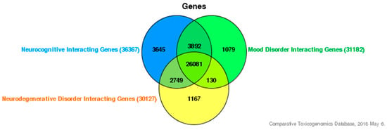
The devastating growth in the worldwide frequency of neurocognitive disorders and its allied difficulties, such as decline in memory, spatial competency, and ability to focus, poses a significant psychological public health problem. Inhibitor of differentiation (ID) proteins are members of a family of [...] Read more.
The devastating growth in the worldwide frequency of neurocognitive disorders and its allied difficulties, such as decline in memory, spatial competency, and ability to focus, poses a significant psychological public health problem. Inhibitor of differentiation (ID) proteins are members of a family of helix-loop-helix (HLH) transcription factors. ID proteins have been demonstrated to be involved in neurodevelopmental and depressive diseases and, thus, may influence neurocognitive deficiencies due to environmental exposure. Previously, it has been demonstrated that environmental factors, such as estrogenic endocrine disruptors (EEDs), have played an essential role in the influence of various neurocognitive disorders such as Alzheimer’s, dementia, and Parkinson’s disease. Based on this increasing number of reports, we consider the impact of these environmental pollutants on ID proteins. Better understanding of how these ID proteins by which EED exposure can affect neurocognitive disorders in populations will prospectively deliver valuable information in the impediment and regulation of these diseases linked with environmental factor exposure. Full article
Show Figures

Figure 1

26 pages, 1187 KiB  
Review
Circadian Rhythm and Alzheimer’s Disease
by Jan Homolak, Monika Mudrovčić, Barbara Vukić and Karlo Toljan
Med. Sci. 2018, 6(3), 52; https://doi.org/10.3390/medsci6030052 - 21 Jun 2018
Cited by 55 | Viewed by 10218
Abstract
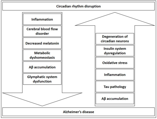
Alzheimer’s disease (AD) is a neurodegenerative disorder with a growing epidemiological importance characterized by significant disease burden. Sleep-related pathological symptomatology often accompanies AD. The etiology and pathogenesis of disrupted circadian rhythm and AD share common factors, which also opens the perspective of viewing [...] Read more.
Alzheimer’s disease (AD) is a neurodegenerative disorder with a growing epidemiological importance characterized by significant disease burden. Sleep-related pathological symptomatology often accompanies AD. The etiology and pathogenesis of disrupted circadian rhythm and AD share common factors, which also opens the perspective of viewing them as a mutually dependent process. This article focuses on the bi-directional relationship between these processes, discussing the pathophysiological links and clinical aspects. Common mechanisms linking both processes include neuroinflammation, neurodegeneration, and circadian rhythm desynchronization. Timely recognition of sleep-specific symptoms as components of AD could lead to an earlier and correct diagnosis with an opportunity of offering treatments at an earlier stage. Likewise, proper sleep hygiene and related treatments ought to be one of the priorities in the management of the patient population affected by AD. This narrative review brings a comprehensive approach to clearly demonstrate the underlying complexities linking AD and circadian rhythm disruption. Most clinical data are based on interventions including melatonin, but larger-scale research is still scarce. Following a pathophysiological reasoning backed by evidence gained from AD models, novel anti-inflammatory treatments and those targeting metabolic alterations in AD might prove useful for normalizing a disrupted circadian rhythm. By restoring it, benefits would be conferred for immunological, metabolic, and behavioral function in an affected individual. On the other hand, a balanced circadian rhythm should provide greater resilience to AD pathogenesis. Full article
Show Figures

Figure 1

13 pages, 1019 KiB  
Review
Results of Beta Secretase-Inhibitor Clinical Trials Support Amyloid Precursor Protein-Independent Generation of Beta Amyloid in Sporadic Alzheimer’s Disease
by Vladimir Volloch and Sophia Rits
Med. Sci. 2018, 6(2), 45; https://doi.org/10.3390/medsci6020045 - 02 Jun 2018
Cited by 23 | Viewed by 4518
Abstract
The present review analyzes the results of recent clinical trials of β secretase inhibition in sporadic Alzheimer’s disease (SAD), considers the striking dichotomy between successes in tests of β-site Amyloid Precursor Protein-Cleaving Enzyme (BACE) inhibitors in healthy subjects and familial Alzheimer’s disease (FAD) [...] Read more.
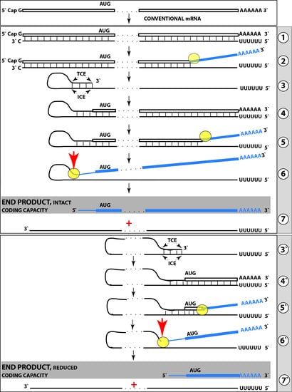
The present review analyzes the results of recent clinical trials of β secretase inhibition in sporadic Alzheimer’s disease (SAD), considers the striking dichotomy between successes in tests of β-site Amyloid Precursor Protein-Cleaving Enzyme (BACE) inhibitors in healthy subjects and familial Alzheimer’s disease (FAD) models versus persistent failures of clinical trials and interprets it as a confirmation of key predictions for a mechanism of amyloid precursor protein (APP)-independent, β secretase inhibition-resistant production of β amyloid in SAD, previously proposed by us. In light of this concept, FAD and SAD should be regarded as distinctly different diseases as far as β-amyloid generation mechanisms are concerned, and whereas β secretase inhibition would be neither applicable nor effective in the treatment of SAD, the β-site APP-Cleaving Enzyme (BACE) inhibitor(s) deemed failed in SAD trials could be perfectly suitable for the treatment of FAD. Moreover, targeting the aspects of Alzheimer’s disease (AD) other than cleavages of the APP by β and α secretases should have analogous impacts in both FAD and SAD. Full article
Show Figures

Graphical abstract

2017

Jump to: 2020, 2018

243 KiB  
Review
Alzheimer’s Disease, Brain Injury, and C.N.S. Nanotherapy in Humans: Sonoporation Augmenting Drug Targeting
by Joseph S. D’Arrigo
Med. Sci. 2017, 5(4), 29; https://doi.org/10.3390/medsci5040029 - 24 Nov 2017
Cited by 2 | Viewed by 4291
Abstract
Owing to the complexity of neurodegenerative diseases, multiple cellular types need to be targeted simultaneously in order for a given therapy to demonstrate any major effectiveness. Ultrasound-sensitive coated microbubbles (in a targeted nanoemulsion) are available. Versatile small-molecule drug(s) targeting multiple pathways of Alzheimer’s [...] Read more.
Owing to the complexity of neurodegenerative diseases, multiple cellular types need to be targeted simultaneously in order for a given therapy to demonstrate any major effectiveness. Ultrasound-sensitive coated microbubbles (in a targeted nanoemulsion) are available. Versatile small-molecule drug(s) targeting multiple pathways of Alzheimer’s disease pathogenesis are known. By incorporating such drug(s) into the targeted lipid-coated microbubble/nanoparticle-derived (LCM/ND) lipid nanoemulsion type, one obtains a multitasking combination therapeutic for translational medicine. This multitasking therapeutic targets cell-surface scavenger receptors (mainly scavenger receptor class B type I (SR-BI)), making it possible for various Alzheimer’s-related cell types to be simultaneously sought for localized drug treatment in vivo. Besides targeting cell-surface SR-BI, the proposed LCM/ND-nanoemulsion combination therapeutic(s) include a characteristic lipid-coated microbubble (LCM) subpopulation (i.e., a stable LCM suspension); such LCM substantially reduce the acoustic power levels needed for accomplishing temporary noninvasive (transcranial) ultrasound treatment, or sonoporation, if additionally desired for the Alzheimer’s patient. Full article
421 KiB  
Review
Cytokine Signaling in Multiple Sclerosis and Its Therapeutic Applications
by Pushpalatha Palle, Kelly L. Monaghan, Sarah M. Milne and Edwin C.K. Wan
Med. Sci. 2017, 5(4), 23; https://doi.org/10.3390/medsci5040023 - 13 Oct 2017
Cited by 55 | Viewed by 9422
Abstract
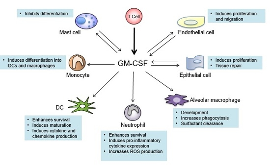
Multiple sclerosis (MS) is one of the most common neurological disorders in young adults. The etiology of MS is not known but it is widely accepted that it is autoimmune in nature. Disease onset is believed to be initiated by the activation of [...] Read more.
Multiple sclerosis (MS) is one of the most common neurological disorders in young adults. The etiology of MS is not known but it is widely accepted that it is autoimmune in nature. Disease onset is believed to be initiated by the activation of CD4+ T cells that target autoantigens of the central nervous system (CNS) and their infiltration into the CNS, followed by the expansion of local and infiltrated peripheral effector myeloid cells that create an inflammatory milieu within the CNS, which ultimately lead to tissue damage and demyelination. Clinical studies have shown that progression of MS correlates with the abnormal expression of certain cytokines. The use of experimental autoimmune encephalomyelitis (EAE) model further delineates the role of these cytokines in neuroinflammation and the therapeutic potential of manipulating their biological activity in vivo. In this review, we will first present an overview on cytokines that may contribute to the pathogenesis of MS or EAE, and provide successful examples and roadblock of translating data obtained from EAE to MS. We will then focus in depth on recent findings that demonstrate the pathological role of granulocyte-macrophage colony-stimulating factor (GM-CSF) in MS and EAE, and briefly discuss the potential of targeting effector myeloid cells as a treatment strategy for MS. Full article
Show Figures

Graphical abstract

536 KiB  
Communication
Molecular Characterization of Peripheral Extracellular Vesicles in Clinically Isolated Syndrome: Preliminary Suggestions from a Pilot Study
by Nicoletta Nuzziello, Maria Blonda, Flavio Licciulli, Sabino Liuni, Antonella Amoruso, Alessio Valletti, Arianna Consiglio, Carlo Avolio and Maria Liguori
Med. Sci. 2017, 5(3), 19; https://doi.org/10.3390/medsci5030019 - 18 Sep 2017
Cited by 5 | Viewed by 3731
Abstract
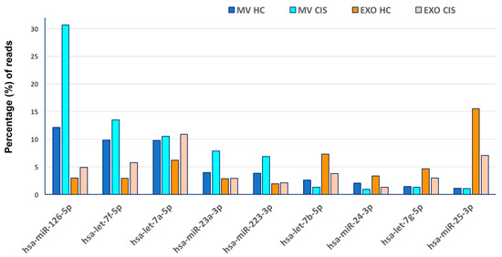
Extracellular vesicles (EVs), nanoparticles originated from different cell types, seem to be implicated in several cellular activities. In the Central Nervous System (CNS), glia and neurons secrete EVs and recent studies have demonstrated that the intercellular communication mediated by EVs has versatile functional [...] Read more.
Extracellular vesicles (EVs), nanoparticles originated from different cell types, seem to be implicated in several cellular activities. In the Central Nervous System (CNS), glia and neurons secrete EVs and recent studies have demonstrated that the intercellular communication mediated by EVs has versatile functional impact in the cerebral homeostasis. This essential role may be due to their proteins and RNAs cargo that possibly modify the phenotypes of the targeted cells. Despite the increasing importance of EVs, little is known about their fluctuations in physiological as well as in pathological conditions. Furthermore, only few studies have investigated the contents of contemporary EVs subgroups (microvesicles, MVs and exosomes, EXOs) with the purpose of discriminating between their features and functional roles. In order to possibly shed light on these issues, we performed a pilot study in which MVs and EXOs extracted from serum samples of a little cohort of subjects (patients with the first clinical evidence of CNS demyelination, also known as Clinically Isolated Syndrome and Healthy Controls) were submitted to deep small-RNA sequencing. Data were analysed by an in-home bioinformatics platform. In line with previous reports, distinct classes of non-coding RNAs have been detected in both the EVs subsets, offering interesting suggestions on their origins and functions. We also verified the feasibility of this extensive molecular approach, thus supporting its valuable use for the analysis of circulating biomarkers (e.g., microRNAs) in order to investigate and monitor specific diseases. Full article
Show Figures

Figure 1

Back to TopTop