


Medicina 2023, 59(2), 299; https://doi.org/10.3390/medicina59020299 - 06 Feb 2023
Cited by 1 | Viewed by 1547
Abstract
►
Show Figures
Background and Objectives: Implant rehabilitation of complete edentulous arches has become more and more popular because of the increased access of the population to this type of treatment. Furthermore, the development of new rehabilitation procedures can be applied in most clinical cases,
[...] Read more.
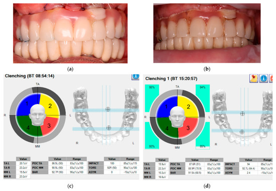
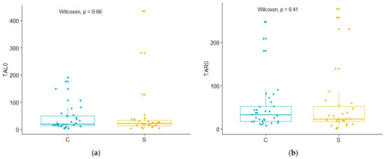
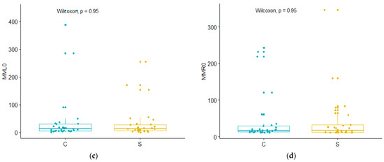
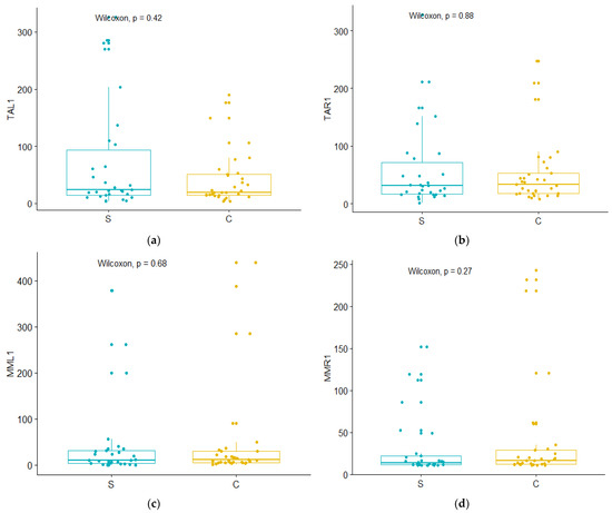
Background and Objectives: Implant rehabilitation of complete edentulous arches has become more and more popular because of the increased access of the population to this type of treatment. Furthermore, the development of new rehabilitation procedures can be applied in most clinical cases, including in those with severe atrophy. Hence, this study aimed to assess the functional changes that can occur in the stomatognathic system after implant rehabilitation procedures. Materials and Methods: A total of 63 patients were accepted in the study. They were divided into a first control (dentate) group (CG) and a second study group (edentulous, SG). For the latter, 30 patients received 204 two-stage implants immediately loaded with provisional prostheses. Surface electromyography (EMG) was assessed at the time of prostheses fixation, while for some patients it was applied six months after the fixation of the fixed prostheses, as well. These supplemental investigated patients formed a third, follow-up study group (FSG). All assessments were performed during the processes of clenching and mastication. The obtained data of the two study groups, SG and FSG, were compared with those of the control group, CG. Results: No statistical differences were found in the electrical muscular activity between the study and control groups during both clenching and mastication (p > 0.05). In addition, there were no differences within the same study group, both initially and after 6 months. The only changes were noticed between static and dynamic values for the right masseter muscle in the follow-up group FSG (p = 0.008). Deviations of the overlapping coefficients were similar for all groups (p = 0.086): for CG, 20.5%, median 11.1 (min. 0, max. 104); for SG, 21.4%, median 12.2 (min. 0, max. 103); for FSG, 36.1%, median 26.9 (min. 0, max. 160). This revealed no neuromuscular adaption to the prostheses. Conclusions: Implant-prosthetic rehabilitation led to an EMG activity that was similar to that of dentate patients immediately after the placement of the fixed implant-supported prostheses. Moreover, the measured values did not change after six months of functioning for all evaluated parameters. This may point to an immediate restoration of the muscle contraction capacity, without the necessity of adaptation over time. The study serves as an argument for the application and reliability of the immediate fixed implant-supported prostheses from the perspective of muscle adaptation and functioning.
Full article
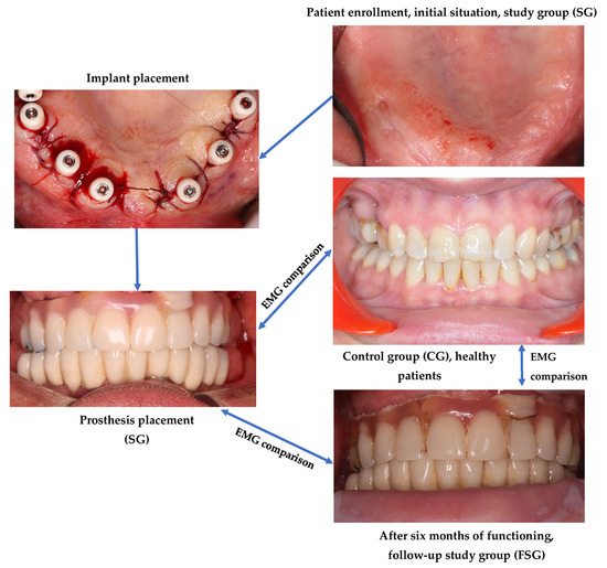
Figure 1


