


Mar. Drugs 2024, 22(2), 53; https://doi.org/10.3390/md22020053 - 23 Jan 2024
Viewed by 1274
Abstract
►
Show Figures
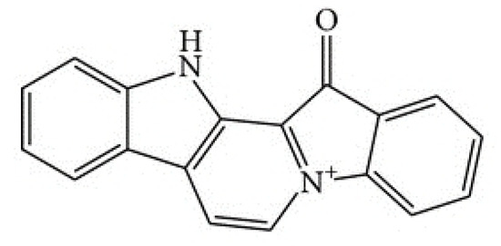
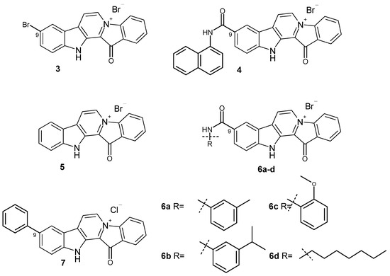
Based on the results of our own preliminary studies, the derivative of the marine alkaloid fascaplysin containing a phenyl substituent at C-9 was selected to evaluate the therapeutic potential in vivo and in vitro. It was shown that this compound has outstandingly high
[...] Read more.
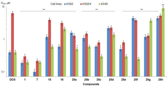
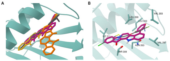
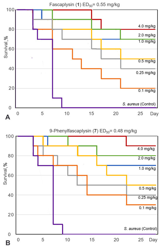
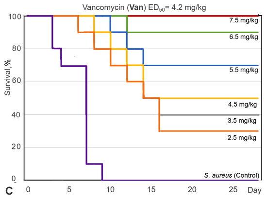
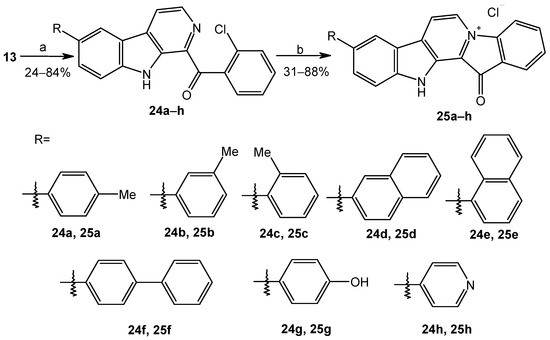
Based on the results of our own preliminary studies, the derivative of the marine alkaloid fascaplysin containing a phenyl substituent at C-9 was selected to evaluate the therapeutic potential in vivo and in vitro. It was shown that this compound has outstandingly high antimicrobial activity against Gram-positive bacteria, including antibiotic-resistant strains in vitro. The presence of a substituent at C-9 of the framework is of fundamental importance, since its replacement to neighboring positions leads to a sharp decrease in the selectivity of the antibacterial action, which indicates the presence of a specific therapeutic target in bacterial cells. On a model of the acute bacterial sepsis in mice, it was shown that the lead compound was more effective than the reference antibiotic vancomycin seven out of nine times. However, ED50 value for 9-phenylfascaplysin (7) was similar for the unsubstituted fascaplysin (1) in vivo, despite the former being significantly more active than the latter in vitro. Similarly, assessments of the anticancer activity of compound 7 against various variants of Ehrlich carcinoma in mice demonstrated its substantial efficacy. To conduct a structure–activity relationship (SAR) analysis and searches of new candidate compounds, we synthesized a series of analogs of 9-phenylfascaplysin with varying aryl substituents. However, these modifications led to the reduced aqueous solubility of fascaplysin derivatives or caused a loss of their antibacterial activity. As a result, further research is required to explore new avenues for enhancing its pharmacokinetic characteristics, the modification of the heterocyclic framework, and optimizing of treatment regimens to harness the remarkable antimicrobial potential of fascaplysin for practical usage.
Full article
Figure 1


