Recent Advances of Examination and Treatment for Retinal and Choroidal Diseases
Share This Topical Collection
Editor
 Dr. Hiroto Terasaki
Dr. Hiroto Terasaki
E-Mail
Website
Collection Editor
Department of Ophthalmology, Kagoshima University Graduate School of Medical and Dental Sciences, Kagoshima, Japan
Interests: diabetic retinopathy; diabetic macular edema; retinal vein occlusion
Topical Collection Information
Dear Colleagues,
In recent years, the management of retinal and choroidal diseases has experienced a major evolution in diagnosis and treatment. In the field of diagnosis, not only optical coherence tomography (OCT), which has made great progress in the past 20 years, but also OCT angiography and color scanning laser ophthalmoscope have become popular worldwide. Thus, multimodal imaging has improved the accuracy of diagnosis and understanding of pathology. The increase in the number of examination devices also means an increase in the number of images of healthy and diseased eyes. Imaging methods have evolved significantly with the spread of artificial intelligence.
Treatment for diseases has also evolved dramatically. No one would disagree that the first and foremost one is anti-vascular endothelial cell growth factor (VEGF) drugs. Anti-VEGF drugs have become the mainstay of treatment for retinal diseases, including age-related macular degeneration (AMD) and diabetic retinopathy and macular edema, and have greatly increased the potential of pharmacotherapy for such diseases.
This Topical Collection invites a wide range of research related to ophthalmic examination, diagnosis, and treatment. Our aim is for this issue to be of help in the management of retinal and choroidal diseases and to assist you in your future research.
Dr. Hiroto Terasaki
Collection Editor
Manuscript Submission Information
Manuscripts should be submitted online at www.mdpi.com by registering and logging in to this website. Once you are registered, click here to go to the submission form. Manuscripts can be submitted until the deadline. All submissions that pass pre-check are peer-reviewed. Accepted papers will be published continuously in the journal (as soon as accepted) and will be listed together on the collection website. Research articles, review articles as well as short communications are invited. For planned papers, a title and short abstract (about 100 words) can be sent to the Editorial Office for announcement on this website.
Submitted manuscripts should not have been published previously, nor be under consideration for publication elsewhere (except conference proceedings papers). All manuscripts are thoroughly refereed through a single-blind peer-review process. A guide for authors and other relevant information for submission of manuscripts is available on the Instructions for Authors page. Journal of Clinical Medicine is an international peer-reviewed open access semimonthly journal published by MDPI.
Please visit the Instructions for Authors page before submitting a manuscript.
The Article Processing Charge (APC) for publication in this open access journal is 2600 CHF (Swiss Francs).
Submitted papers should be well formatted and use good English. Authors may use MDPI's
English editing service prior to publication or during author revisions.
Keywords
- optical coherence tomography (OCT)
- OCT angiography
- color scanning laser ophthalmoscope (SLO)
- anti-VEGF therapy
- age-related macular degeneration (AMD)
- diabetic retinopathy
- diabetic macular edema
- retinal vein occlusion
- central serous chorioretinopathy
Published Papers (11 papers)
Open AccessComment
Does Subretinal Fluid Influence Choroidal Thickness (ChT) and Structure in Preeclampsia with Serous Retinal Detachment? Comment on Fukui et al. Changes in Choroidal Thickness and Structure in Preeclampsia with Serous Retinal Detachment. J. Clin. Med. 2023, 12, 609
by
Andrea Valerio Marino, Aniello La Marca, Martina De Luca, Eleonora D’Aniello and Marco Gioia
Cited by 3 | Viewed by 649
Abstract
We read with great interest the article by Fukui A et al. concerning the “Changes in Choroidal Thickness (ChT) and Structure in Preeclampsia with Serous Retinal Detachment” [...]
Full article
Open AccessArticle
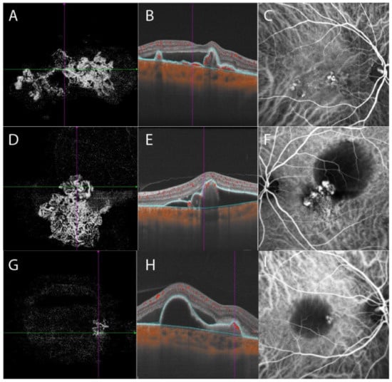
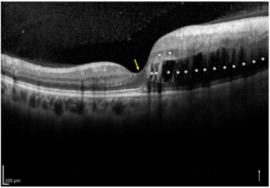
Different Morphology of Branching Neovascular Network in Polypoidal Choroidal Vasculopathy: A Swept-Source Optical Coherence Tomography Angiography Study
by
Lulu Chen, Mingzhen Yuan, Lu Sun and Youxin Chen
Cited by 3 | Viewed by 1451
Abstract
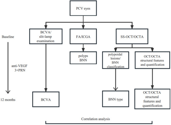
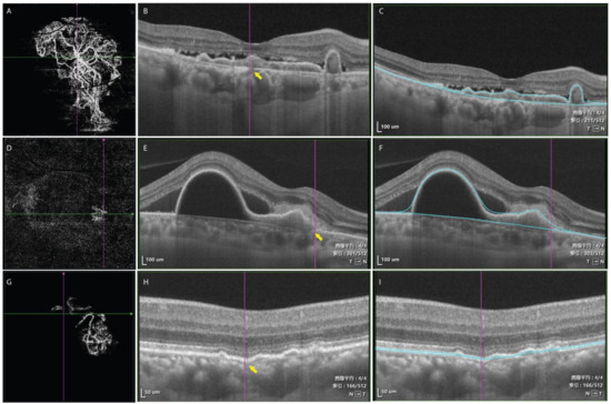
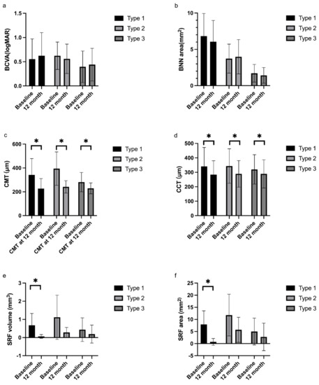
Purpose: To evaluate the classification system of branching neovascular network (BNN) morphology in polypoidal choroidal vasculopathy (PCV) patients based on swept-source optical coherence tomography (SS-OCT) and swept-source optical coherence tomography angiography (SS-OCTA), and analyze the morphological features in each group as potential prognostic
[...] Read more.
Purpose: To evaluate the classification system of branching neovascular network (BNN) morphology in polypoidal choroidal vasculopathy (PCV) patients based on swept-source optical coherence tomography (SS-OCT) and swept-source optical coherence tomography angiography (SS-OCTA), and analyze the morphological features in each group as potential prognostic features.
Methods: A total of 32 PCV eyes were included in this retrospective study. SS-OCT and SS-OCTA images of 6 mm × 6 mm centered on the foveal of each eye were analyzed. PCV cases were classified into three types (“trunk”, “glomeruli”, and “stick” type) based on the morphological features of BNN. OCT and OCTA features were compared among the three groups. The correlation of OCT/OCTA features with visual acuity at 12 months after anti-VEGF treatment was also analyzed.
Results: Type 1 group had the largest BNN area and the largest numbers of polypoidal lesions. Type 2 group has the largest pigment epithelial detachment (PED) area, PED volume, subretinal fluid (SRF) area, and SRF volume. Type 3 group had better baseline BCVA, the smallest BNN area, the smallest PED size, and the smallest SRF size. Type 1 was also featured by a clear break on Bruch’s membrane which corresponded to the origin of neovascular tissue. BCVA at 12 months was not significantly different among groups. Baseline BCVA and baseline central macular thickness were correlated with the final BCVA.
Conclusions: The current classification system based on BNN morphology on SS-OCTA was highly applicable and revealed distinct characteristics in each group. The BNN type was not correlated with BCVA at 12 months after treatment.
Full article
►▼
Show Figures
Open AccessArticle
Changes in Choroidal Thickness and Structure in Preeclampsia with Serous Retinal Detachment
by
Ayumi Fukui, Hiroshi Tanaka, Nobuhiro Terao, Kenji Nagata, Akifumi Matsumoto, Natsuki Kusada, Kentaro Kojima and Chie Sotozono
Cited by 4 | Viewed by 1391
Abstract
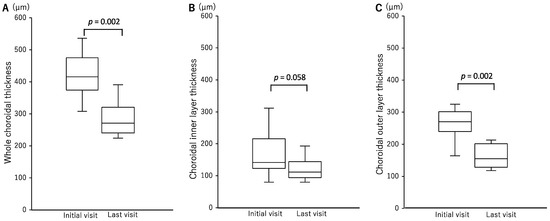
Preeclampsia is a pregnancy-specific syndrome characterized by hypertension and proteinuria. We retrospectively investigated the clinical features, including choroidal layer thickness and luminal area to stromal area ratio, in a case series of preeclampsia with serous retinal detachment (SRD). The subjects were pregnant women
[...] Read more.
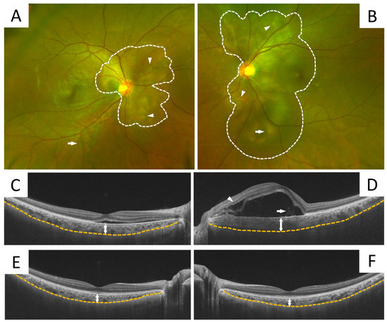
Preeclampsia is a pregnancy-specific syndrome characterized by hypertension and proteinuria. We retrospectively investigated the clinical features, including choroidal layer thickness and luminal area to stromal area ratio, in a case series of preeclampsia with serous retinal detachment (SRD). The subjects were pregnant women with SRD during hospitalization for preeclampsia from October 2014 to June 2021. Based on medical records, affected eyes, time of onset, fundus examination findings, and subfoveal choroidal thickness (SCT), the choroidal layer thickness and choroidal vascular index (CVI) in each patient was examined. Thirteen eyes from seven patients (mean age 30.7 ± 4.7 years) were included in the study. In all cases, SRD improved without topical ocular treatment. The mean SCT at the initial visit was 424.4 ± 70.5 μm, and all patients had choroidal thickening, which significantly decreased to 286.0 ± 57.9 μm (
p < 0.01) at the last visit. The mean choroidal inner layer was 162.7 ± 69.4 μm at the initial visit and 122.3 ± 35.5 μm at the final follow-up visit (
p = 0.06), showing no significant difference; however, the mean choroidal outer layer was 261.7 ± 47.6 μm at the initial visit and 163.7 ± 37.1 μm at the final follow-up visit (
p < 0.01), thus showing a significant decrease. The mean CVI was 67.2 ± 1.3% at the initial visit, yet it had significantly decreased to 65.4 ± 1.1% (
p < 0.01) at the final follow-up visit. The findings of this study show that SRD with preeclampsia is associated with increased thickening of the choroidal outer layer, especially in the choroidal luminal area.
Full article
►▼
Show Figures
Open AccessArticle
Impact of Treating Age-Related Macular Degeneration before Visual Function Is Impaired
by
Risa Aichi, Norihiro Nagai, Kishiko Ohkoshi and Yoko Ozawa
Viewed by 1141
Abstract
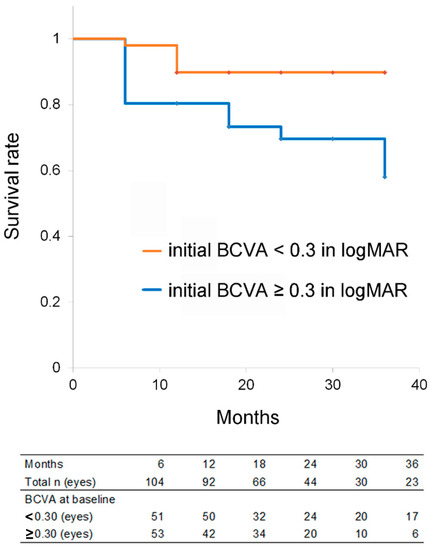
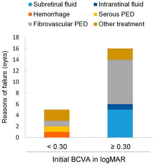
Visual outcomes of age-related macular degeneration (AMD) have substantially improved via anti-vascular endothelial growth factor (anti-VEGF) therapy. However, the treatment effects vary among individuals. Medical charts of 104 eyes (104 patients) with AMD, treated with anti-VEGF drugs and followed up for 12–36 months,
[...] Read more.
Visual outcomes of age-related macular degeneration (AMD) have substantially improved via anti-vascular endothelial growth factor (anti-VEGF) therapy. However, the treatment effects vary among individuals. Medical charts of 104 eyes (104 patients) with AMD, treated with anti-VEGF drugs and followed up for 12–36 months, were retrospectively analyzed. Logistic regression analyses adjusted for age showed that eyes with an initial best-corrected visual acuity (BCVA) < 0.3 in the logarithm of the minimum angle of resolution (logMAR) were a positive predictor (odds ratio = 3.172; 95% confidence interval [CI] = 1.029–9.783;
p = 0.045), and the presence of initial fibrovascular pigment epithelial detachment (PED) was a negative predictor (0.222; 0.078–0.637;
p = 0.005) of maintained or improved BCVA at the final visit. Kaplan–Meier survival analysis showed that eyes with an initial BCVA < 0.3 (Cox hazard ratio = 2.947; 95% CI = 1.047–8.289;
p = 0.041) had a better survival rate after adjusting for age when failure was defined as a BCVA reduction ≥ 0.2 of logMAR. Eyes with an initial BCVA < 0.3 belonged to younger patients; more frequently had subretinal fluid as an exudative change; and less frequently had intraretinal fluid, submacular hemorrhage, and fibrovascular PED. Initiating anti-VEGF treatment before BCVA declines and advanced lesions develop would afford better visual outcomes for AMD eyes in the real-world clinic, although further analyses are required.
Full article
►▼
Show Figures
Open AccessArticle
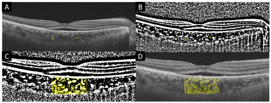
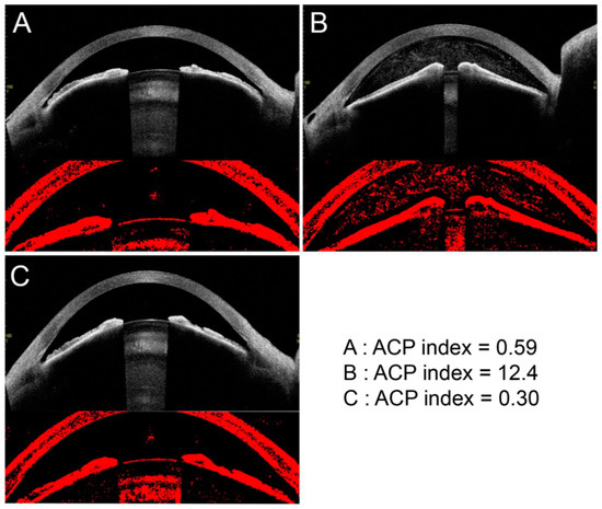
Quantification of Anterior Chamber Particles Using Anterior Segment Optical Coherence Tomography in Angle-Closure Glaucoma Patients after Laser Iridotomy
by
Naoya Yoshihara, Hiroto Terasaki, Hideki Shiihara, Ryoh Funatsu, Takehiro Yamashita and Taiji Sakamoto
Viewed by 1354
Abstract
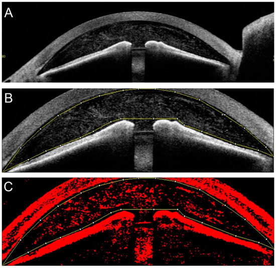
Purpose: To determine whether the degree of particle density in the anterior chamber can be evaluated objectively and quantitatively by anterior segment optical coherence tomography (AS-OCT) in cases after laser iridotomy (LI). Methods: This was a retrospective observational study. All of the subjects
[...] Read more.
Purpose: To determine whether the degree of particle density in the anterior chamber can be evaluated objectively and quantitatively by anterior segment optical coherence tomography (AS-OCT) in cases after laser iridotomy (LI). Methods: This was a retrospective observational study. All of the subjects who received LI for angle-closure glaucoma between January 2018 and May 2019 at Kagoshima University Hospital were studied. AS-OCT recordings were made before, immediately after, and one week after LI in 22 eyes of 14 consecutive patients. The anterior chamber particle (ACP) index was defined as the ratio of the number of particles in the anterior chamber to the total area of the anterior chamber. The ACP index was determined by binarization of the AS-OCT images and analysis with the ImageJ program. Results: The mean age of the participants was 75.4 ± 8.9 years, with a range of 61–91 years. The ACP index before the LI was 0.78 ± 0.68, and it was significantly increased to 7.72 ± 2.64 immediately after the LI (paired
t-test,
p < 0.01). The ACP index returned to the pre-LI density of 0.92 ± 0.48 one week after the LI. Conclusions: We successfully quantified the degree of anterior chamber particles accumulation by analyzing images obtained by AS-OCT. This simple and repeatable technique should be useful because the particles, including inflammatory cells, in the anterior chamber can be evaluated non-invasively and objectively.
Full article
►▼
Show Figures
Open AccessArticle
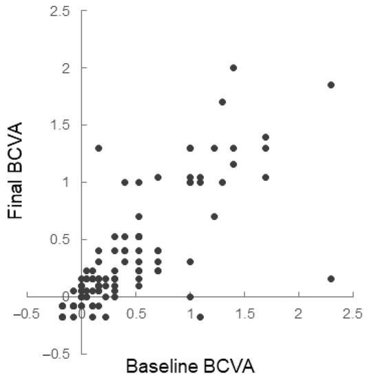
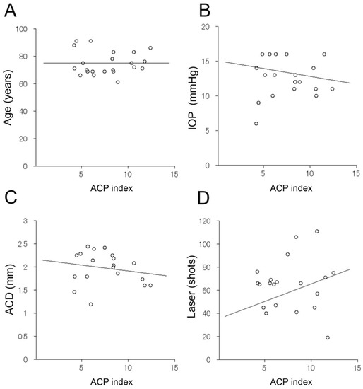
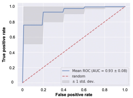
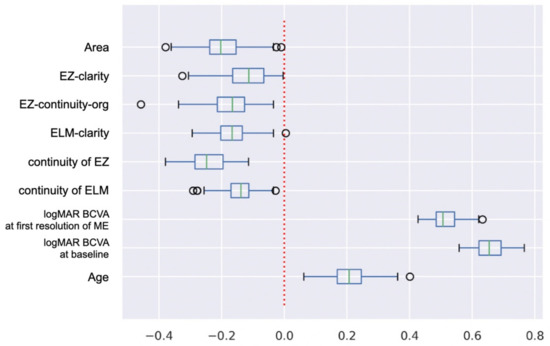
Which Explanatory Variables Contribute to the Classification of Good Visual Acuity over Time in Patients with Branch Retinal Vein Occlusion with Macular Edema Using Machine Learning?
by
Yoshitsugu Matsui, Kazuya Imamura, Shinichiro Chujo, Yoko Mase, Hisashi Matsubara, Masahiko Sugimoto, Hiroharu Kawanaka and Mineo Kondo
Cited by 1 | Viewed by 1817
Abstract
This study’s goal is to determine the accuracy of a linear classifier that predicts the prognosis of patients with macular edema (ME) due to a branch retinal vein occlusion during the maintenance phase of antivascular endothelial growth factor (anti-VEGF) therapy. The classifier was
[...] Read more.
This study’s goal is to determine the accuracy of a linear classifier that predicts the prognosis of patients with macular edema (ME) due to a branch retinal vein occlusion during the maintenance phase of antivascular endothelial growth factor (anti-VEGF) therapy. The classifier was created using the clinical information and optical coherence tomographic (OCT) findings obtained up to the time of the first resolution of ME. In total, 66 eyes of 66 patients received an initial intravitreal injection of anti-VEGF followed by repeated injections with the pro re nata (PRN) regimen for 12 months. The patients were divided into two groups: those with and those without good vision during the PRN phase. The mean AUC of the classifier was 0.93, and the coefficients of the explanatory variables were: best-corrected visual acuity (BCVA) at baseline was 0.66, BCVA at first resolution of ME was 0.51, age was 0.21, the average brightness of the ellipsoid zone (EZ) was −0.12, the intactness of the external limiting membrane (ELM) was −0.14, the average brightness of the ELM was −0.17, the brightness value of EZ was −0.17, the area of the outer segments of the photoreceptors was −0.20, and the intactness of the EZ was −0.24. This algorithm predicted the prognosis over time for individual patients during the PRN phase.
Full article
►▼
Show Figures
Open AccessArticle
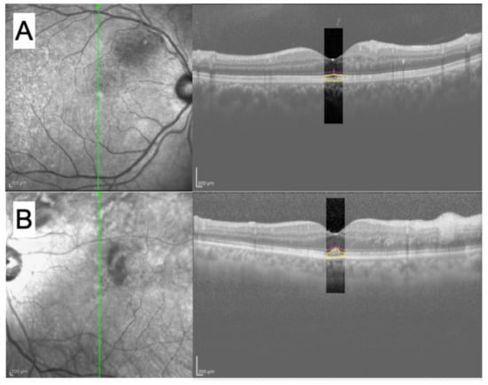
Foveal Intraretinal Fluid Localization Affects the Visual Prognosis of Branch Retinal Vein Occlusion
by
Hirofumi Sasajima, Masahiro Zako, Rio Maeda, Kenta Murotani, Hidetoshi Ishida and Yoshiki Ueta
Cited by 2 | Viewed by 1512
Abstract
We investigated whether baseline foveal intraretinal fluid (IRF) localization affects the visual prognosis of branch retinal vein occlusion (BRVO). Fifty eyes from 50 patients were included in this retrospective study. We classified the eyes with IRF involving and not involving the central foveola
[...] Read more.
We investigated whether baseline foveal intraretinal fluid (IRF) localization affects the visual prognosis of branch retinal vein occlusion (BRVO). Fifty eyes from 50 patients were included in this retrospective study. We classified the eyes with IRF involving and not involving the central foveola on the vertical optical coherence tomography (OCT) image at the initial visit into both-sides (
n = 17) and one-side IRF (
n = 33) groups, respectively. Multiple regression analyses demonstrated that not only the baseline logarithm of the minimum angle of resolution (logMAR) best-corrected visual acuity (BCVA) but also the IRF localization significantly correlated with the 12-month logMAR BCVA (
p = 0.04 and
p = 0.001, respectively), indicating that eyes with better baseline logMAR BCVA and one-side IRF have a significantly better visual prognosis in BRVO. The foveal ellipsoid zone band was significantly more disrupted (
p < 0.001) in the both-sides IRF (47.1%) group than in the one-side IRF (3.0%) group. No eyes with decimal BCVA less than 0.5 were detected in the one-side IRF group at 12 months. Thus, baseline foveal IRF localization on vertical OCT images can be considered a novel biomarker for the visual prognosis of BRVO.
Full article
►▼
Show Figures
Open AccessArticle
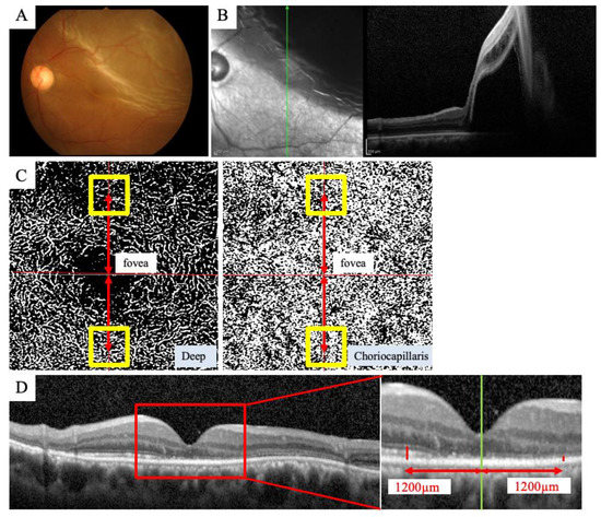
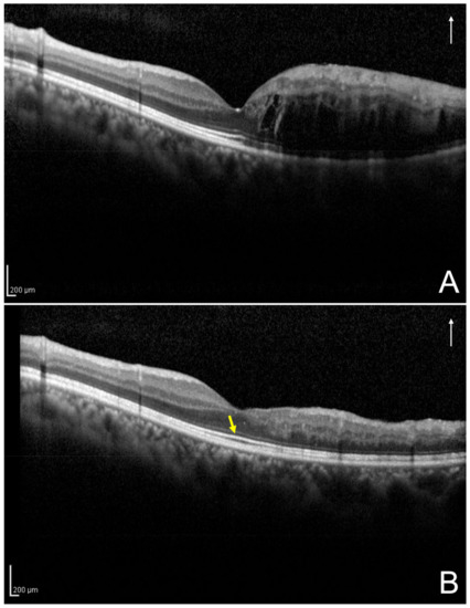
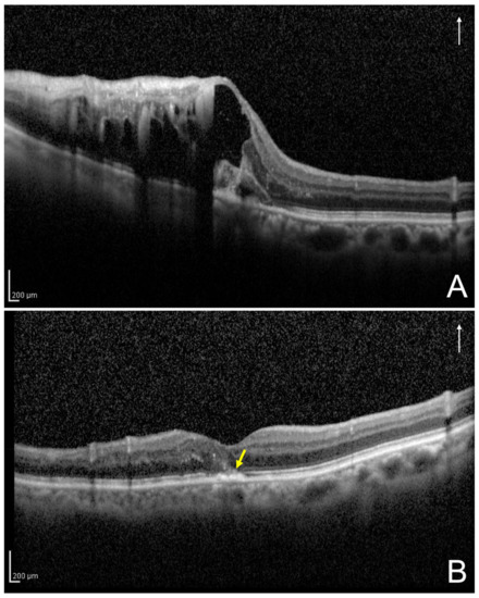
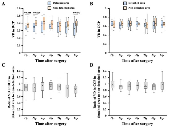
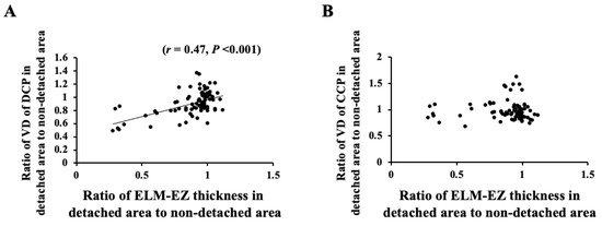
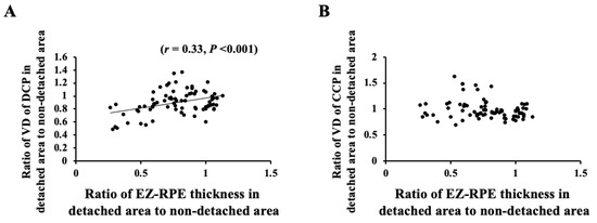
Differences in Vascular Density between Detached and Nondetached Areas in Eyes with Rhegmatogenous Retinal Detachment
by
Mariko Sato and Takeshi Iwase
Cited by 1 | Viewed by 1294
Abstract
We examined the vessel density (VD) of the deep capillary plexus (DCP) and choriocapillaris plexus (CCP) by optical coherence tomography (OCT) angiography in eyes with rhegmatogenous retinal detachment, which had similar amounts of detached and nondetached areas in the macula region, and then
[...] Read more.
We examined the vessel density (VD) of the deep capillary plexus (DCP) and choriocapillaris plexus (CCP) by optical coherence tomography (OCT) angiography in eyes with rhegmatogenous retinal detachment, which had similar amounts of detached and nondetached areas in the macula region, and then determined the morphology by OCT until 6 months after surgery. A total of 13 eyes of 13 patients whose average age was 55.8 ± 12.3 years and were successfully treated were enrolled in this study. Throughout the postoperative period, the VD of the DCP in the detached area decreased significantly compared to that in the nondetached area. Conversely, there was no significant difference in the VD of the CCP between the detached and the nondetached areas. The ratio of VD of both the DCP and CCP in the detached area to the in the nondetached area did not show significant changes during the follow-up period of 6 months. The ratio of VD of the DCP in the detached area to that in the nondetached area correlated significantly with the ratio of the external limiting membrane–ellipsoid zone (
r = 0.57,
p < 0.001) and ellipsoid zone–retinal pigment epithelium (
r = 0.39,
p < 0.001) thickness in the detached area to that in the nondetached area. A well-preserved DCP blood flow could result in the restoration of the outer retina.
Full article
►▼
Show Figures
Open AccessArticle
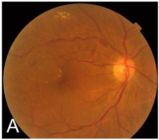
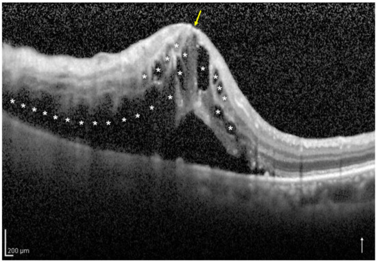
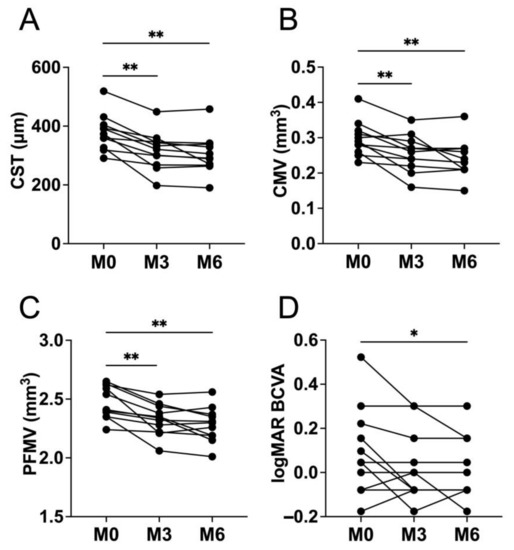
Direct Photocoagulation for Treating Microaneurysms with Hyperreflective Ring in Eyes with Refractory Macular Edema Associated with Branch Retinal Vein Occlusion
by
Hirofumi Sasajima, Masahiro Zako, Yoshiki Ueta, Hideo Tate, Chisato Otaki, Kenta Murotani, Takafumi Suzuki, Hidetoshi Ishida, Yoshihiro Hashimoto and Naoko Tachi
Cited by 1 | Viewed by 1978
Abstract
Microaneurysms (MAs) with hyperreflective rings are sometimes detected in eyes with refractory macular edema (ME) associated with branch retinal vein occlusion (BRVO) for more than 12 months after onset when examined using optical coherence tomography (OCT). We proposed that these MAs could result
[...] Read more.
Microaneurysms (MAs) with hyperreflective rings are sometimes detected in eyes with refractory macular edema (ME) associated with branch retinal vein occlusion (BRVO) for more than 12 months after onset when examined using optical coherence tomography (OCT). We proposed that these MAs could result in refractory ME secondary to BRVO and hypothesized that OCT-guided direct photocoagulation of MAs could result in a reduction in refractory ME. Eleven eyes (from eleven different patients) with refractory ME associated with BRVO for more than 12 months following initial treatment were included. The mean number of MAs in each eye at baseline was 3.5 ± 2.0 (range, 1–8). The mean central subfield thickness, central macular volume, and parafoveal macular volume significantly decreased 6 months following initial direct photocoagulation when compared with those at baseline (baseline = 378.7 ± 61.8 μm, post-treatment = 304.2 ± 66.7 μm,
p = 0.0005; baseline = 0.3 ± 0.049 mm
3, post-treatment = 0.24 ± 0.053 mm
3,
p = 0.001; and baseline = 2.5 ± 0.14 mm
3, post-treatment = 2.28 ± 0.15 mm
3,
p = 0.001, respectively). Moreover, the mean best-corrected visual acuity significantly improved 6 months following initial direct photocoagulation when compared with that at baseline (baseline = 0.096 ± 0.2 logarithm of the minimum angle of resolution (logMAR), post-treatment = 0.0077 ± 0.14 logMAR,
p = 0.031). Direct photocoagulation could be suggested as a treatment option for refractory ME associated with BRVO in MAs with a hyperreflective ring on OCT.
Full article
►▼
Show Figures
Open AccessFeature PaperArticle
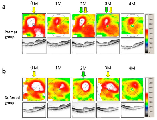
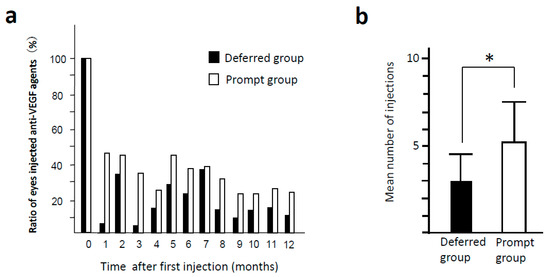
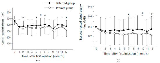
The Impact of Interval between Recurrence and Reinjection in Anti-VEGF Therapy for Diabetic Macular Edema in Pro Re Nata Regimen
by
Yoshihiro Takamura, Teruyo Kida, Hidetaka Noma, Makoto Inoue, Shigeo Yoshida, Taiji Nagaoka, Kousuke Noda, Yutaka Yamada, Masakazu Morioka, Makoto Gozawa, Takehiro Matsumura and Masaru Inatani
Cited by 3 | Viewed by 2386
Abstract
Background: Pro re nata (PRN) regimen using anti-vascular endothelial growth factor (VEGF) agent is popular for the treatment of diabetic macular edema (DME). We investigated the influence of waiting time (WT) and interval between the date of recurrence of edema and re-injection on
[...] Read more.
Background: Pro re nata (PRN) regimen using anti-vascular endothelial growth factor (VEGF) agent is popular for the treatment of diabetic macular edema (DME). We investigated the influence of waiting time (WT) and interval between the date of recurrence of edema and re-injection on treatment efficacy. Methods: This retrospective study conducted at 7 sites in Japan enrolled patients who received intravitreal injection of ranibizumab (IVR) and aflibercept (IVA) in 1+PRN regimen. Enrolled patients were divided into 2 groups: prompt group (less than 1 week) and deferred group (3 weeks or more). Central retinal thickness (CRT) and best corrected visual acuity (BCVA) were measured every month for 1 year. Results: CRT in the deferred group was significantly higher than that in the prompt group at 2, 5, 6, 7, and 12 months (
p < 0.05). BCVA in the prompt group was significantly better than that in the deferred group at 7, 10, and 12 months (
p < 0.05). Conclusion: The prompt group was superior in anatomical and functional improvement of DME in anti-VEGF therapy than the deferred group. Our data suggests that shorter WT is recommended for better visual prognosis in the treatment for DME.
Full article
►▼
Show Figures


