


J. Clin. Med. 2023, 12(17), 5508; https://doi.org/10.3390/jcm12175508 - 24 Aug 2023
Cited by 1 | Viewed by 1087
Abstract
►
Show Figures
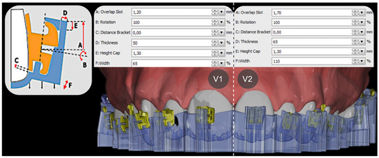
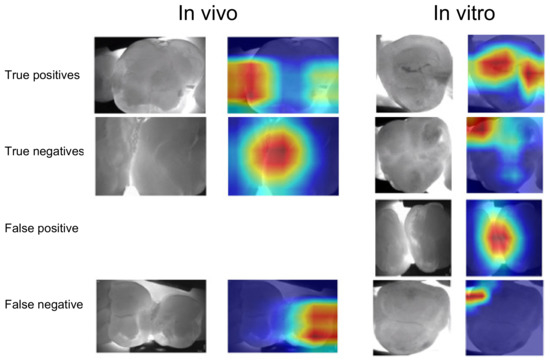
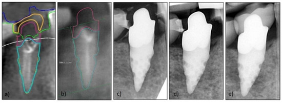
An accurate impression is vital during prosthodontic rehabilitation. Digital scanning has become an alternative to conventional impressions. This study compares conventional preliminary impression techniques with digital scanning, evaluating the efficiency, treatment comfort, and trueness. Impressions of 28 patients were taken using conventional and
[...] Read more.
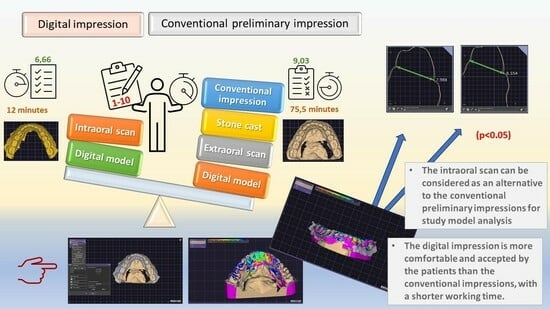
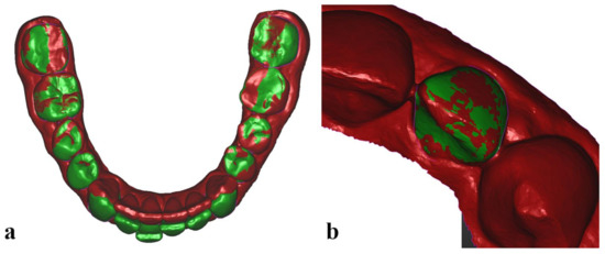
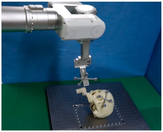
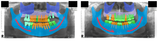
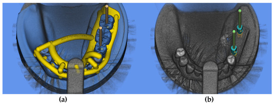
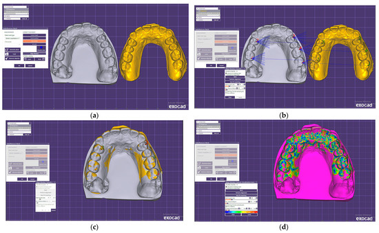
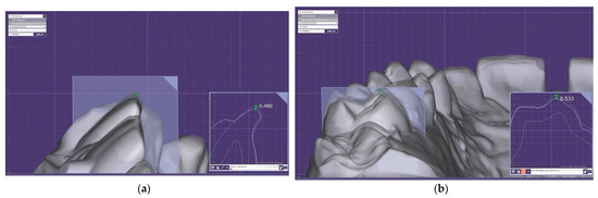
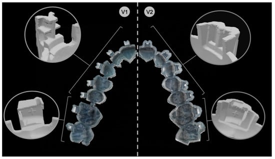
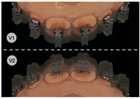
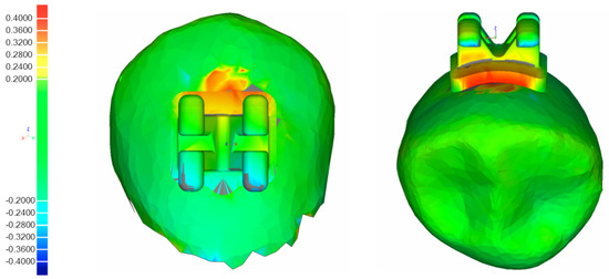
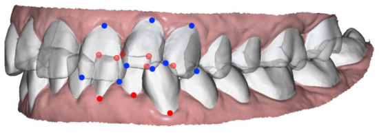
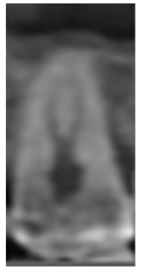
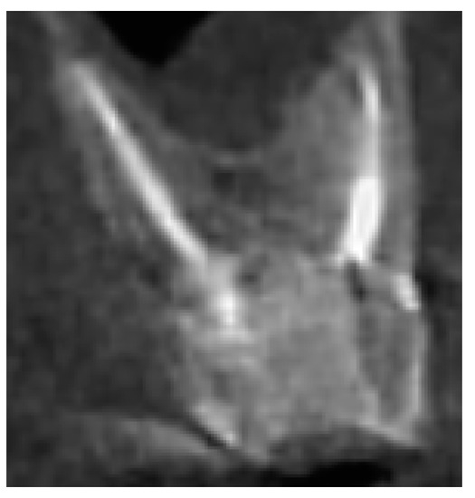
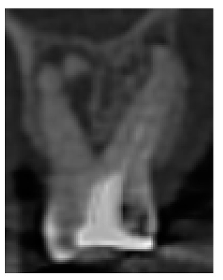
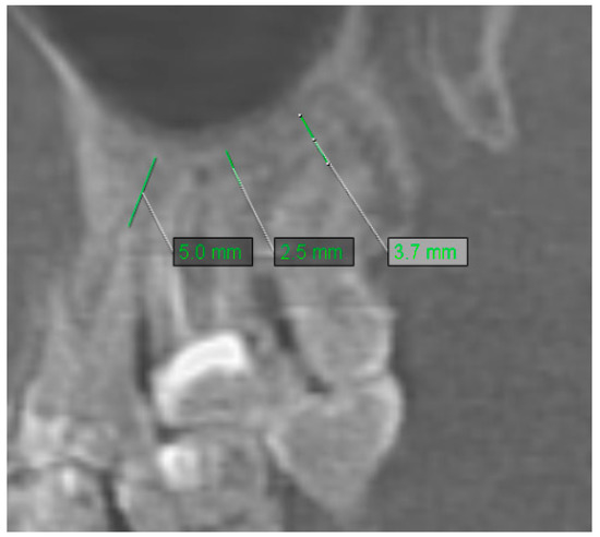
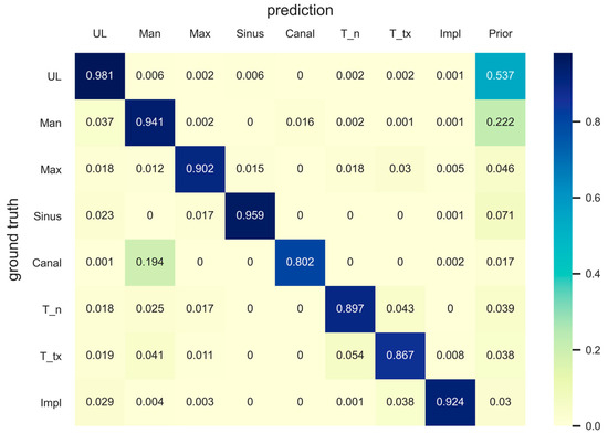
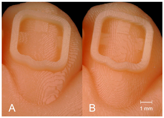
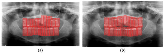
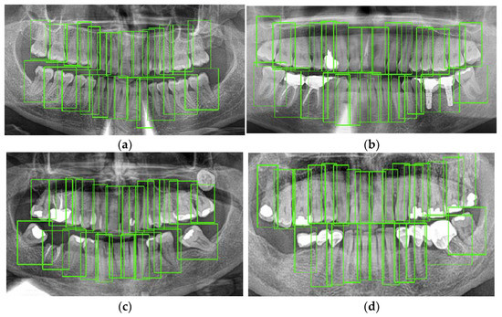
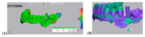
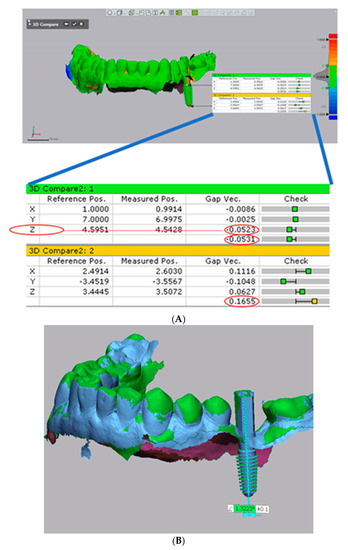
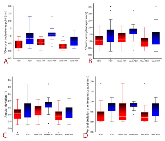
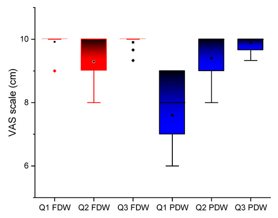
An accurate impression is vital during prosthodontic rehabilitation. Digital scanning has become an alternative to conventional impressions. This study compares conventional preliminary impression techniques with digital scanning, evaluating the efficiency, treatment comfort, and trueness. Impressions of 28 patients were taken using conventional and digital techniques. The efficiency of both impression techniques was evaluated by measuring the mean working time. A visual analog scale questionnaire (1–10) was used to appreciate the participants’ perceptions of discomfort. Morphometric measurements, which were carried out to determine the differences between the casts, were made on the buccolingual cross sections of teeth 11 and 31 and the distolingual and mesiobuccal cusp tips of each first molar. The total treatment time was 75.5 min for conventional and 12 min for digital impressions. The patients scored a mean discomfort assessment of 6.66 for conventional and 9.03 for digital scanning. No significant differences existed between the examined areas (p < 0.05, Wilcoxon and Mann–Whitney tests) of the digital casts obtained by both techniques. The intraoral scan can be considered as an alternative to conventional preliminary impressions for performing study model analysis during orthodontic treatment planning. The digital impression is more comfortable and accepted by the patients than the conventional impression and has a shorter working time.
Full article
Graphical abstract