


J. Clin. Med. 2022, 11(6), 1466; https://doi.org/10.3390/jcm11061466 - 08 Mar 2022
Cited by 2 | Viewed by 1676
Abstract
►
Show Figures
Percutaneous iliosacral screw fixation is a widely accepted method of stabilizing the posterior pelvic ring. Recently developed tools such as 3D-navigated fluoroscopy and computed navigation seem to prevent a surgeon from conducting screw misplacement. The study aimed to comparatively assess the introduction of
[...] Read more.
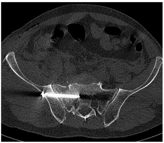
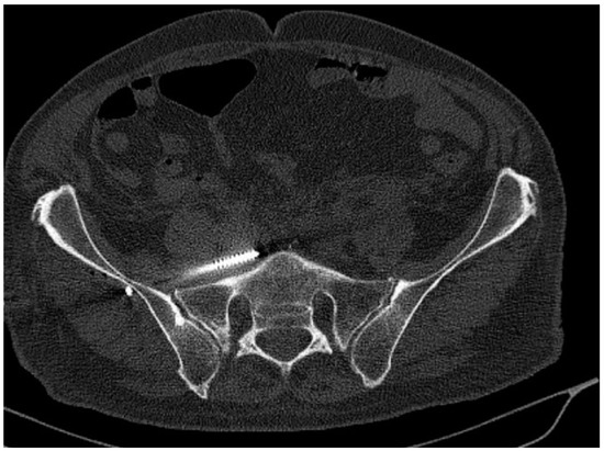
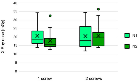
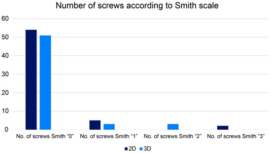
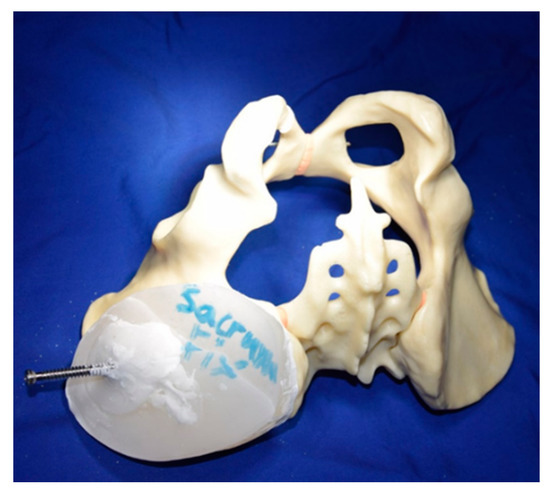
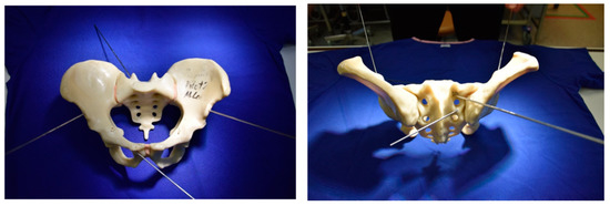
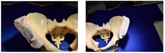
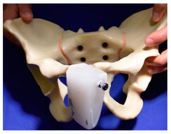
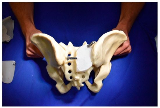
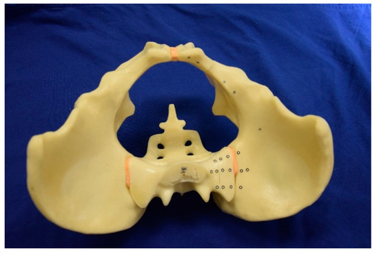
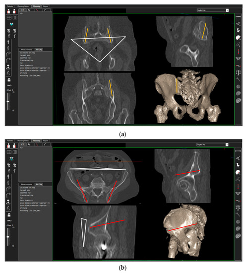
Percutaneous iliosacral screw fixation is a widely accepted method of stabilizing the posterior pelvic ring. Recently developed tools such as 3D-navigated fluoroscopy and computed navigation seem to prevent a surgeon from conducting screw misplacement. The study aimed to comparatively assess the introduction of sacroiliac screw placement using 2D and 3D fluoroscopy in terms of accuracy and radiation exposure. Iliosacral screws were introduced in 37 patients using 2D (group N1) and in 36 patients using 3D fluoroscopy (group N2) techniques. Overall, 61 and 56 screws were introduced in groups N1 and N2, respectively. Screw placement accuracy was assessed using postoperative computed tomography and Smith’s scale. Intraoperative radiation exposure was also assessed. No differences were noted between groups in terms of screw positioning accuracy and radiation dose. Both 2D and 3D fluoroscopy provide good visualization for safely placing percutaneous iliosacral joint screws. Using 3D fluoroscopy-based navigation in comparison with 2D fluoroscopy is not advantageous.
Full article
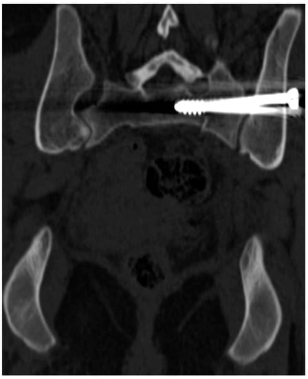
Figure 1
