


J. Clin. Med. 2023, 12(19), 6242; https://doi.org/10.3390/jcm12196242 - 27 Sep 2023
Viewed by 1524
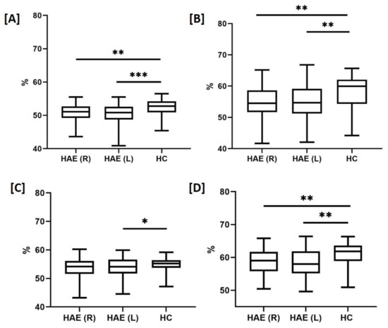
Abstract
►
Show Figures
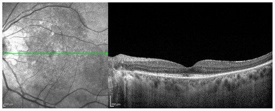
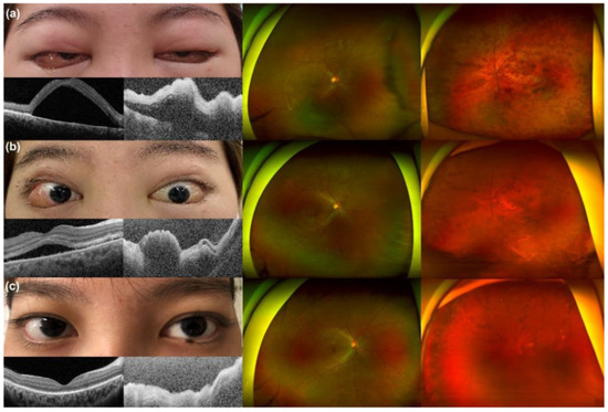
Vogt–Koyanagi–Harada (VKH) is a rare multisystem inflammatory disease affecting the eyes, ears, brain, skin, and hair. The Coronavirus Disease 2019 (COVID-19) is a new contagious infection that might trigger the onset of VKH disease, as previously proposed for other viruses. Moreover, after the
[...] Read more.
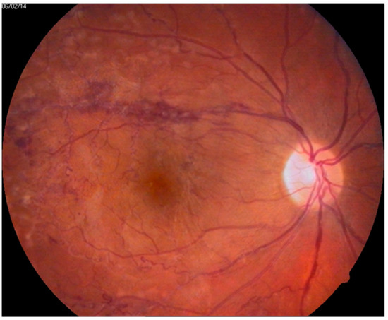
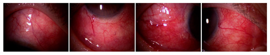
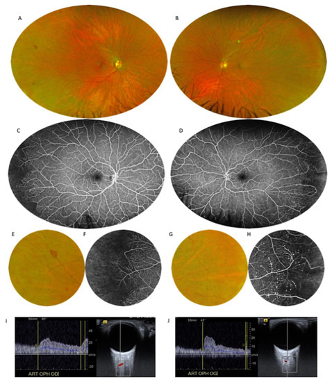
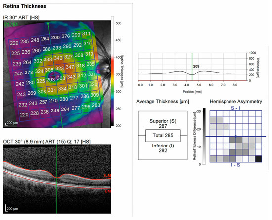
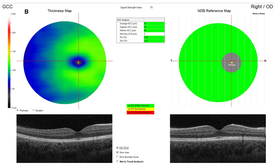
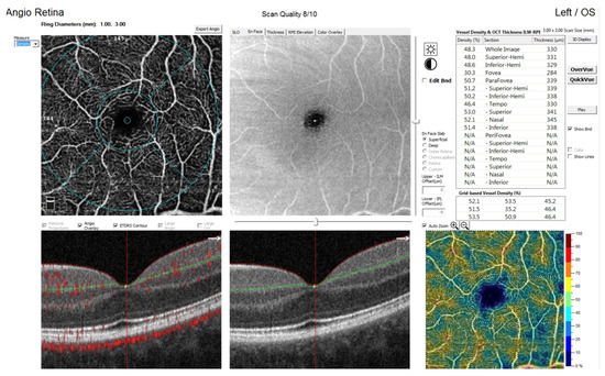
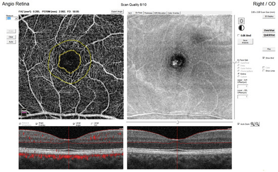
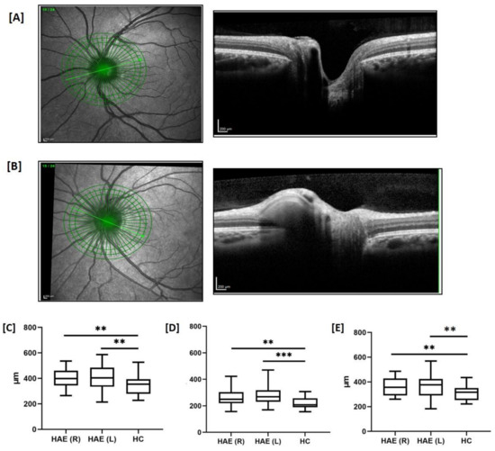
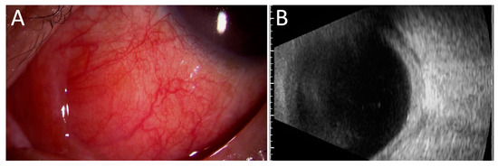
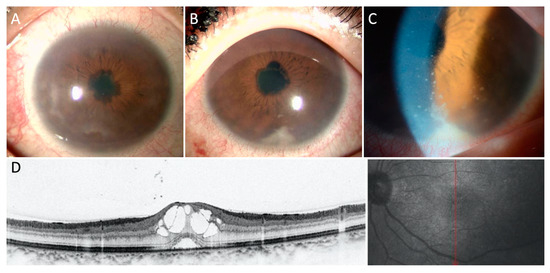
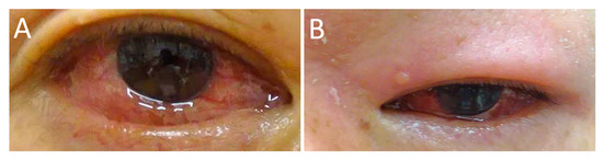
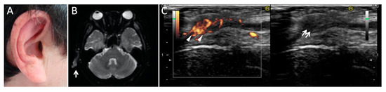
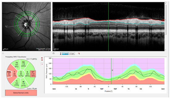
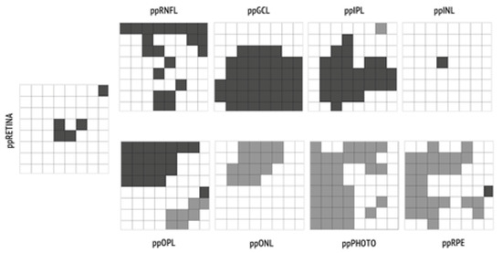
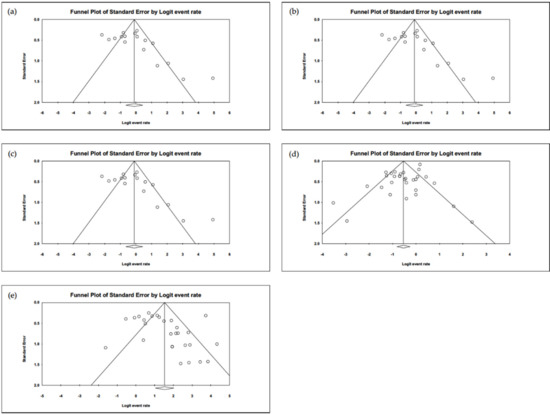
Vogt–Koyanagi–Harada (VKH) is a rare multisystem inflammatory disease affecting the eyes, ears, brain, skin, and hair. The Coronavirus Disease 2019 (COVID-19) is a new contagious infection that might trigger the onset of VKH disease, as previously proposed for other viruses. Moreover, after the mass vaccination against SARS-CoV-2 worldwide, cases of VKH disease associated with COVID-19 vaccination have been reported. We present an overview of VKH and a comprehensive literature revision of all the VKH cases described after COVID-19 infection and vaccination, adding our experience. No differences have been found considering epidemiology and clinical findings of the disease compared to those reported in the no-COVID era. All of the patients promptly responded to systemic and local corticosteroid therapy with a good final visual prognosis. Different possible pathogenetic mechanisms underlying the onset of VKH after COVID-19 vaccination are discussed, while the presence of the HLA DR4 antigen as a genetic predisposition for the onset of the disease after COVID-19 infection and vaccination is proposed. VKH disease is one of the most frequently reported uveitic entities after COVID-19 vaccination, but a good response to therapy should not discourage vaccination. Nevertheless, ophthalmologists should be alerted to the possibility of VKH occurrence or relapse after COVID-19 vaccination, especially in genetically predisposed subjects.
Full article
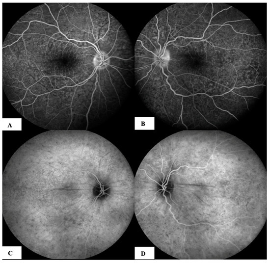
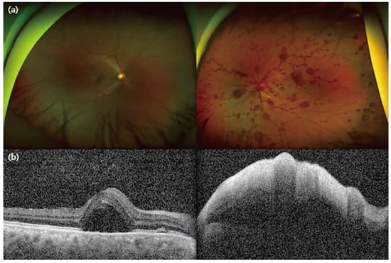
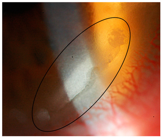
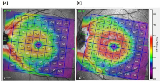
Figure 1
