ijms-logo

Journal Browser

Journal Browser

Sphingolipid Signaling in Health and Disease (Closed)

A topical collection in International Journal of Molecular Sciences (ISSN 1422-0067). This collection belongs to the section "Molecular Pharmacology".

Viewed by 58654

Editor


E-Mail Website
Collection Editor
Institute of Pharmacology, University of Bern, Inselspital, CH-3010 Bern, Switzerland
Interests: sphingolipid signaling; prostaglandins; chronic inflammatory kidney diseases
Special Issues, Collections and Topics in MDPI journals

Topical Collection Information

Dear colleagues,

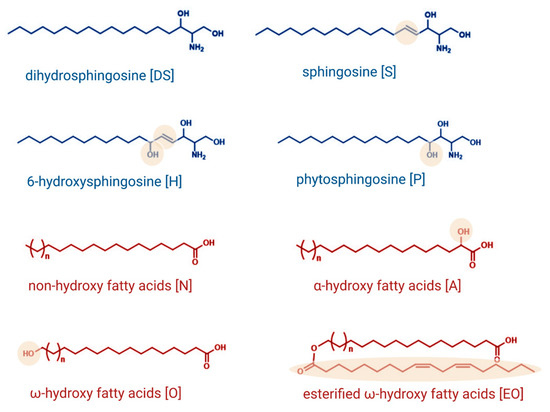
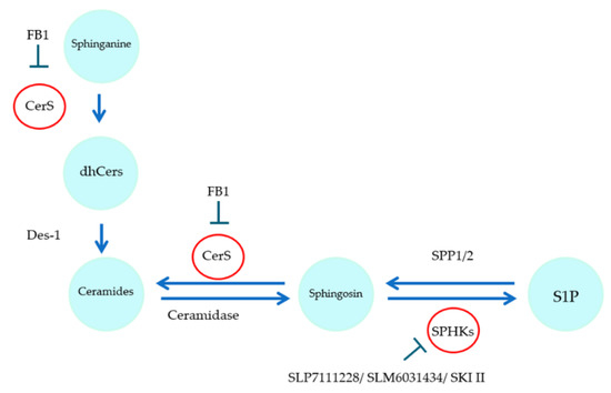
Sphingolipids have long been known as important structural components of biological membranes. This view has drastically changed after groundbreaking discoveries in the early 1990s, which showed that certain sphingolipid subspecies are bioactive and are able to induce signal transduction and regulate fundamental cellular responses such as cell proliferation and survival, differentiation, apoptosis, and migration. Evidence is now increasing that the dysregulation of sphingolipid signaling contributes to the pathogenesis of multiple diseases, including inflammation, pain, cancer, diabetes, cardiovascular dysfunction, organ fibrosis, and various neurodegenerative diseases. Nevertheless, the exact molecular mechanisms used by sphingolipids to mediate the cellular effects are still not completely understood. Also, the regulation of those enzymes that catalyze the generation and interconversion of sphingolipids, although being cloned and known for decades, are not well described.

This Topical Collection is dedicated to shedding light on the molecular mechanisms used by bioactive sphingolipids, including sphingosine, ceramides, glycosphingolipids, and phosphorylated sphingoids, and to better characterize their involvement in physiological, pathophysiological, and pathological conditions. Furthermore, advances in the development of new drugs and strategies to manipulate sphingolipid-regulating enzymes and receptors for therapeutic applications are welcome.

Prof. Andrea Huwiler
Collection Editor

Manuscript Submission Information

Manuscripts should be submitted online at www.mdpi.com by registering and logging in to this website. Once you are registered, click here to go to the submission form. Manuscripts can be submitted until the deadline. All submissions that pass pre-check are peer-reviewed. Accepted papers will be published continuously in the journal (as soon as accepted) and will be listed together on the collection website. Research articles, review articles as well as short communications are invited. For planned papers, a title and short abstract (about 100 words) can be sent to the Editorial Office for announcement on this website.

Submitted manuscripts should not have been published previously, nor be under consideration for publication elsewhere (except conference proceedings papers). All manuscripts are thoroughly refereed through a single-blind peer-review process. A guide for authors and other relevant information for submission of manuscripts is available on the Instructions for Authors page. International Journal of Molecular Sciences is an international peer-reviewed open access semimonthly journal published by MDPI.

Please visit the Instructions for Authors page before submitting a manuscript. There is an Article Processing Charge (APC) for publication in this open access journal. For details about the APC please see here. Submitted papers should be well formatted and use good English. Authors may use MDPI's English editing service prior to publication or during author revisions.

Keywords

  • sphingolipids
  • ceramide
  • sphingosine-1-phosphate
  • glycosphingolipids
  • disease
  • physiology

Published Papers (11 papers)

2023

Jump to: 2021, 2020, 2019

5 pages, 191 KiB  
Editorial
Topical Collection: New Insights on Sphingolipids in Health and Disease
by Andrea Huwiler
Int. J. Mol. Sci. 2023, 24(11), 9528; https://doi.org/10.3390/ijms24119528 - 31 May 2023
Viewed by 680
Abstract
The last two decades have boosted research on sphingolipids as bioactive and signaling molecules [...] Full article
21 pages, 2345 KiB  
Review
Sphingosine 1-Phosphate as Essential Signaling Molecule in Inflammatory Skin Diseases
by Burkhard Kleuser and Wolfgang Bäumer
Int. J. Mol. Sci. 2023, 24(2), 1456; https://doi.org/10.3390/ijms24021456 - 11 Jan 2023
Cited by 14 | Viewed by 2949
Abstract
Sphingolipids are crucial molecules of the mammalian epidermis. The formation of skin-specific ceramides contributes to the formation of lipid lamellae, which are important for the protection of the epidermis from excessive water loss and protect the skin from the invasion of pathogens and [...] Read more.
Sphingolipids are crucial molecules of the mammalian epidermis. The formation of skin-specific ceramides contributes to the formation of lipid lamellae, which are important for the protection of the epidermis from excessive water loss and protect the skin from the invasion of pathogens and the penetration of xenobiotics. In addition to being structural constituents of the epidermal layer, sphingolipids are also key signaling molecules that participate in the regulation of epidermal cells and the immune cells of the skin. While the importance of ceramides with regard to the proliferation and differentiation of skin cells has been known for a long time, it has emerged in recent years that the sphingolipid sphingosine 1-phosphate (S1P) is also involved in processes such as the proliferation and differentiation of keratinocytes. In addition, the immunomodulatory role of this sphingolipid species is becoming increasingly apparent. This is significant as S1P mediates a variety of its actions via G-protein coupled receptors. It is, therefore, not surprising that dysregulation in the signaling pathways of S1P is involved in the pathophysiological conditions of skin diseases. In the present review, the importance of S1P in skin cells, as well as the immune cells of the skin, is elaborated. In particular, the role of the molecule in inflammatory skin diseases will be discussed. This is important because interfering with S1P signaling pathways may represent an innovative option for the treatment of inflammatory skin diseases. Full article
Show Figures

Figure 1

2021

Jump to: 2023, 2020, 2019

14 pages, 2122 KiB  
Article
S1P Stimulates Erythropoietin Production in Mouse Renal Interstitial Fibroblasts by S1P1 and S1P3 Receptor Activation and HIF-2α Stabilization
by Redona Hafizi, Faik Imeri, Roland H. Wenger and Andrea Huwiler
Int. J. Mol. Sci. 2021, 22(17), 9467; https://doi.org/10.3390/ijms22179467 - 31 Aug 2021
Cited by 8 | Viewed by 3019
Abstract
Erythropoietin (Epo) is the critical hormone for erythropoiesis. In adults, Epo is mainly produced by a subset of interstitial fibroblasts in the kidney, with minor amounts being produced in the liver and the brain. In this study, we used the immortalized renal interstitial [...] Read more.
Erythropoietin (Epo) is the critical hormone for erythropoiesis. In adults, Epo is mainly produced by a subset of interstitial fibroblasts in the kidney, with minor amounts being produced in the liver and the brain. In this study, we used the immortalized renal interstitial fibroblast cell line FAIK F3-5 to investigate the ability of the bioactive sphingolipid sphingosine 1-phosphate (S1P) to stimulate Epo production and to reveal the mechanism involved. Stimulation of cells with exogenous S1P under normoxic conditions (21% O2) led to a dose-dependent increase in Epo mRNA and protein levels and subsequent release of Epo into the medium. S1P also enhanced the stabilization of HIF-2α, a key transcription factor for Epo expression. S1P-stimulated Epo mRNA and protein expression was abolished by HIF-2α mRNA knockdown or by the HIF-2 inhibitor compound 2. Furthermore, the approved S1P receptor modulator FTY720, and its active form FTY720-phosphate, both exerted a similar effect on Epo expression as S1P. The effect of S1P on Epo was antagonized by the selective S1P1 and S1P3 antagonists NIBR-0213 and TY-52156, but not by the S1P2 antagonist JTE-013. Moreover, inhibitors of the classical MAPK/ERK, the p38-MAPK, and inhibitors of protein kinase (PK) C and D all blocked the effect of S1P on Epo expression. Finally, the S1P and FTY720 effects were recapitulated in the Epo-producing human neuroblastoma cell line Kelly, suggesting that S1P receptor-dependent Epo synthesis is of general relevance and not species-specific. In summary, these data suggest that, in renal interstitial fibroblasts, which are the primary source of plasma Epo, S1P1 and 3 receptor activation upregulates Epo under normoxic conditions. This may have a therapeutic impact on disease situations such as chronic kidney disease, where Epo production is impaired, causing anemia, but it may also have therapeutic value as Epo can mediate additional tissue-protective effects in various organs. Full article
Show Figures

Figure 1

2020

Jump to: 2023, 2021, 2019

17 pages, 4837 KiB  
Article
Morpholino Analogues of Fingolimod as Novel and Selective S1P1 Ligands with In Vivo Efficacy in a Mouse Model of Experimental Antigen-Induced Encephalomyelitis
by Bisera Stepanovska, Aleksandra Zivkovic, Gaby Enzmann, Silvia Tietz, Thomas Homann, Burkhard Kleuser, Britta Engelhardt, Holger Stark and Andrea Huwiler
Int. J. Mol. Sci. 2020, 21(18), 6463; https://doi.org/10.3390/ijms21186463 - 04 Sep 2020
Cited by 13 | Viewed by 2961
Abstract
Multiple sclerosis (MS) is a chronic, inflammatory, autoimmune disease of the central nervous system (CNS) which is associated with lower life expectancy and disability. The experimental antigen-induced encephalomyelitis (EAE) in mice is a useful animal model of MS, which allows exploring the etiopathogenetic [...] Read more.
Multiple sclerosis (MS) is a chronic, inflammatory, autoimmune disease of the central nervous system (CNS) which is associated with lower life expectancy and disability. The experimental antigen-induced encephalomyelitis (EAE) in mice is a useful animal model of MS, which allows exploring the etiopathogenetic mechanisms and testing novel potential therapeutic drugs. A new therapeutic paradigm for the treatment of MS was introduced in 2010 through the sphingosine 1-phosphate (S1P) analogue fingolimod (FTY720, Gilenya®), which acts as a functional S1P1 antagonist on T lymphocytes to deplete these cells from the blood. In this study, we synthesized two novel structures, ST-1893 and ST-1894, which are derived from fingolimod and chemically feature a morpholine ring in the polar head group. These compounds showed a selective S1P1 activation profile and a sustained S1P1 internalization in cultures of S1P1-overexpressing Chinese hamster ovary (CHO)-K1 cells, consistent with a functional antagonism. In vivo, both compounds induced a profound lymphopenia in mice. Finally, these substances showed efficacy in the EAE model, where they reduced clinical symptoms of the disease, and, on the molecular level, they reduced the T-cell infiltration and several inflammatory mediators in the brain and spinal cord. In summary, these data suggest that S1P1-selective compounds may have an advantage over fingolimod and siponimod, not only in MS but also in other autoimmune diseases. Full article
Show Figures

Figure 1

20 pages, 3512 KiB  
Article
A Sphingosine 1-Phosphate Gradient Is Linked to the Cerebral Recruitment of T Helper and Regulatory T Helper Cells during Acute Ischemic Stroke
by Alexandra Lucaciu, Hannah Kuhn, Sandra Trautmann, Nerea Ferreirós, Helmuth Steinmetz, Josef Pfeilschifter, Robert Brunkhorst, Waltraud Pfeilschifter, Julien Subburayalu and Rajkumar Vutukuri
Int. J. Mol. Sci. 2020, 21(17), 6242; https://doi.org/10.3390/ijms21176242 - 28 Aug 2020
Cited by 17 | Viewed by 5427
Abstract
Emerging evidence suggests a complex relationship between sphingosine 1-phosphate (S1P) signaling and stroke. Here, we show the kinetics of S1P in the acute phase of ischemic stroke and highlight accompanying changes in immune cells and S1P receptors (S1PR). Using a C57BL/6 [...] Read more.
Emerging evidence suggests a complex relationship between sphingosine 1-phosphate (S1P) signaling and stroke. Here, we show the kinetics of S1P in the acute phase of ischemic stroke and highlight accompanying changes in immune cells and S1P receptors (S1PR). Using a C57BL/6 mouse model of middle cerebral artery occlusion (MCAO), we assessed S1P concentrations in the brain, plasma, and spleen. We found a steep S1P gradient from the spleen towards the brain. Results obtained by qPCR suggested that cells expressing the S1PR type 1 (S1P1+) were the predominant population deserting the spleen. Here, we report the cerebral recruitment of T helper (TH) and regulatory T (TREG) cells to the ipsilateral hemisphere, which was associated with differential regulation of cerebral S1PR expression patterns in the brain after MCAO. This study provides insight that the S1P-S1PR axis facilitates splenic T cell egress and is linked to the cerebral recruitment of S1PR+ TH and TREG cells. Further insights by which means the S1P-S1PR-axis orchestrates neuronal positioning may offer new therapeutic perspectives after ischemic stroke. Full article
Show Figures

Figure 1

11 pages, 733 KiB  
Review
The Role of Lipid Metabolism in COVID-19 Virus Infection and as a Drug Target
by Mohamed Abu-Farha, Thangavel Alphonse Thanaraj, Mohammad G. Qaddoumi, Anwar Hashem, Jehad Abubaker and Fahd Al-Mulla
Int. J. Mol. Sci. 2020, 21(10), 3544; https://doi.org/10.3390/ijms21103544 - 17 May 2020
Cited by 187 | Viewed by 25365
Abstract
The current Coronavirus disease 2019 or COVID-19 pandemic has infected over two million people and resulted in the death of over one hundred thousand people at the time of writing this review. The disease is caused by severe acute respiratory syndrome coronavirus 2 [...] Read more.
The current Coronavirus disease 2019 or COVID-19 pandemic has infected over two million people and resulted in the death of over one hundred thousand people at the time of writing this review. The disease is caused by severe acute respiratory syndrome coronavirus 2 (SARS-CoV-2). Even though multiple vaccines and treatments are under development so far, the disease is only slowing down under extreme social distancing measures that are difficult to maintain. SARS-COV-2 is an enveloped virus that is surrounded by a lipid bilayer. Lipids are fundamental cell components that play various biological roles ranging from being a structural building block to a signaling molecule as well as a central energy store. The role lipids play in viral infection involves the fusion of the viral membrane to the host cell, viral replication, and viral endocytosis and exocytosis. Since lipids play a crucial function in the viral life cycle, we asked whether drugs targeting lipid metabolism, such as statins, can be utilized against SARS-CoV-2 and other viruses. In this review, we discuss the role of lipid metabolism in viral infection as well as the possibility of targeting lipid metabolism to interfere with the viral life cycle. Full article
Show Figures

Figure 1

21 pages, 4616 KiB  
Article
Sorafenib Treatment and Modulation of the Sphingolipid Pathway Affect Proliferation and Viability of Hepatocellular Carcinoma In Vitro
by Katja Jakobi, Sandra Beyer, Alexander Koch, Dominique Thomas, Stephanie Schwalm, Stefan Zeuzem, Josef Pfeilschifter and Georgios Grammatikos
Int. J. Mol. Sci. 2020, 21(7), 2409; https://doi.org/10.3390/ijms21072409 - 31 Mar 2020
Cited by 7 | Viewed by 2845
Abstract
Hepatocellular carcinoma (HCC) shows a remarkable heterogeneity and is recognized as a chemoresistant tumor with dismal prognosis. In previous studies, we observed significant alterations in the serum sphingolipids of patients with HCC. This study aimed to investigate the in vitro effects of sorafenib, [...] Read more.
Hepatocellular carcinoma (HCC) shows a remarkable heterogeneity and is recognized as a chemoresistant tumor with dismal prognosis. In previous studies, we observed significant alterations in the serum sphingolipids of patients with HCC. This study aimed to investigate the in vitro effects of sorafenib, which is the most widely used systemic HCC medication, on the sphingolipid pathway as well as the effects of inhibiting the sphingolipid pathway in HCC. Huh7.5 and HepG2 cells were stimulated with sorafenib, and inhibitors of the sphingolipid pathway and cell proliferation, viability, and concentrations of bioactive metabolites were assessed. We observed a significant downregulation of cell proliferation and viability and a simultaneous upregulation of dihydroceramides upon sorafenib stimulation. Interestingly, fumonisin B1 (FB1) and the general sphingosine kinase inhibitor SKI II were able to inhibit cell proliferation more prominently in HepG2 and Huh7.5 cells, whereas there were no consistent effects on the formation of dihydroceramides, thus implying an involvement of distinct metabolic pathways. In conclusion, our study demonstrates a significant downregulation of HCC proliferation upon sorafenib, FB1, and SKI II treatment, whereas it seems they exert antiproliferative effects independently from sphingolipids. Certainly, further data would be required to elucidate the potential of FB1 and SKI II as putative novel therapeutic targets in HCC. Full article
Show Figures

Figure 1

13 pages, 2213 KiB  
Article
Ceramide Kinase Is Upregulated in Metastatic Breast Cancer Cells and Contributes to Migration and Invasion by Activation of PI 3-Kinase and Akt
by Stephanie Schwalm, Martin Erhardt, Isolde Römer, Josef Pfeilschifter, Uwe Zangemeister-Wittke and Andrea Huwiler
Int. J. Mol. Sci. 2020, 21(4), 1396; https://doi.org/10.3390/ijms21041396 - 19 Feb 2020
Cited by 24 | Viewed by 4089
Abstract
Ceramide kinase (CerK) is a lipid kinase that converts the proapoptotic ceramide to ceramide 1-phosphate, which has been proposed to have pro-malignant properties and regulate cell responses such as proliferation, migration, and inflammation. We used the parental human breast cancer cell line MDA-MB-231 [...] Read more.
Ceramide kinase (CerK) is a lipid kinase that converts the proapoptotic ceramide to ceramide 1-phosphate, which has been proposed to have pro-malignant properties and regulate cell responses such as proliferation, migration, and inflammation. We used the parental human breast cancer cell line MDA-MB-231 and two single cell progenies derived from lung and bone metastasis upon injection of the parental cells into immuno-deficient mice. The lung and the bone metastatic cell lines showed a marked upregulation of CerK mRNA and activity when compared to the parental cell line. The metastatic cells also had increased migratory and invasive activity, which was dose-dependently reduced by the selective CerK inhibitor NVP-231. A similar reduction of migration was seen when CerK was stably downregulated with small hairpin RNA (shRNA). Conversely, overexpression of CerK in parental MDA-MB-231 cells enhanced migration, and this effect was also observed in the non-metastatic cell line MCF7 upon CerK overexpression. On the molecular level, CerK overexpression increased the activation of protein kinase Akt. The increased migration of CerK overexpressing cells was mitigated by the CerK inhibitor NVP-231, by inhibition of the phosphoinositide 3-kinase (PI3K)/Akt pathway and the Rho kinase, but not by inhibition of the classical extracellular signal-regulated kinase (ERK) pathway. Altogether, our data demonstrate for the first time that CerK promotes migration and invasion of metastatic breast cancer cells and that targeting of CerK has potential to counteract metastasis in breast cancer. Full article
Show Figures

Graphical abstract

19 pages, 4033 KiB  
Article
Downregulation of S1P Lyase Improves Barrier Function in Human Cerebral Microvascular Endothelial Cells Following an Inflammatory Challenge
by Bisera Stepanovska, Antonia I. Lange, Stephanie Schwalm, Josef Pfeilschifter, Sina M. Coldewey and Andrea Huwiler
Int. J. Mol. Sci. 2020, 21(4), 1240; https://doi.org/10.3390/ijms21041240 - 13 Feb 2020
Cited by 14 | Viewed by 4048
Abstract
Sphingosine 1-phosphate (S1P) is a key bioactive lipid that regulates a myriad of physiological and pathophysiological processes, including endothelial barrier function, vascular tone, vascular inflammation, and angiogenesis. Various S1P receptor subtypes have been suggested to be involved in the regulation of these processes, [...] Read more.
Sphingosine 1-phosphate (S1P) is a key bioactive lipid that regulates a myriad of physiological and pathophysiological processes, including endothelial barrier function, vascular tone, vascular inflammation, and angiogenesis. Various S1P receptor subtypes have been suggested to be involved in the regulation of these processes, whereas the contribution of intracellular S1P (iS1P) through intracellular targets is little explored. In this study, we used the human cerebral microvascular endothelial cell line HCMEC/D3 to stably downregulate the S1P lyase (SPL-kd) and evaluate the consequences on endothelial barrier function and on the molecular factors that regulate barrier tightness under normal and inflammatory conditions. The results show that in SPL-kd cells, transendothelial electrical resistance, as a measure of barrier integrity, was regulated in a dual manner. SPL-kd cells had a delayed barrier build up, a shorter interval of a stable barrier, and, thereafter, a continuous breakdown. Contrariwise, a protection was seen from the rapid proinflammatory cytokine-mediated barrier breakdown. On the molecular level, SPL-kd caused an increased basal protein expression of the adherens junction molecules PECAM-1, VE-cadherin, and β-catenin, increased activity of the signaling kinases protein kinase C, AMP-dependent kinase, and p38-MAPK, but reduced protein expression of the transcription factor c-Jun. However, the only factors that were significantly reduced in TNFα/SPL-kd compared to TNFα/control cells, which could explain the observed protection, were VCAM-1, IL-6, MCP-1, and c-Jun. Furthermore, lipid profiling revealed that dihydro-S1P and S1P were strongly enhanced in TNFα-treated SPL-kd cells. In summary, our data suggest that SPL inhibition is a valid approach to dampenan inflammatory response and augmente barrier integrity during an inflammatory challenge. Full article
Show Figures

Graphical abstract

2019

Jump to: 2023, 2021, 2020

11 pages, 1297 KiB  
Article
Feeding Stimulates Sphingosine-1-Phosphate Mobilization in Mouse Hypothalamus
by Valentina Vozella, Natalia Realini, Alessandra Misto and Daniele Piomelli
Int. J. Mol. Sci. 2019, 20(16), 4008; https://doi.org/10.3390/ijms20164008 - 17 Aug 2019
Cited by 3 | Viewed by 3338
Abstract
Previous studies have shown that the sphingolipid-derived mediator sphingosine-1-phosphate (S1P) reduces food intake by activating G protein-coupled S1P receptor-1 (S1PR1) in the hypothalamus. Here, we examined whether feeding regulates hypothalamic mobilization of S1P and other sphingolipid-derived messengers. We prepared lipid extracts from the [...] Read more.
Previous studies have shown that the sphingolipid-derived mediator sphingosine-1-phosphate (S1P) reduces food intake by activating G protein-coupled S1P receptor-1 (S1PR1) in the hypothalamus. Here, we examined whether feeding regulates hypothalamic mobilization of S1P and other sphingolipid-derived messengers. We prepared lipid extracts from the hypothalamus of C57Bl6/J male mice subjected to one of four conditions: free feeding, 12 h fasting, and 1 h or 6 h refeeding. Liquid chromatography/tandem mass spectrometry was used to quantify various sphingolipid species, including sphinganine (SA), sphingosine (SO), and their bioactive derivatives SA-1-phosphate (SA1P) and S1P. In parallel experiments, transcription of S1PR1 (encoded in mice by the S1pr1 gene) and of key genes of sphingolipid metabolism (Sptlc2, Lass1, Sphk1, Sphk2) was measured by RT-PCR. Feeding increased levels of S1P (in pmol-mg−1 of wet tissue) and SA1P. This response was accompanied by parallel changes in SA and dihydroceramide (d18:0/18:0), and was partially (SA1P) or completely (S1P) reversed by fasting. No such effects were observed with other sphingolipid species targeted by our analysis. Feeding also increased transcription of Sptlc2, Lass1, Sphk2, and S1pr1. Feeding stimulates mobilization of endogenous S1PR1 agonists S1P and SA1P in mouse hypothalamus, via a mechanism that involves transcriptional up-regulation of de novo sphingolipid biosynthesis. The results support a role for sphingolipid-mediated signaling in the central control of energy balance. Full article
Show Figures

Figure 1

12 pages, 2095 KiB  
Article
A Role for Neutral Sphingomyelinase in Wound Healing Induced by Keratinocyte Proliferation upon 1α, 25-Dihydroxyvitamin D3 Treatment
by Federica Filomena Patria, Maria Rachele Ceccarini, Michela Codini, Carmela Conte, Luana Perioli, Tommaso Beccari and Elisabetta Albi
Int. J. Mol. Sci. 2019, 20(15), 3634; https://doi.org/10.3390/ijms20153634 - 25 Jul 2019
Cited by 14 | Viewed by 2707
Abstract
The skin has many functions, such as providing a barrier against injury and pathogens, protecting from ultraviolet light, and regulating body temperature. Mechanical causes and many different pathologies can lead to skin damage. Therefore, it is important for the skin to be always [...] Read more.
The skin has many functions, such as providing a barrier against injury and pathogens, protecting from ultraviolet light, and regulating body temperature. Mechanical causes and many different pathologies can lead to skin damage. Therefore, it is important for the skin to be always adaptable and renewable and for cells to undergo proliferation. Here, we demonstrate that 1α, 25-dihydroxyvitamin D3 (VD3) stimulates keratinocyte proliferation, leading to wound closure in a simulation model of injury. Functionally, our results show that VD3 acts by stimulating cyclin D1, a cyclin that promotes the G1/S transition of the cell cycle. The study on the mechanism underlying cyclin D1 expression upon VD3 stimulation clearly demonstrates a key role of neutral sphingomyelinase. The enzyme, whose gene and protein expression is stimulated by VD3, is itself able to induce effects on cyclin D1 and wound healing similar to those obtained with VD3. These results could be very useful in the future to better understand wound mechanisms and improve therapeutic interventions. Full article
Show Figures

Figure 1

Back to TopTop