


Int. J. Mol. Sci. 2024, 25(5), 2871; https://doi.org/10.3390/ijms25052871 - 01 Mar 2024
Viewed by 592
Abstract
►
Show Figures
c-Met is a tyrosine-kinase receptor, and its aberrant activation plays critical roles in tumorigenesis, invasion, and metastatic spread in many human tumors. PHA-665752 (PHA) is an inhibitor of c-Met and has antitumor effects on many hematological malignancies and solid cancers. However, the activation
[...] Read more.
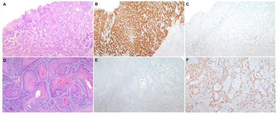
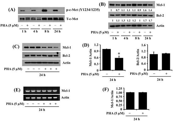
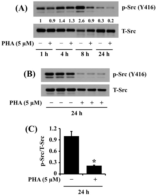
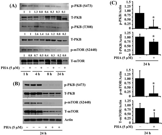
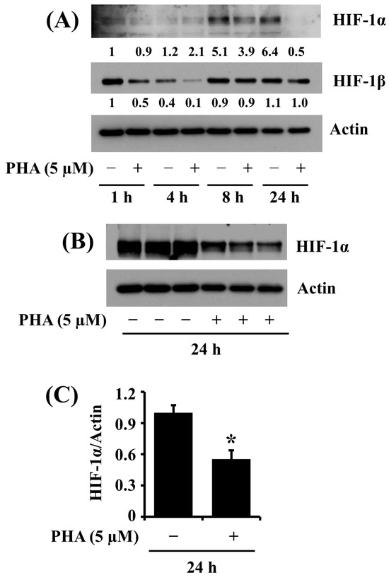
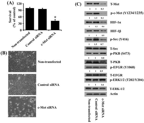
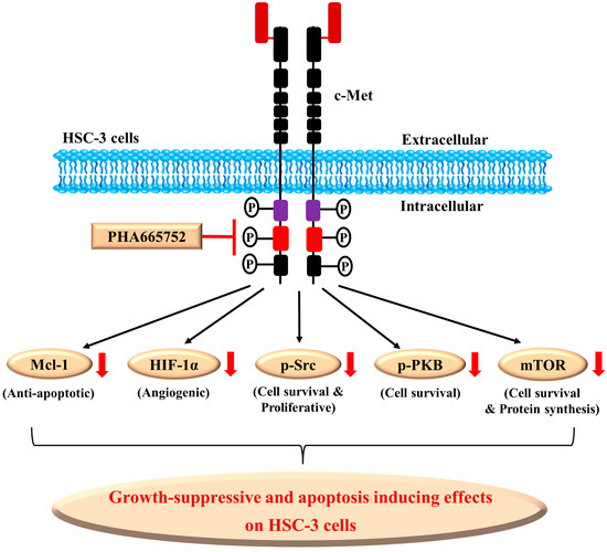
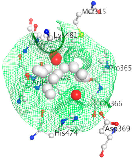
c-Met is a tyrosine-kinase receptor, and its aberrant activation plays critical roles in tumorigenesis, invasion, and metastatic spread in many human tumors. PHA-665752 (PHA) is an inhibitor of c-Met and has antitumor effects on many hematological malignancies and solid cancers. However, the activation and expression of c-Met and its role and the antitumor effect of PHA on human oral squamous cell carcinoma (OSCC) cells remain unclear. Here, we investigated the activation and expression of c-Met and the effects of PHA on the growth of a highly tumorigenic HSC-3 human OSCC cell line with high c-Met phosphorylation and expression. Of note, c-Met was highly expressed and phosphorylated on Y1234/1235 in HSC-3 cells, and PHA treatment significantly suppressed the growth and induced apoptosis of these cells. Moreover, PHA that inhibited the phosphorylation (activation) of c-Met further caused the reduced phosphorylation and expression levels of Src, protein kinase B (PKB), mammalian target of rapamycin (mTtor), and myeloid cell leukemia-1 (Mcl-1) in HSC-3 cells. In addition, the antiangiogenic property of PHA in HSC-3 cells was shown, as evidenced by the drug’s suppressive effect on the expression of hypoxia-inducible factor-1α (HIF-1α), a critical tumor angiogenic transcription factor. Importantly, genetic ablation of c-Met caused the reduced growth of HSC-3 cells and decreased Src phosphorylation and HIF-1α expression. Together, these results demonstrate that c-Met is highly activated in HSC-3 human oral cancer cells, and PHA exhibits strong antigrowth, proapoptotic, and antiangiogenic effects on these cells, which are mediated through regulation of the phosphorylation and expression of multiple targets, including c-Met, Src, PKB, mTOR, Mcl-1, and HIF-1α.
Full article
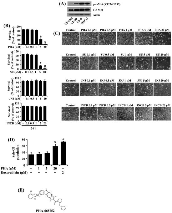
Figure 1


