


Int. J. Mol. Sci. 2023, 24(13), 10577; https://doi.org/10.3390/ijms241310577 - 24 Jun 2023
Cited by 2 | Viewed by 1279
Abstract
►
Show Figures
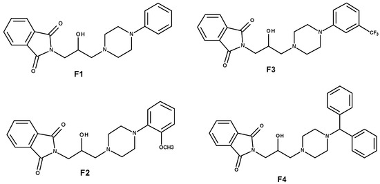
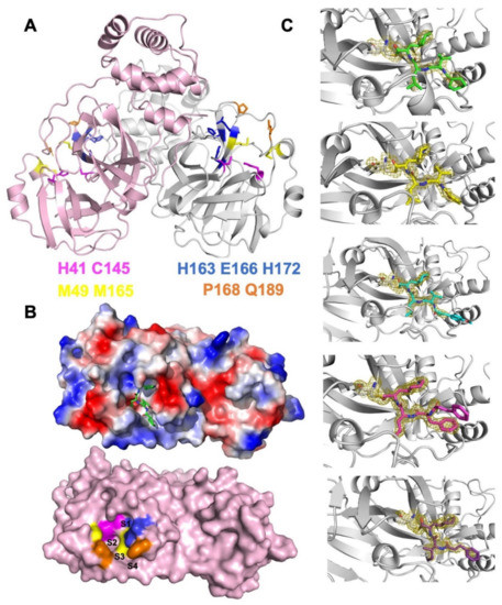
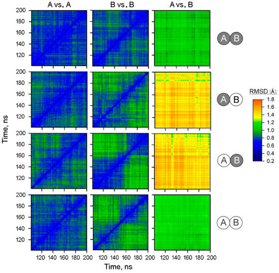
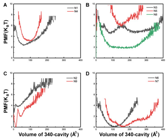
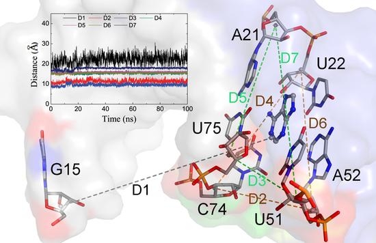
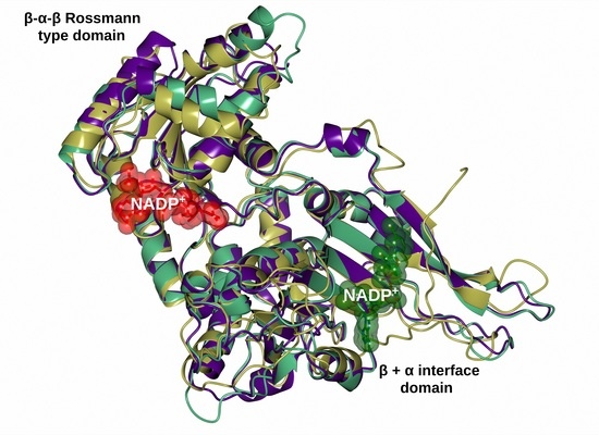
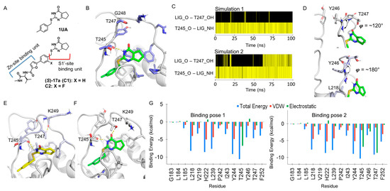
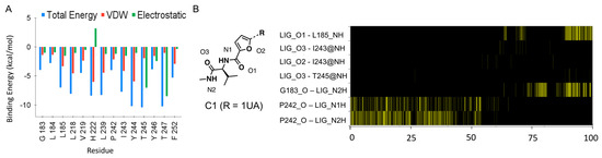
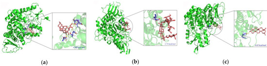
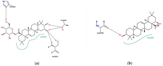
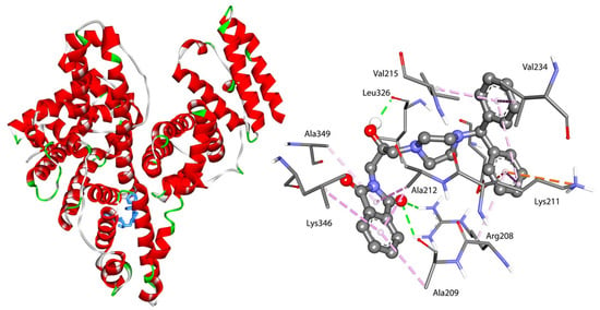
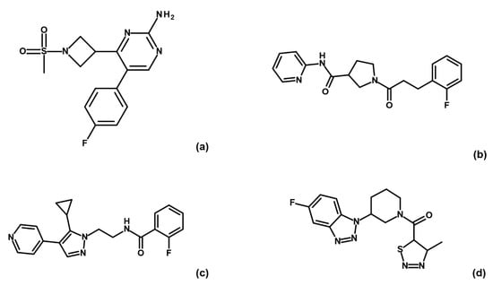
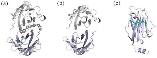
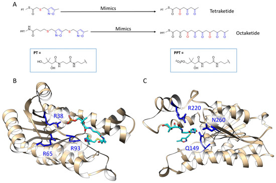
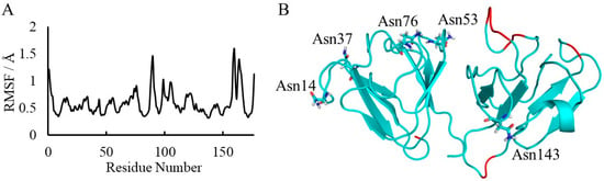
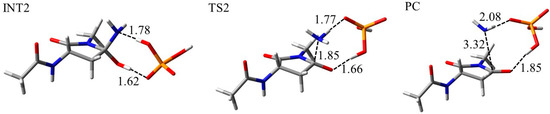
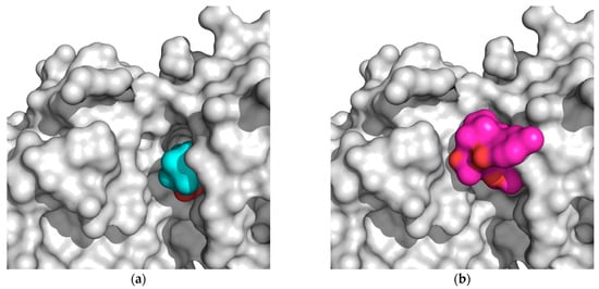
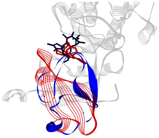
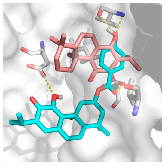
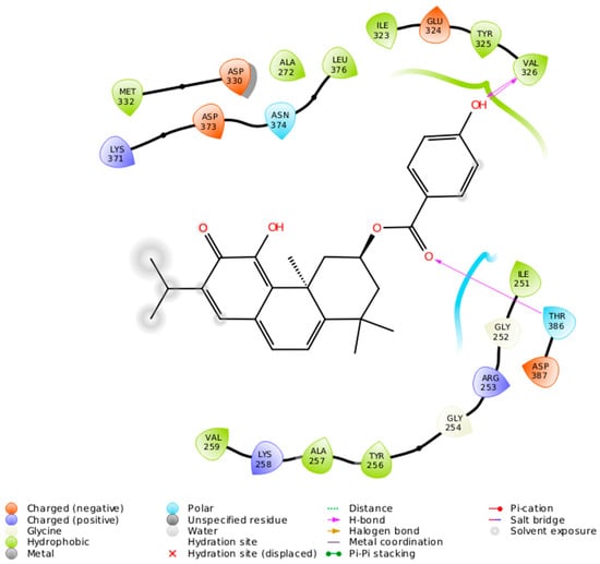
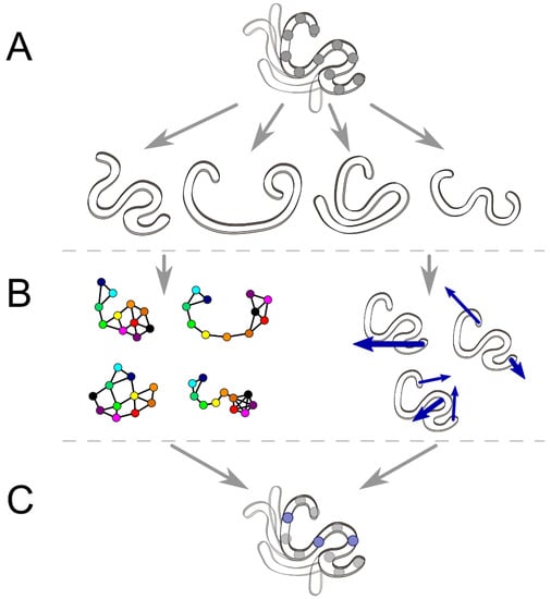
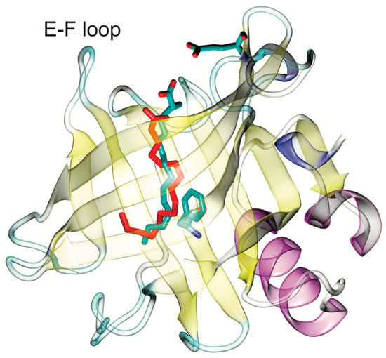
The specificity loop of Matrix Metalloproteinases (MMPs) is known to regulate recognition of their substrates, and the S1′−site surrounded by the loop is a unique place to address the selectivity of ligands toward each MMP. Molecular dynamics (MD) simulations of apo−MMP−13 and its
[...] Read more.
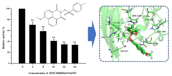
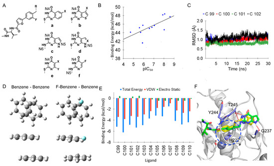
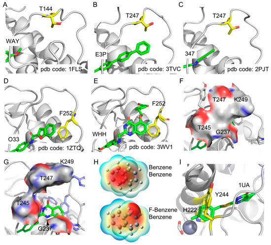
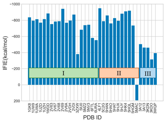
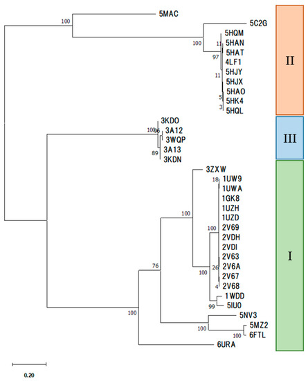
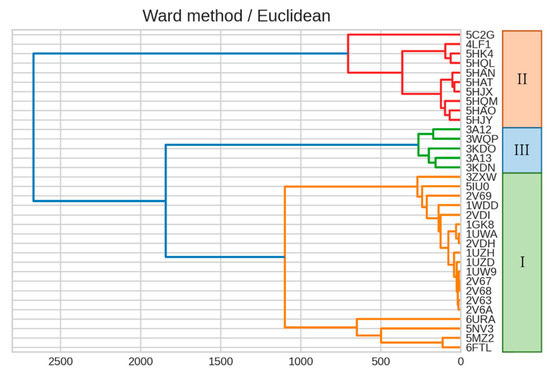
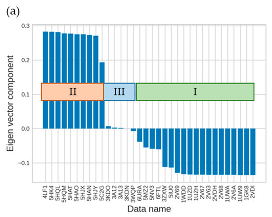
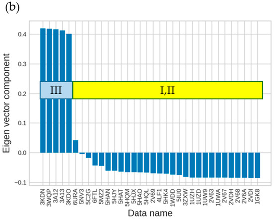
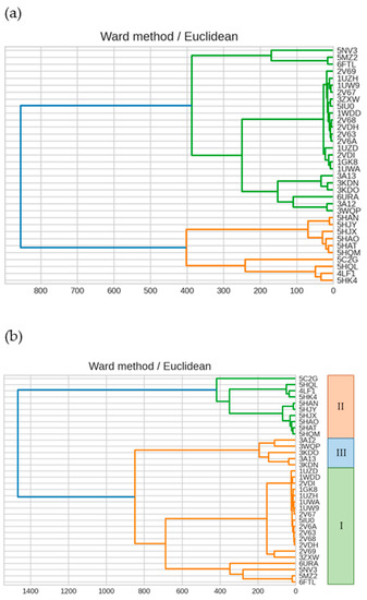
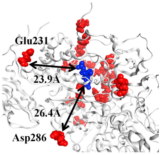
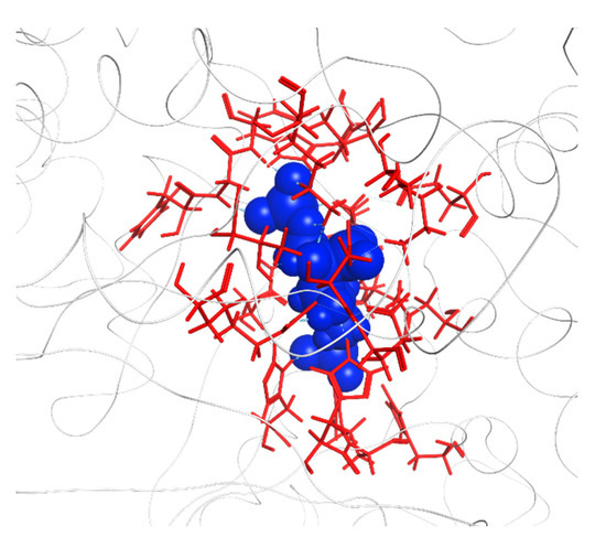
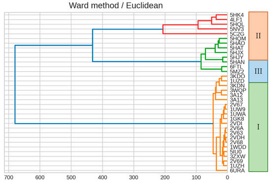
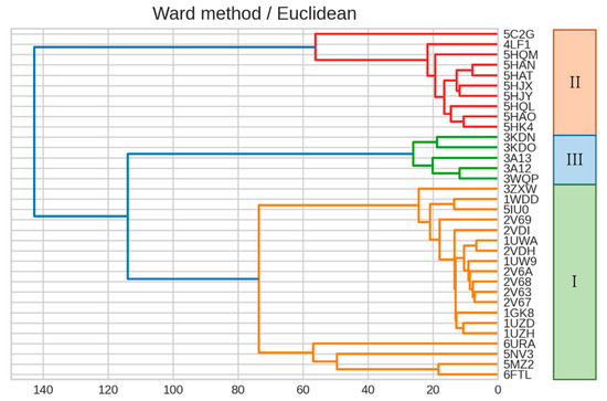
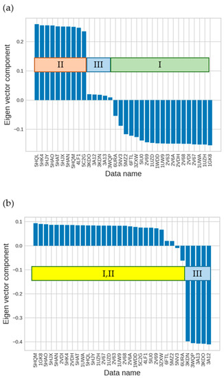
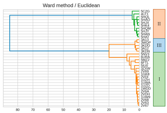
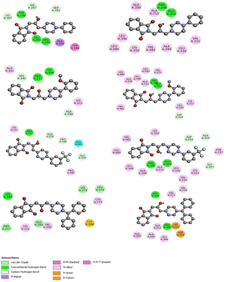
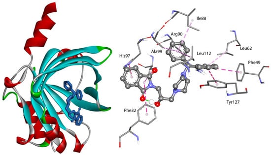
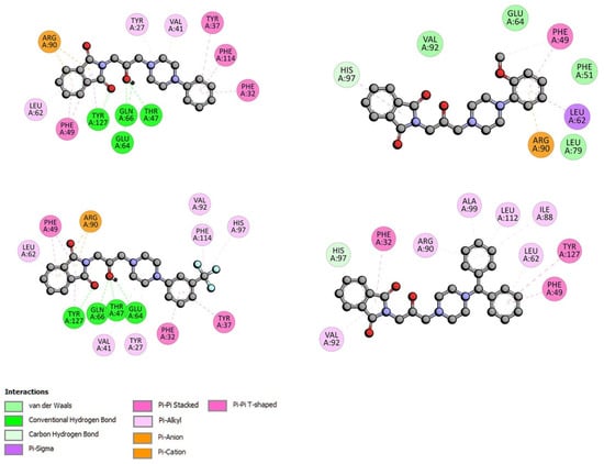
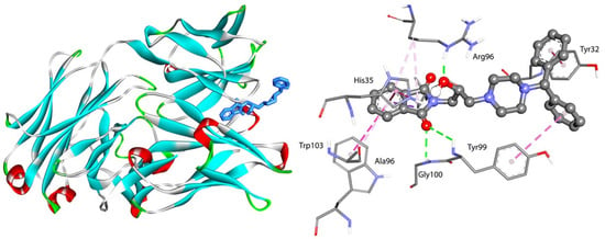
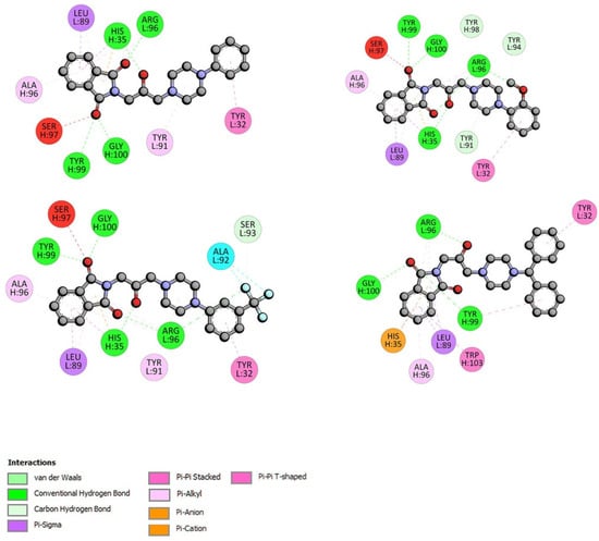
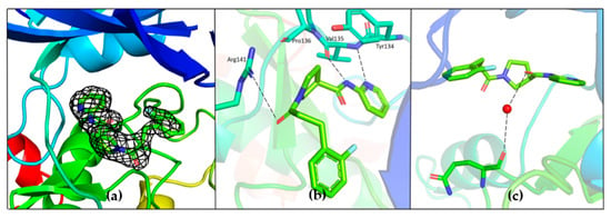
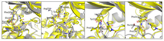
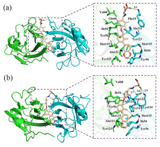
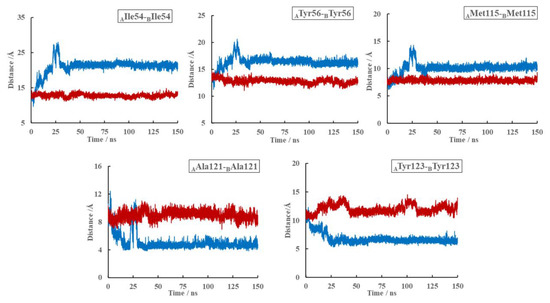
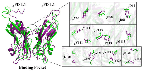
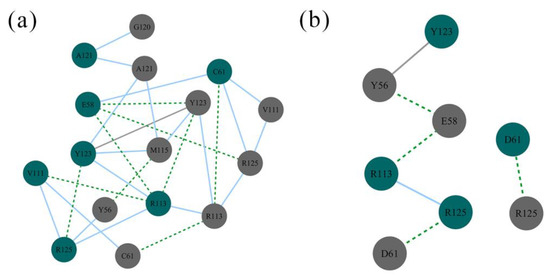
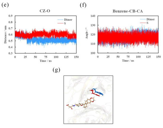
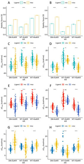
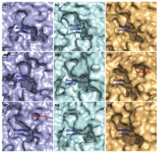
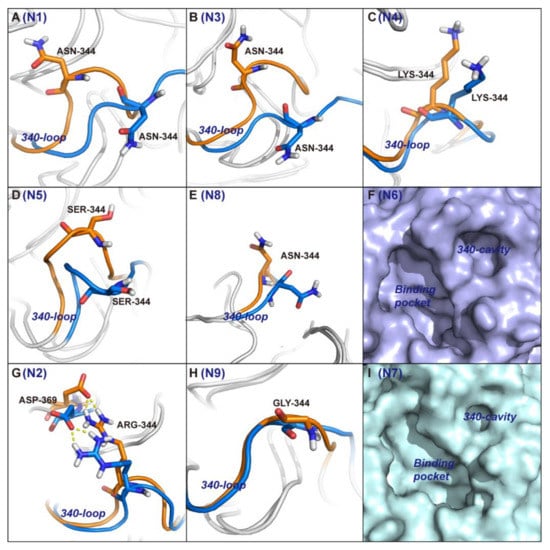
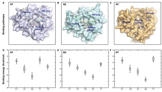
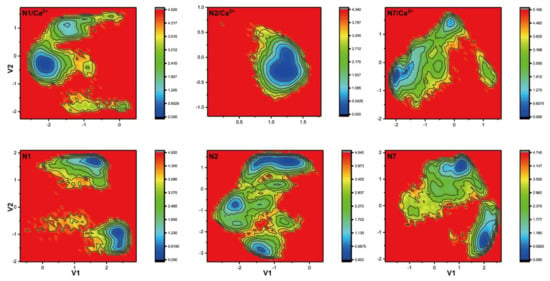
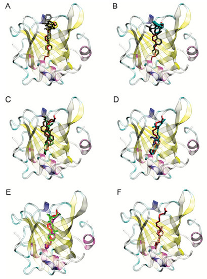
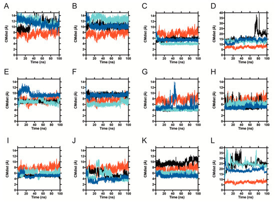
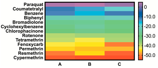
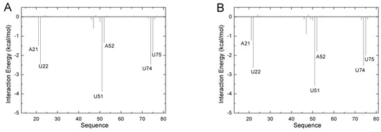
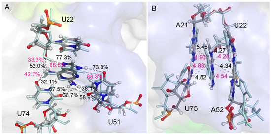
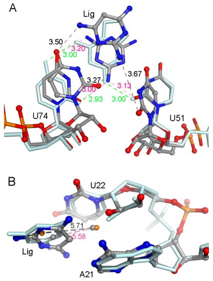
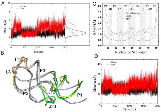
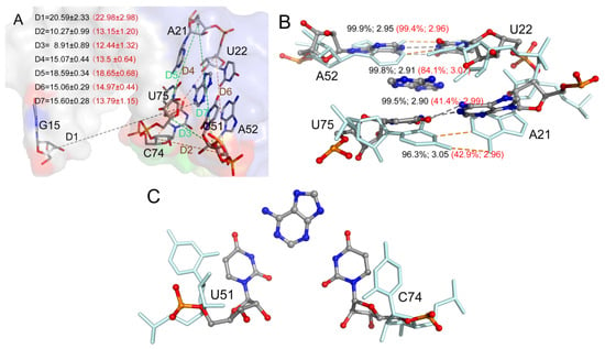
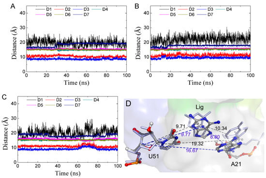
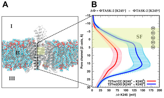
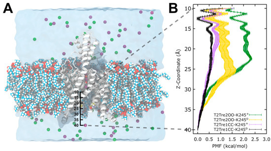
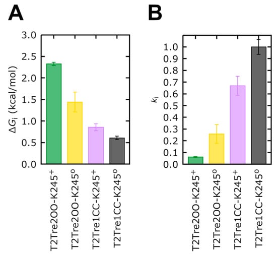
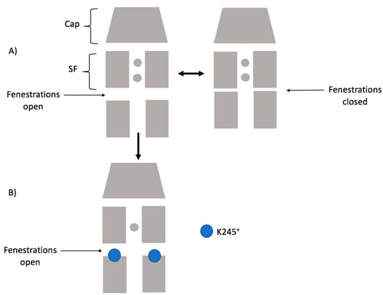
The specificity loop of Matrix Metalloproteinases (MMPs) is known to regulate recognition of their substrates, and the S1′−site surrounded by the loop is a unique place to address the selectivity of ligands toward each MMP. Molecular dynamics (MD) simulations of apo−MMP−13 and its complex forms with various ligands were conducted to identify the role of the specificity loop for the ligand binding to MMP−13. The MD simulations showed the dual role of T247 as a hydrogen bond donor to the ligand, as well as a contributor to the formation of the van der Waal surface area, with T245 and K249 on the S1′−site. The hydrophobic surface area mediated by T247 blocks the access of water molecules to the S1′−site of MMP−13 and stabilizes the ligand in the site. The F252 residue is flexible in order to search for the optimum location in the S1′−site of the apo−MMP−13, but once a ligand binds to the S1′−site, it can form offset π−π or edge−to−π stacking interactions with the ligand. Lastly, H222 and Y244 provide the offset π−π and π−CH(Cβ) interactions on each side of the phenyl ring of the ligand, and this sandwiched interaction could be critical for the ligand binding to MMP−13.
Full article
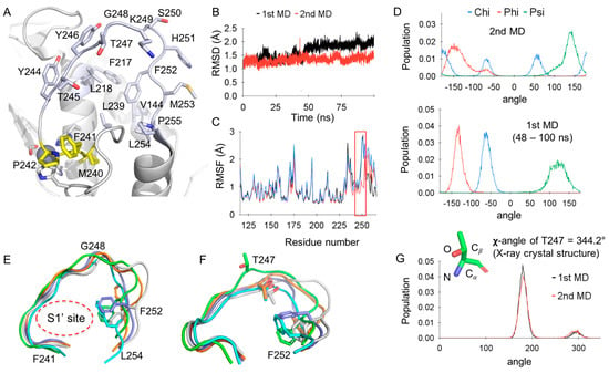
Figure 1


