


Int. J. Mol. Sci. 2023, 24(24), 17335; https://doi.org/10.3390/ijms242417335 - 11 Dec 2023
Viewed by 995
Abstract
►
Show Figures
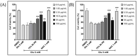
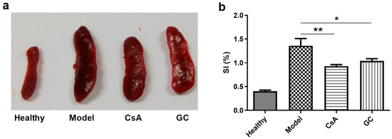
Glycyrol (GC) is one natural active product. Imiquimod-induced psoriasis-like Balb/c mouse models were established. The model mice were intraperitoneally injected with cyclosporine A (CsA) and GC for 8 days followed by a series of biological detections. GC had little toxicity according to the
[...] Read more.
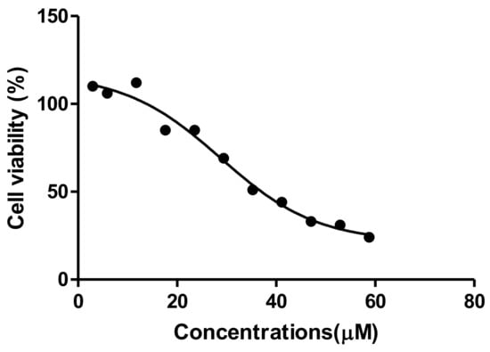
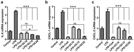
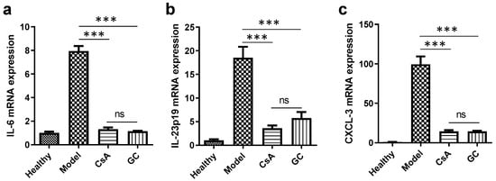
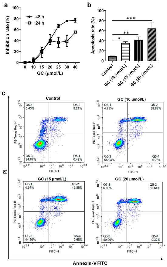
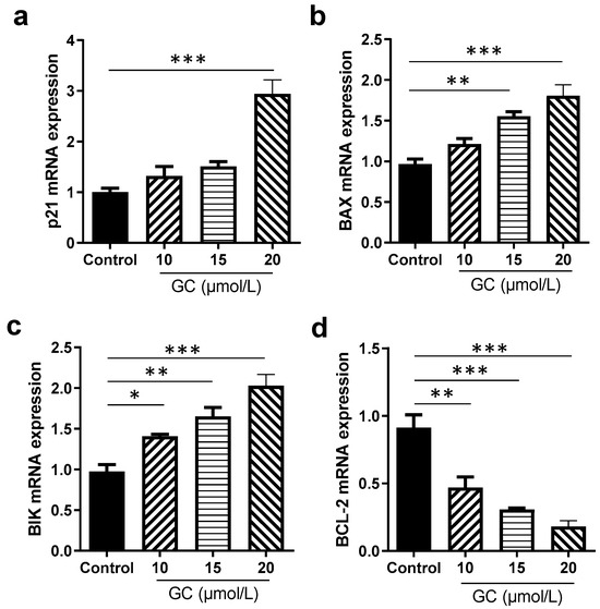
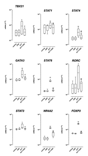
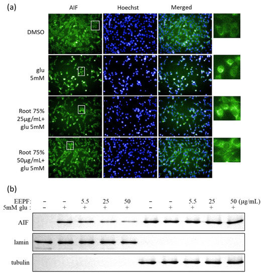
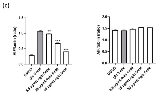
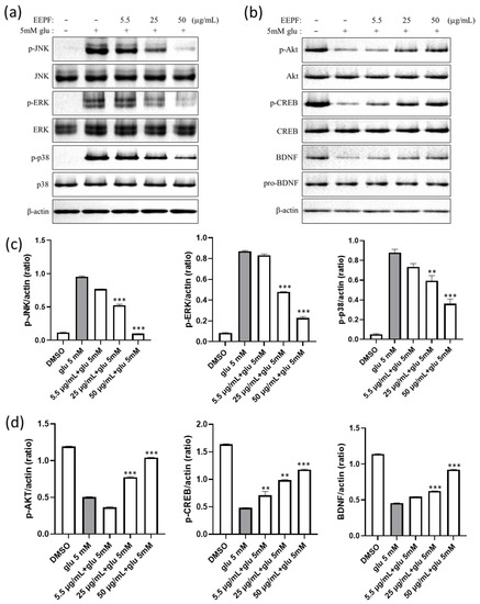
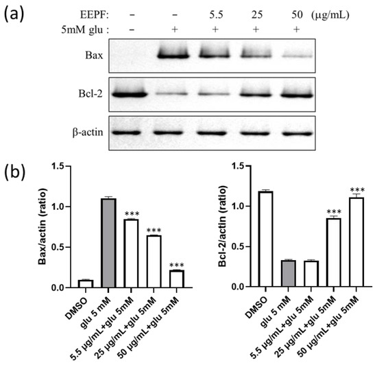
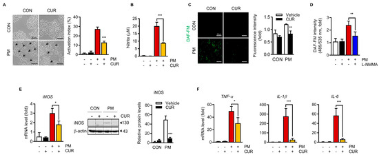
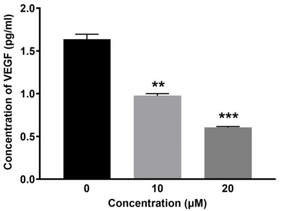
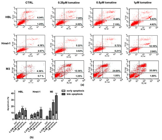
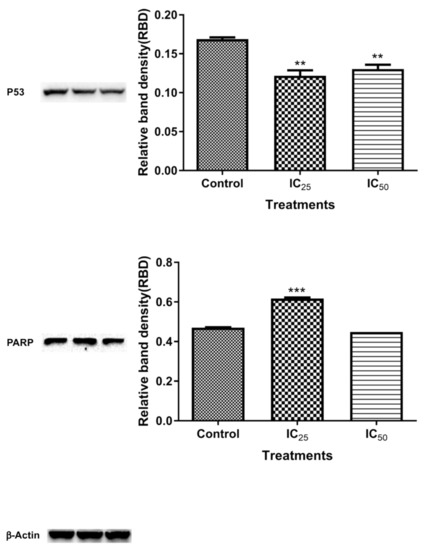
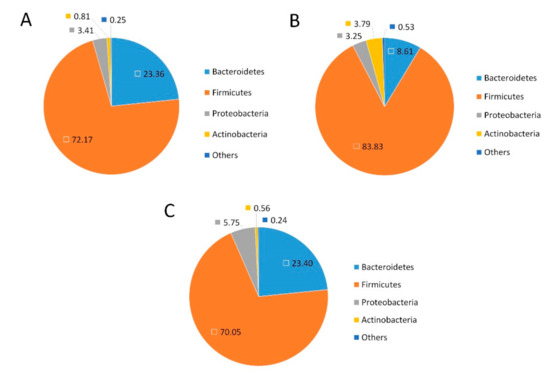
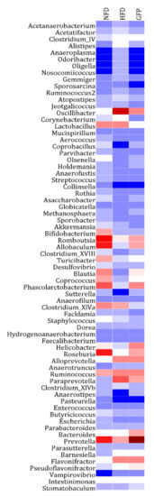
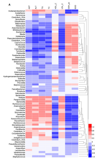
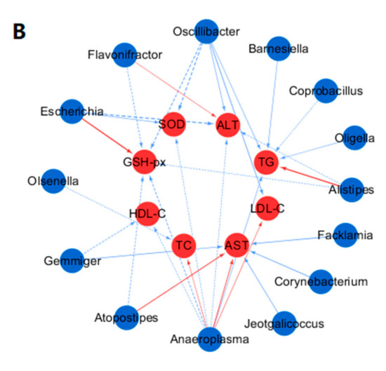
Glycyrol (GC) is one natural active product. Imiquimod-induced psoriasis-like Balb/c mouse models were established. The model mice were intraperitoneally injected with cyclosporine A (CsA) and GC for 8 days followed by a series of biological detections. GC had little toxicity according to the levels of peripheral blood cells, hemoglobin, blood urea nitrogen (BUN), and serum creatinine (CRE), while CsA significantly increased the levels of BUN and CRE. GC decreased the splenic index and reduced the expressions of IL-6, IL-23, and CXCL-3 in the model mice and IL-6, CXCL-1, and CXCL-2 in the inflammatory HaCaT cells. The half inhibition concentration (IC50) of GC on HaCaT cells was 29.72 μmol/L, resulting in improved apoptosis, enhanced expressions of p21, BAX, and BIK, and reduced expressions of BCL-2. GC is an immunosuppressive agent against psoriasis-like symptoms by anti-inflammatory effects, which provides a strategy for the discovery of anti-psoriatic natural products.
Full article
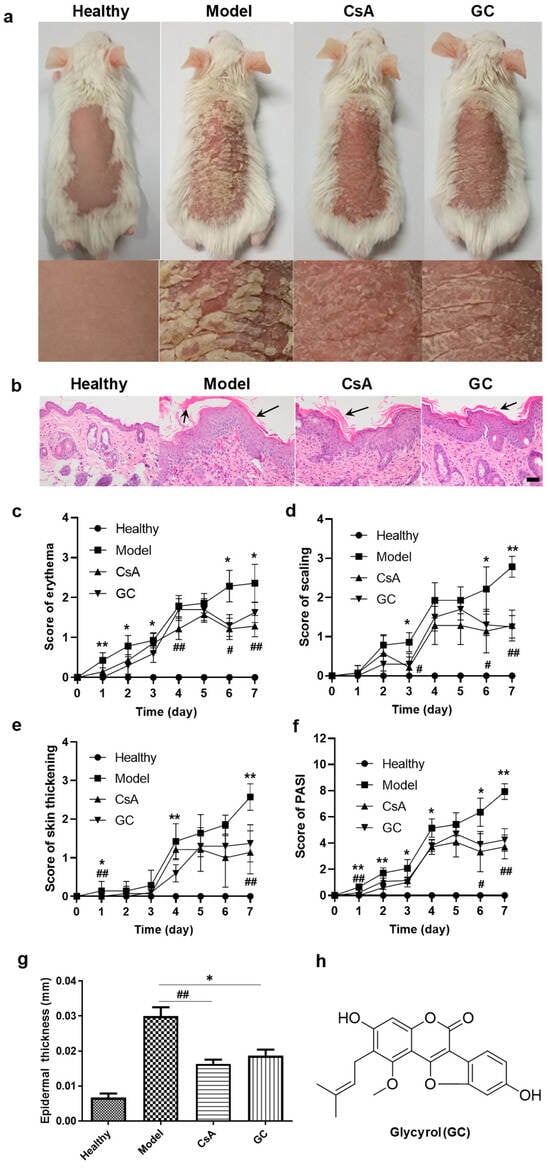
Figure 1


