


Int. J. Mol. Sci. 2024, 25(6), 3131; https://doi.org/10.3390/ijms25063131 - 08 Mar 2024
Viewed by 540
Abstract
►
Show Figures
RTX toxins are important virulence factors produced by a wide range of Gram-negative bacteria. They are secreted as water-soluble proteins that are able to bind to the host cell membrane and insert hydrophobic segments into the lipid bilayer that ultimately contribute to the
[...] Read more.
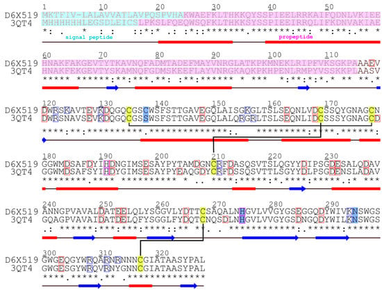
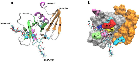
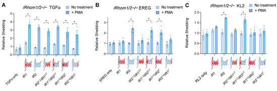
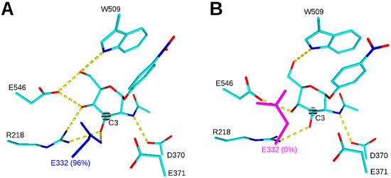
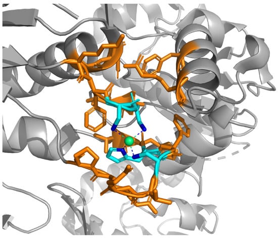
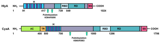
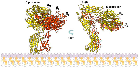
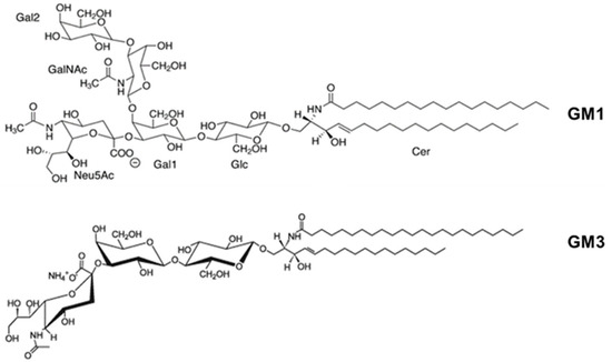
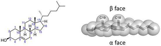
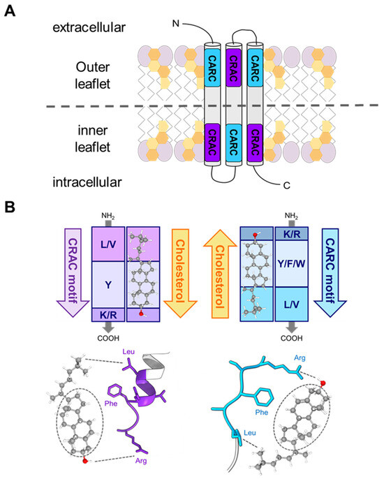
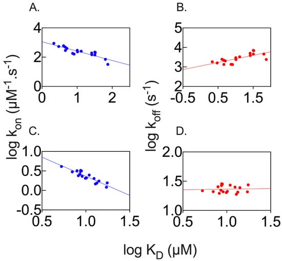
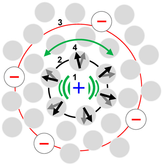
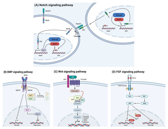
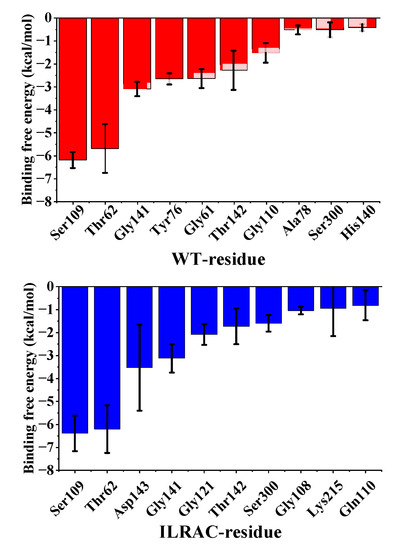
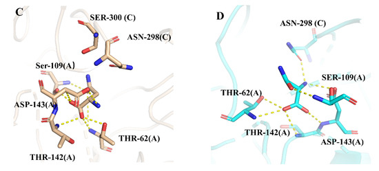
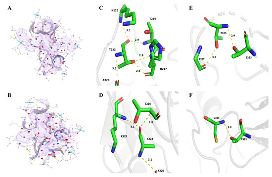
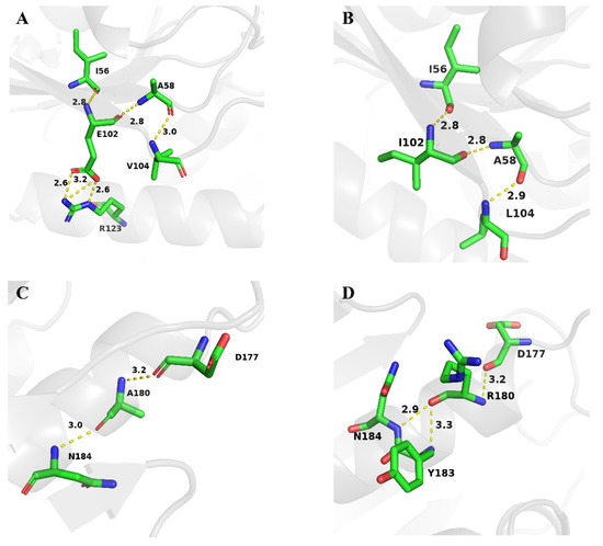
RTX toxins are important virulence factors produced by a wide range of Gram-negative bacteria. They are secreted as water-soluble proteins that are able to bind to the host cell membrane and insert hydrophobic segments into the lipid bilayer that ultimately contribute to the formation of transmembrane pores. Ion diffusion through these pores leads then to cytotoxic and cytolytic effects on the hosts. Several reports have evidenced that the binding of several RTX toxins to the target cell membrane may take place through a high-affinity interaction with integrins of the β2 family that is highly expressed in immune cells of the myeloid lineage. However, at higher toxin doses, cytotoxicity by most RTX toxins has been observed also on β2-deficient cells in which toxin binding to the cell membrane has been proposed to occur through interaction with glycans of glycosylated lipids or proteins present in the membrane. More recently, cumulative pieces of evidence show that membrane cholesterol is essential for the mechanism of action of several RTX toxins. Here, we summarize the most important aspects of the RTX toxin interaction with the target cell membrane, including the cholesterol dependence, the recent identification in the sequences of several RTX toxins of linear motifs coined as the Cholesterol Recognition/interaction Amino acid Consensus (CRAC), and the reverse or mirror CARC motif, which is involved in the toxin–cholesterol interaction.
Full article
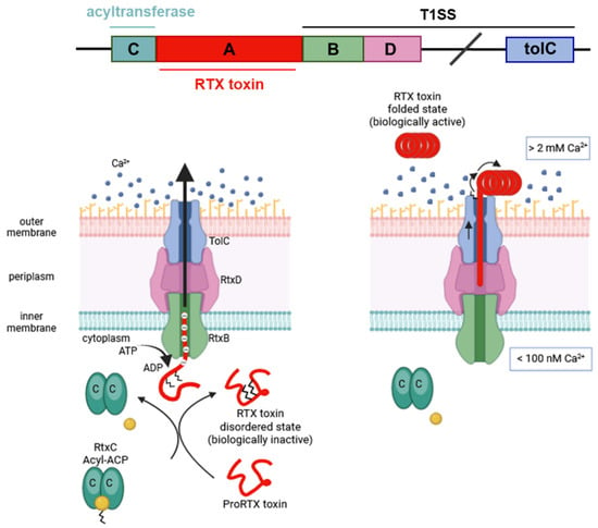
Figure 1


