Morphological Approaches in Biomolecular Sciences
(Closed)
Share This Topical Collection
Editor
 Dr. Gobbi Pietro
Dr. Gobbi Pietro
E-Mail
Website
Collection Editor
Electron Microscopy Laboratories Director, Morphology, Physiology and Environmental Biology Division, Department of Biomolecular Sciences (DiSB), Urbino University Carlo Bo, 61029 Urbino, Italy
Interests: human anatomy; cell biology; nanomaterials; electron microscopy; biomedical technologies
Topical Collection Information
Dear Colleagues,
The morphological approach in biomolecular sciences represents an approach that encourages the study of fine morphology in many fields of interest to biomolecular and functional researchers. Electron microscopy, confocal microscopy, light microscopy, and histochemical or cytochemical approaches enable the greatest refinement of biological (normal and pathological) models in nanobiomaterials or nanopathology.
The main aim of this Topical Collection is to collate important research on the following topics: morphological relationships between molecules and structures at tissue or cellular levels, fine cellular effects induced by micro or nano carriers, and subcellular morphological alterations suggested as diagnostic tools. Specially, we encourage the original research and review articles covering molecular data and molecular mechanisms. The possibilities of combining the morphological aspects of living matter with interactions at the molecular level are practically infinite.
Dr. Gobbi Pietro
Collection Editor
Manuscript Submission Information
Manuscripts should be submitted online at www.mdpi.com by registering and logging in to this website. Once you are registered, click here to go to the submission form. Manuscripts can be submitted until the deadline. All submissions that pass pre-check are peer-reviewed. Accepted papers will be published continuously in the journal (as soon as accepted) and will be listed together on the collection website. Research articles, review articles as well as short communications are invited. For planned papers, a title and short abstract (about 100 words) can be sent to the Editorial Office for announcement on this website.
Submitted manuscripts should not have been published previously, nor be under consideration for publication elsewhere (except conference proceedings papers). All manuscripts are thoroughly refereed through a single-blind peer-review process. A guide for authors and other relevant information for submission of manuscripts is available on the Instructions for Authors page. International Journal of Molecular Sciences is an international peer-reviewed open access semimonthly journal published by MDPI.
Please visit the Instructions for Authors page before submitting a manuscript.
There is an Article Processing Charge (APC) for publication in this
open access journal. For details about the APC please see here.
Submitted papers should be well formatted and use good English. Authors may use MDPI's
English editing service prior to publication or during author revisions.
Keywords
- morphology
- ultrastructure
- microscopy
- histochemistry
- citochemistry
- nanoscience
- pathology and nanopathology
- diagnostic microscopic tools
Published Papers (5 papers)
Open AccessReview
Two-Photon and Multiphoton Microscopy in Anterior Segment Diseases of the Eye
by
Merrelynn Hong, Shu Zhen Chong, Yun Yao Goh and Louis Tong
Viewed by 724
Abstract
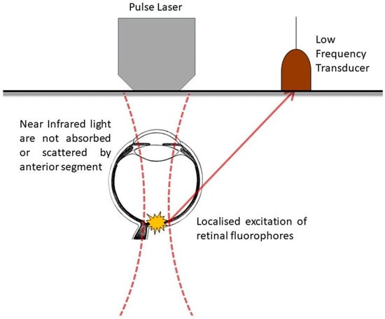
Two-photon excitation microscopy (TPM) and multiphoton fluorescence microscopy (MPM) are advanced forms of intravital high-resolution functional microscopy techniques that allow for the imaging of dynamic molecular processes and resolve features of the biological tissues of interest. Due to the cornea’s optical properties and
[...] Read more.
Two-photon excitation microscopy (TPM) and multiphoton fluorescence microscopy (MPM) are advanced forms of intravital high-resolution functional microscopy techniques that allow for the imaging of dynamic molecular processes and resolve features of the biological tissues of interest. Due to the cornea’s optical properties and the uniquely accessible position of the globe, it is possible to image cells and tissues longitudinally to investigate ocular surface physiology and disease. MPM can also be used for the in vitro investigation of biological processes and drug kinetics in ocular tissues. In corneal immunology, performed via the use of TPM, cells thought to be intraepithelial dendritic cells are found to resemble tissue-resident memory T cells, and reporter mice with labeled plasmacytoid dendritic cells are imaged to understand the protective antiviral defenses of the eye. In mice with limbal progenitor cells labeled by reporters, the kinetics and localization of corneal epithelial replenishment are evaluated to advance stem cell biology. In studies of the conjunctiva and sclera, the use of such imaging together with second harmonic generation allows for the delineation of matrix wound healing, especially following glaucoma surgery. In conclusion, these imaging models play a pivotal role in the progress of ocular surface science and translational research.
Full article
►▼
Show Figures
Open AccessArticle
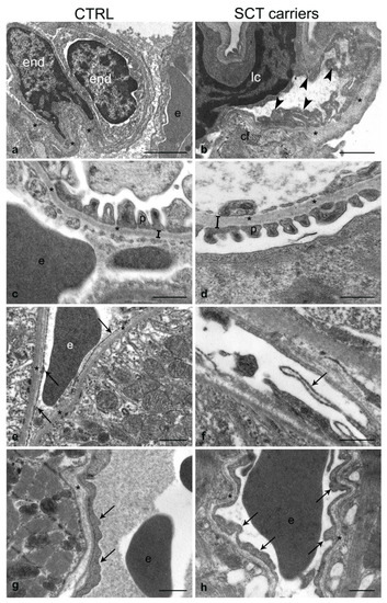
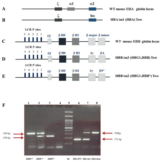
Multi-Organ Morphological Findings in a Humanized Murine Model of Sickle Cell Trait
by
Marcello Trucas, Sabrina Burattini, Susanna Porcu, Michela Simbula, Maria Serafina Ristaldi, Marta Anna Kowalik, Maria Pina Serra, Pietro Gobbi, Michela Battistelli, Andrea Perra and Marina Quartu
Viewed by 1346
Abstract
Sickle cell disease (SCD) is caused by the homozygous beta-globin gene mutation that can lead to ischemic multi-organ damage and consequently reduce life expectancy. On the other hand, sickle cell trait (SCT), the heterozygous beta-globin gene mutation, is still considered a benign condition.
[...] Read more.
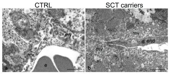
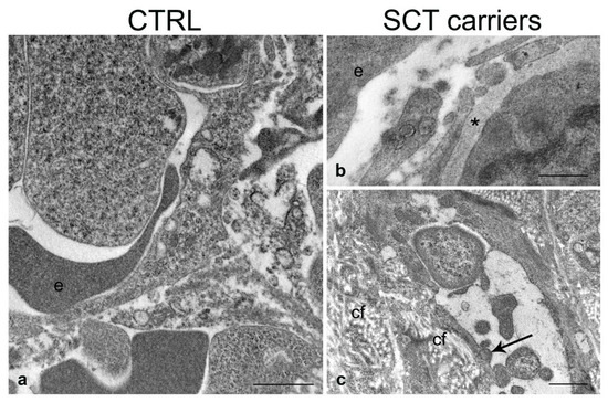
Sickle cell disease (SCD) is caused by the homozygous beta-globin gene mutation that can lead to ischemic multi-organ damage and consequently reduce life expectancy. On the other hand, sickle cell trait (SCT), the heterozygous beta-globin gene mutation, is still considered a benign condition. Although the mechanisms are not well understood, clinical evidence has recently shown that specific pathological symptoms can also be recognized in SCT carriers. So far, there are still scant data regarding the morphological modifications referable to possible multi-organ damage in the SCT condition. Therefore, after genotypic and hematological characterization, by conventional light microscopy and transmission electron microscopy (TEM), we investigated the presence of tissue alterations in 13 heterozygous Townes mice, one of the best-known animal models that, up to now, was used only for the study of the homozygous condition. We found that endothelial alterations, as among which the thickening of vessel basal lamina, are ubiquitous in the lung, liver, kidney, and spleen of SCT carrier mice. The lung shows the most significant alterations, with a distortion of the general tissue architecture, while the heart is the least affected. Collectively, our findings contribute novel data to the histopathological modifications at microscopic and ultrastructural levels, underlying the heterozygous beta-globin gene mutation, and indicate the translational suitability of the Townes model to characterize the features of multiple organ involvement in the SCT carriers.
Full article
►▼
Show Figures
Open AccessArticle
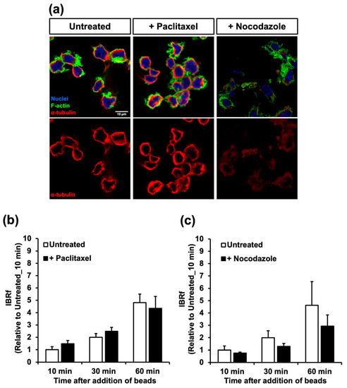
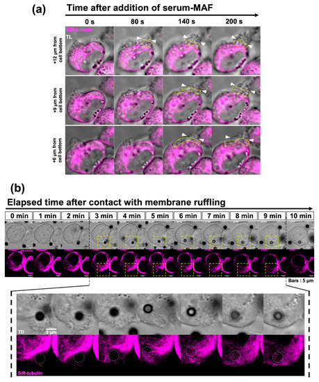
Morphological Evidence for Novel Roles of Microtubules in Macrophage Phagocytosis
by
Yoshika Seta, Kumpei Kawakatsu, Shiori Degawa, Toshiyuki Goto and Takahito Nishikata
Cited by 1 | Viewed by 2268
Abstract
Although the phagocytic activity of macrophages has long been studied, the involvement of microtubules in the process is not well understood. In this study, we improved the fixation protocol and revealed a dynamically rearranging microtubule network in macrophages, consisting of a basal meshwork,
[...] Read more.
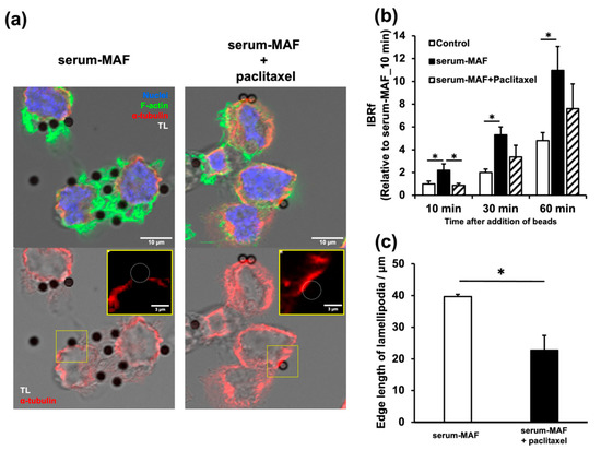
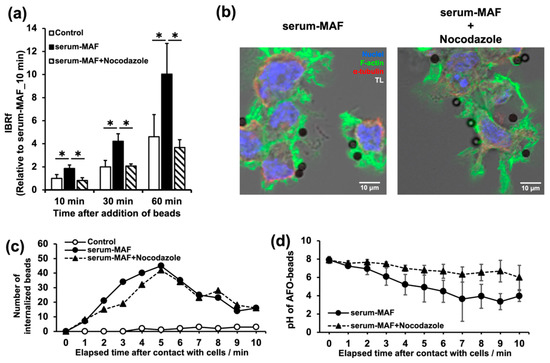
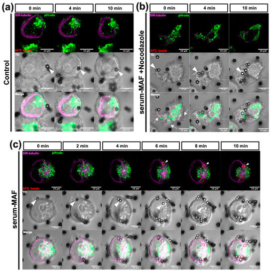
Although the phagocytic activity of macrophages has long been studied, the involvement of microtubules in the process is not well understood. In this study, we improved the fixation protocol and revealed a dynamically rearranging microtubule network in macrophages, consisting of a basal meshwork, thick bundles at the cell edge, and astral microtubules. Some astral microtubules extended beneath the cell cortex and continued to form bundles at the cell edge. These microtubule assemblies were mutually exclusive of actin accumulation during membrane ruffling. Although the stabilization of microtubules with paclitaxel did not affect the resting stage of the macrophages, it reduced the phagocytic activity and membrane ruffling of macrophages activated with serum-MAF, which induced rapid phagocytosis. In contrast, the destabilization of microtubules with nocodazole enhanced membrane ruffling and the internalization of phagocytic targets suggesting an inhibitory effect of the microtubule network on the remodeling of the actin network. Meanwhile, the microtubule network was necessary for phagosome maturation. Our detailed analyses of cytoskeletal filaments suggest a phagocytosis control system involving Ca
2+ influx, the destabilization of microtubules, and activation of actin network remodeling, followed by the translocation and acidification of phagosomes on the microtubule bundles.
Full article
►▼
Show Figures
Open AccessReview
Super-Resolution Microscopy to Study Interorganelle Contact Sites
by
Jon Ander Nieto-Garai, June Olazar-Intxausti, Itxaso Anso, Maier Lorizate, Oihana Terrones and Francesc-Xabier Contreras
Cited by 4 | Viewed by 2871
Abstract
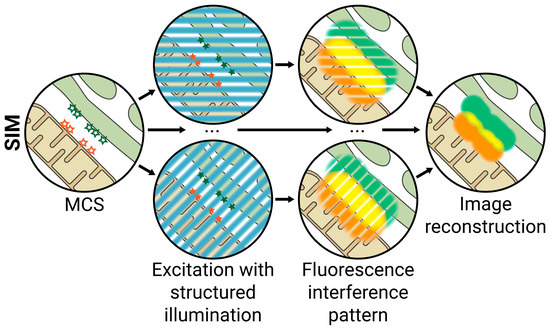
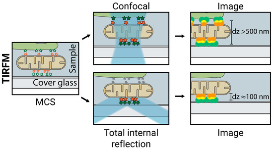
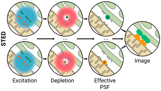
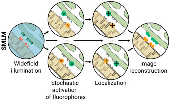
Interorganelle membrane contact sites (MCS) are areas of close vicinity between the membranes of two organelles that are maintained by protein tethers. Recently, a significant research effort has been made to study MCS, as they are implicated in a wide range of biological
[...] Read more.
Interorganelle membrane contact sites (MCS) are areas of close vicinity between the membranes of two organelles that are maintained by protein tethers. Recently, a significant research effort has been made to study MCS, as they are implicated in a wide range of biological functions, such as organelle biogenesis and division, apoptosis, autophagy, and ion and phospholipid homeostasis. Their composition, characteristics, and dynamics can be studied by different techniques, but in recent years super-resolution fluorescence microscopy (SRFM) has emerged as a powerful tool for studying MCS. In this review, we first explore the main characteristics and biological functions of MCS and summarize the different approaches for studying them. Then, we center on SRFM techniques that have been used to study MCS. For each of the approaches, we summarize their working principle, discuss their advantages and limitations, and explore the main discoveries they have uncovered in the field of MCS.
Full article
►▼
Show Figures
Open AccessArticle
A Multidisciplinary Approach in Examining the Susceptibility to Microbial Attack of Polyacrylic and Polyurethane Resins Used in Art Restoration
by
Raffaella Campana, Luigia Sabatini, Luca Giorgi, Giulia Pettinari, Laura Valentini and Pietro Gobbi
Cited by 6 | Viewed by 1389
Abstract
The synthetic polymers used to protect artworks from deterioration process can be colonized by the fungi and bacteria responsible for the biodeterioration process. In this study, the susceptibility of synthetic polyacrylics and polyurethane resins to microorganisms (
Aspergillus niger ATCC 9642,
Aureobasidium pullulans
[...] Read more.
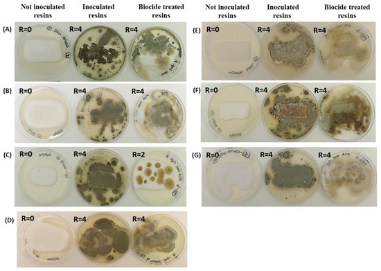
The synthetic polymers used to protect artworks from deterioration process can be colonized by the fungi and bacteria responsible for the biodeterioration process. In this study, the susceptibility of synthetic polyacrylics and polyurethane resins to microorganisms (
Aspergillus niger ATCC 9642,
Aureobasidium pullulans ATCC 15233,
Chaetomium globosum ATCC 6205,
Cladosporium cladosporioides ATCC 16022,
Alternaria alternata BC01,
Penicillium citrinum LS1 and
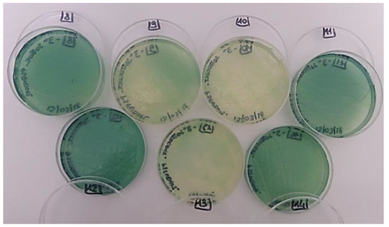
Pseudomonas aeruginosa ATCC 9027) was investigated. The microbial attack was simulated alone and with a biocide and the related growth was observed up to 21 days for bacteria and 28 days for fungi. The polyacrylic and polyurethane resins were subjected to microbial attack, regardless of the biocide treatment, with a fungal growth from 60% to the complete coverage of the plate surface.
Penicillium citrinum showed the greatest adaptation ability and was found in all the examined resins.
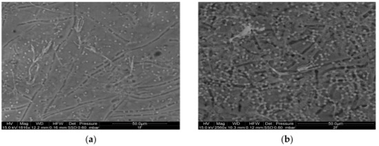
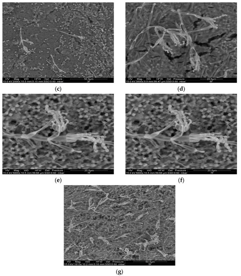
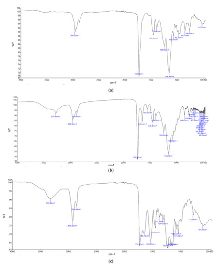
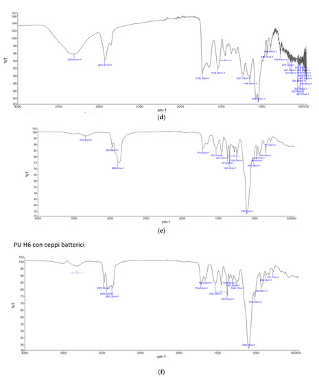
P. aeruginosa was visible in all the different resins, regardless of the presence of biocide. An environmental scanning electron microscope (ESEM) revealed the presence of fungal conidia and hyphae in the inoculated resins and the Fourier transform IR spectroscopy (FTIR-ATR) indicated chemical transformations in the IR spectra, particularly the hydrolysis of esters, with some differences between the polyacrylic and polyurethane resins, which were probably due to their different chemical features. Overall, our data stress that the chemical, physical and biological deterioration caused by microorganisms capable of degrading synthetic polymers is still a problem in art restoration and that new strategies must be considered to counteract this phenomenon.
Full article
►▼
Show Figures


