


Int. J. Mol. Sci. 2024, 25(7), 3943; https://doi.org/10.3390/ijms25073943 - 01 Apr 2024
Viewed by 617
Abstract
►
Show Figures
Refolding multi-disulfide bonded proteins expressed in E. coli into their native structure is challenging. Nevertheless, because of its cost-effectiveness, handiness, and versatility, the E. coli expression of viral envelope proteins, such as the RBD (Receptor-Binding Domain) of the influenza Hemagglutinin protein, could significantly
[...] Read more.
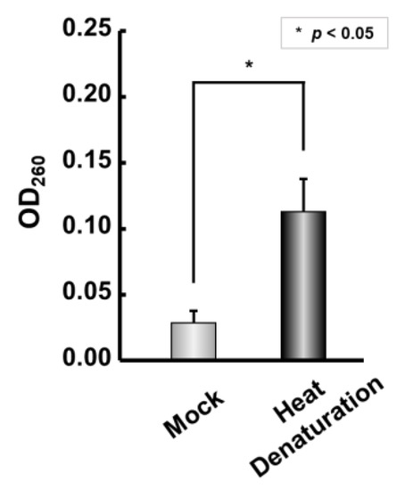
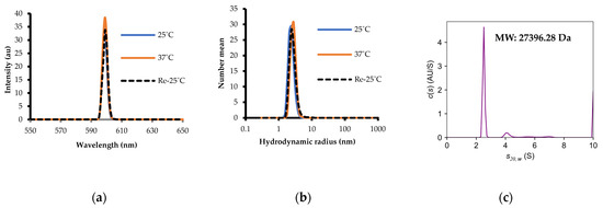
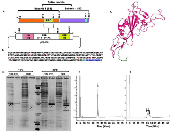
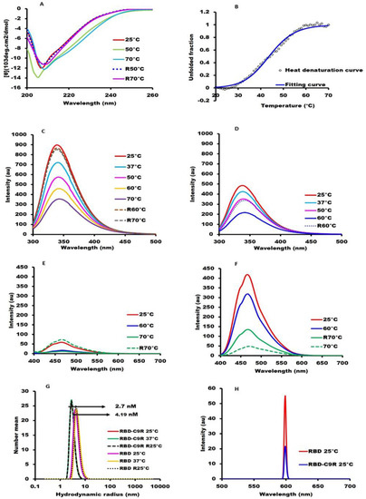
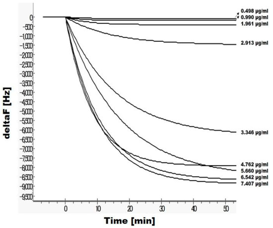
Refolding multi-disulfide bonded proteins expressed in E. coli into their native structure is challenging. Nevertheless, because of its cost-effectiveness, handiness, and versatility, the E. coli expression of viral envelope proteins, such as the RBD (Receptor-Binding Domain) of the influenza Hemagglutinin protein, could significantly advance research on viral infections. Here, we show that H1N1-PR8-RBD (27 kDa, containing four cysteines forming two disulfide bonds) expressed in E. coli and was purified with nickel affinity chromatography, and reversed-phase HPLC was successfully refolded into its native structure, as assessed with several biophysical and biochemical techniques. Analytical ultracentrifugation indicated that H1N1-PR8-RBD was monomeric with a hydrodynamic radius of 2.5 nm. Thermal denaturation, monitored with DSC and CD at a wavelength of 222 nm, was cooperative with a midpoint temperature around 55 °C, strongly indicating a natively folded protein. In addition, the 15N-HSQC NMR spectrum exhibited several 1H-15N resonances indicative of a beta-sheeted protein. Our results indicate that a significant amount (40 mg/L) of pure and native H1N1-PR8-RBD can be produced using an E. coli expression system with our refolding procedure, offering potential insights into the molecular characterization of influenza virus infection.
Full article
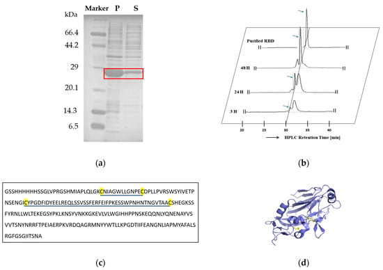
Figure 1

