State-of-the-Art Molecular Mechanisms and Pathophysiology of Cardiovascular and Cerebrovascular Diseases in Italy
(Closed)
Share This Topical Collection
Editor
 Dr. Lucia La Sala
Dr. Lucia La Sala
E-Mail
Website
Collection Editor
Department of Cardiovascular and Metabolic Diseases, IRCCS Multimedica, Via Fantoli 16/15, 20135 Milan, Italy
Interests: glycemic variability; redox systems; diabetes; insulin resistance; prediabetes; microRNA; extracellular vesicles; obesity; innate immune response; cutaneous and visceral adipose tissues; inflammation; signaling pathways
Special Issues, Collections and Topics in MDPI journals
Topical Collection Information
Dear Colleagues,
This regional project aims to collect high-quality research articles, review articles, and communications in the molecular research on cardiovascular and cerebrovascular diseases from the Italy. Cardiovascular diseases (CVD) represent the main cause of morbidity and mortality in industrialized and developing countries, which are considered a real emergency worldwide.
The prevention of cardiovascular diseases, through a series of combined actions aimed to the interventions in risk factors and therapeutics able to reduce their burden, has unequivocally demonstrated a benefit in the reduction of adverse events with a reduction of healthcare costs. The known pathologies with a strong impact on cardiovascular and cerebrovascular functions, diabetes mellitus (DM) and some conditions frequently associated with it - such as obesity, high blood pressure, insulin resistance (IR) – represents the first cause of increased risk of cardiovascular events: more than half the mortality and a vast amount of morbidity in people with DM is related to CVD, including stroke disease and peripheral disease.
We encourage the submission of manuscripts that provide novel and mechanistic molecular insights and papers that report significant advances in the field. The topics of interest for this regional collection include but are not limited to:
- Abnormal heart rhythms;
- Arrhythmias;
- Aorta disease;
- Marfan syndrome;
- Congenital heart disease;
- Coronary artery disease;
- Deep vein thrombosis;
- Heart attack;
- Heart failure;
- Heart muscle disease;
- Heart valve disease;
- Pericardial disease;
- Peripheral vascular disease;
- Rheumatic heart disease;
- Stroke;
- Vascular disease;
- Aneurysms;
- Vascular malformations;
- Transient ischemic attack;
- Cardiovascular complications of diabetes.
Dr. Lucia La Sala
Collection Editor
Manuscript Submission Information
Manuscripts should be submitted online at www.mdpi.com by registering and logging in to this website. Once you are registered, click here to go to the submission form. Manuscripts can be submitted until the deadline. All submissions that pass pre-check are peer-reviewed. Accepted papers will be published continuously in the journal (as soon as accepted) and will be listed together on the collection website. Research articles, review articles as well as short communications are invited. For planned papers, a title and short abstract (about 100 words) can be sent to the Editorial Office for announcement on this website.
Submitted manuscripts should not have been published previously, nor be under consideration for publication elsewhere (except conference proceedings papers). All manuscripts are thoroughly refereed through a single-blind peer-review process. A guide for authors and other relevant information for submission of manuscripts is available on the Instructions for Authors page. International Journal of Molecular Sciences is an international peer-reviewed open access semimonthly journal published by MDPI.
Please visit the Instructions for Authors page before submitting a manuscript.
There is an Article Processing Charge (APC) for publication in this
open access journal. For details about the APC please see here.
Submitted papers should be well formatted and use good English. Authors may use MDPI's
English editing service prior to publication or during author revisions.
Keywords
- cardiovascular diseases
- cerebrovascular diseases
- cardio-neuro-vascular
- molecular pathology
- microRNAs
- diabetes
- cardiovascular complications of diabetes
Published Papers (3 papers)
Open AccessArticle
Role of Uric Acid in Vascular Remodeling: Cytoskeleton Changes and Migration in VSMCs
by
Elisa Russo, Maria Bertolotto, Valentina Zanetti, Daniela Picciotto, Pasquale Esposito, Federico Carbone, Fabrizio Montecucco, Roberto Pontremoli, Giacomo Garibotto, Francesca Viazzi and Daniela Verzola
Cited by 3 | Viewed by 1514
Abstract
The mechanisms by which hyperuricemia induces vascular dysfunction and contributes to cardiovascular disease are still debated. Phenotypic transition is a property of vascular smooth muscle cells (VSMCs) involved in organ damage. The aim of this study was to investigate the effects of uric
[...] Read more.
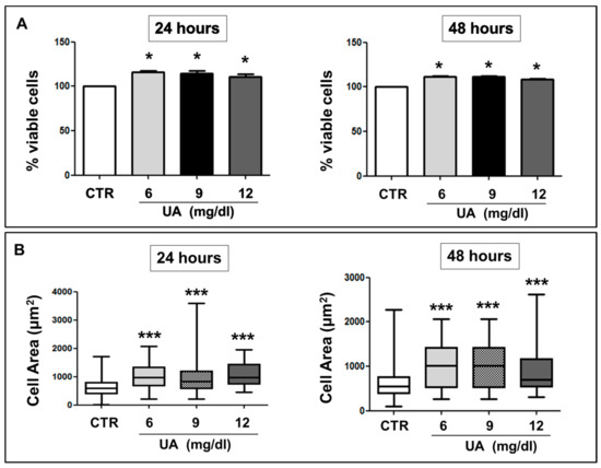
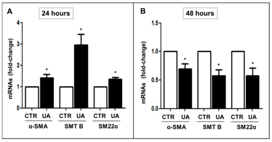
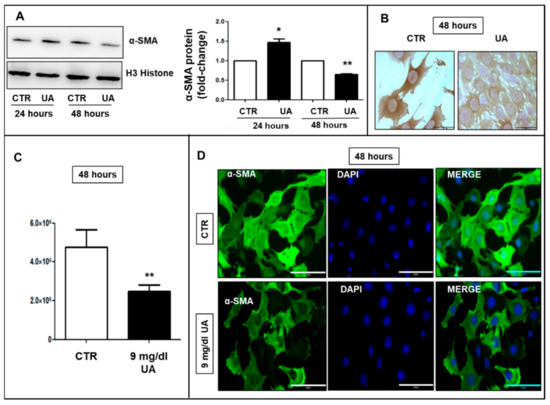
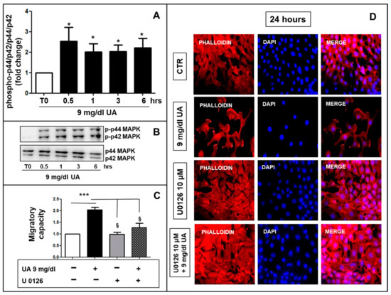
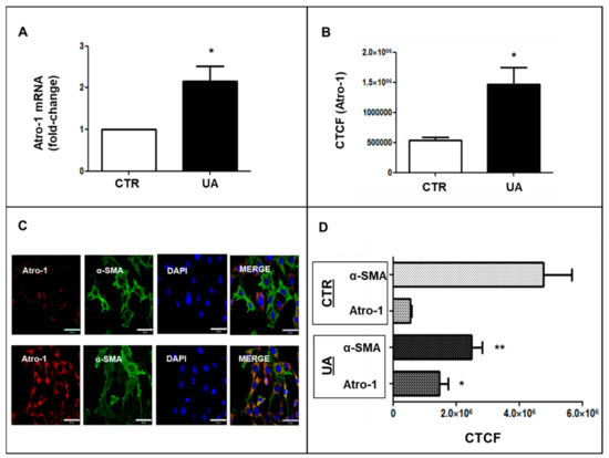
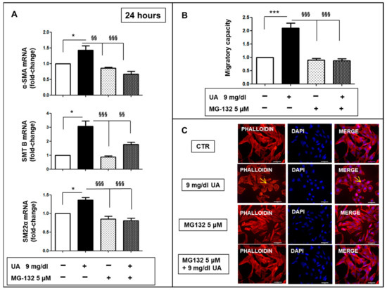
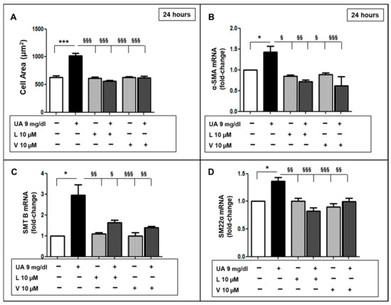
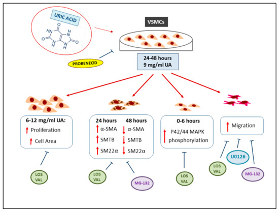
The mechanisms by which hyperuricemia induces vascular dysfunction and contributes to cardiovascular disease are still debated. Phenotypic transition is a property of vascular smooth muscle cells (VSMCs) involved in organ damage. The aim of this study was to investigate the effects of uric acid (UA) on changes in the VSMC cytoskeleton, cell migration and the signals involved in these processes. MOVAS, a mouse VSMC line, was incubated with 6, 9 and 12 mg/dL of UA, angiotensin receptor blockers (ARBs), proteasome and MEK-inhibitors. Migration property was assessed in a micro-chemotaxis chamber and by phalloidin staining. Changes in cytoskeleton proteins (Smoothelin B (SMTB), alpha-Smooth Muscle Actin (αSMA), Smooth Muscle 22 Alpha (SM22α)), Atrogin-1 and MAPK activation were determined by Western blot, immunostaining and quantitative reverse transcription PCR. UA exposition modified SMT, αSMA and SM22α levels (
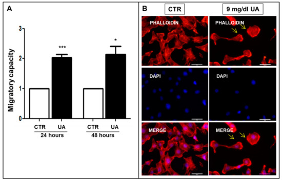
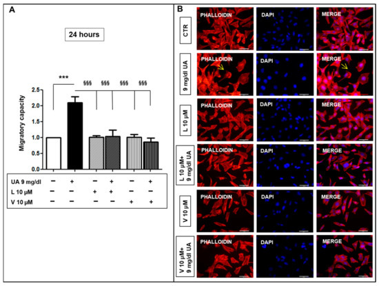
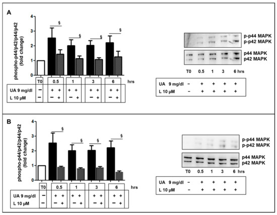
p < 0.05) and significantly upregulated Atrogin-1 and MAPK activation. UA-treated VSMCs showed an increased migratory rate as compared to control cells (
p < 0.001) and a re-arrangement of F-actin. Probenecid, proteasome inhibition and ARBs prevented the development of dysfunctional VSMC. This study shows, for the first time, that UA-induced cytoskeleton changes determine an increase in VSMC migratory rate, suggesting UA as a key player in vascular remodeling.
Full article
►▼
Show Figures
Open AccessCase Report
Distant Recurrence of a Cerebral Cavernous Malformation in the Vicinity of a Developmental Venous Anomaly: Case Report of Local Oxy-Inflammatory Events
by
Andrea Bianconi, Luca Francesco Salvati, Andrea Perrelli, Chiara Ferraris, Armando Massara, Massimiliano Minardi, Gelsomina Aruta, Miriam Rosso, Barbara Massa Micon, Diego Garbossa and Saverio Francesco Retta
Cited by 2 | Viewed by 1893
Abstract
Background: Cerebral cavernous malformations (CCMs) are a major type of cerebrovascular lesions of proven genetic origin that occur in either sporadic (sCCM) or familial (fCCM) forms, the latter being inherited as an autosomal dominant condition linked to loss-of-function mutations in three known CCM
[...] Read more.
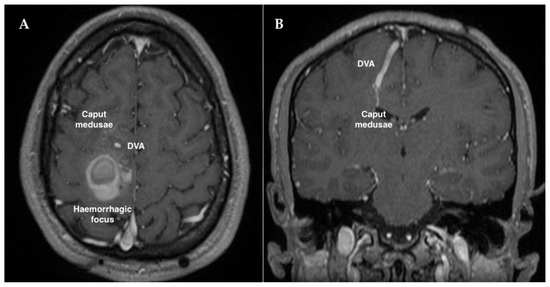
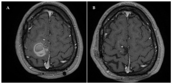
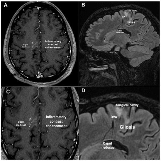
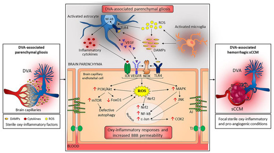
Background: Cerebral cavernous malformations (CCMs) are a major type of cerebrovascular lesions of proven genetic origin that occur in either sporadic (sCCM) or familial (fCCM) forms, the latter being inherited as an autosomal dominant condition linked to loss-of-function mutations in three known CCM genes. In contrast to fCCMs, sCCMs are rarely linked to mutations in CCM genes and are instead commonly and peculiarly associated with developmental venous anomalies (DVAs), suggesting distinct origins and common pathogenic mechanisms. Case report: A hemorrhagic sCCM in the right frontal lobe of the brain was surgically excised from a symptomatic 3 year old patient, preserving intact and pervious the associated DVA. MRI follow-up examination performed periodically up to 15 years after neurosurgery intervention demonstrated complete removal of the CCM lesion and no residual or relapse signs. However, 18 years after surgery, the patient experienced acute episodes of paresthesia due to a distant recurrence of a new hemorrhagic CCM lesion located within the same area as the previous one. A new surgical intervention was, therefore, necessary, which was again limited to the CCM without affecting the pre-existing DVA. Subsequent follow-up examination by contrast-enhanced MRI evidenced a persistent pattern of signal-intensity abnormalities in the bed of the DVA, including hyperintense gliotic areas, suggesting chronic inflammatory conditions. Conclusions: This case report highlights the possibility of long-term distant recurrence of hemorrhagic sCCMs associated with a DVA, suggesting that such recurrence is secondary to focal sterile inflammatory conditions generated by the DVA.
Full article
►▼
Show Figures
Open AccessReview
Molecular Approaches and Echocardiographic Deformation Imaging in Detecting Myocardial Fibrosis
by
Andrea Sonaglioni, Gian Luigi Nicolosi, Elisabetta Rigamonti, Michele Lombardo and Lucia La Sala
Cited by 13 | Viewed by 2292
Abstract
The pathological remodeling of myocardial tissue is the main cause of heart diseases. Several processes are involved in the onset of heart failure, and the comprehension of the mechanisms underlying the pathological phenotype deserves special attention to find novel procedures to identify the
[...] Read more.
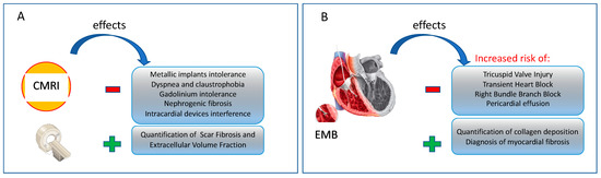
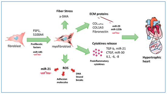
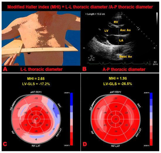
The pathological remodeling of myocardial tissue is the main cause of heart diseases. Several processes are involved in the onset of heart failure, and the comprehension of the mechanisms underlying the pathological phenotype deserves special attention to find novel procedures to identify the site of injury and develop novel strategies, as well as molecular druggable pathways, to counteract the high degree of morbidity associated with it. Myocardial fibrosis (MF) is recognized as a critical trigger for disruption of heart functionality due to the excessive accumulation of extracellular matrix proteins, in response to an injury. Its diagnosis remains focalized on invasive techniques, such as endomyocardial biopsy (EMB), or may be noninvasively detected by cardiac magnetic resonance imaging (CMRI). The detection of MF by non-canonical markers remains a challenge in clinical practice. During the last two decades, two-dimensional (2D) speckle tracking echocardiography (STE) has emerged as a new non-invasive imaging modality, able to detect myocardial tissue abnormalities without specifying the causes of the underlying histopathological changes. In this review, we highlighted the clinical utility of 2D-STE deformation imaging for tissue characterization, and its main technical limitations and criticisms. Moreover, we focalized on the importance of coupling 2D-STE examination with the molecular approaches in the clinical decision-making processes, in particular when the 2D-STE does not reflect myocardial dysfunction directly. We also attempted to examine the roles of epigenetic markers of MF and hypothesized microRNA-based mechanisms aiming to understand how they match with the clinical utility of echocardiographic deformation imaging for tissue characterization and MF assessment.
Full article
►▼
Show Figures


