Radiology-Driven Projects: Science, Networks, and Healthcare
Share This Topical Collection
Editor
 Prof. Dr. Norbert Hosten
Prof. Dr. Norbert Hosten
E-Mail
Website
Guest Editor
Department of Radiology, Universitätsmedizin Greifswald, 17475 Greifswald, Germany
Interests: diagnostic radiology; neuroradiology; teleradiology; digital imaging (MRI and CT); population-based imaging
Topical Collection Information
Dear Colleagues,
In the last decade, radiological imaging has ventured far beyond providing diagnoses to individual patients treated by individual clinicians. Digital imaging has been a prerequisite for this. It was enabled very early, with the first issue of the DICOM standard “Digital Imaging and Communications” in 1985. The early digital competence originating there was used by radiology in various ways: for whole-body MR imaging of large healthy populations providing insights into the natural history of disease; for connecting radiology departments in hospitals and private practices, thus providing radiological services nationally and internationally; for using imaging in networks focusing on single diseases, stroke and breast cancer among them. Use of digital imaging outside of treating individual patients is also a powerful enabler of Artificial Intelligence (AI) in radiology.
This Special Issue of Healthcare is dedicated to offering an overview of these activities. Topics which will be included include (1) teleradiological networks; (2) population-based MR imaging projects; (3) screening programs making use of imaging, e.g., for patients at high risk of breast cancer and lung cancer screening programs; (4) networks making use of CT to care for patients with stroke and other diseases; (5) imaging used in other branches of science; (6) international research networks.
Groups from all specialties are encouraged to submit original research, project reports, short reports, reviews, and opinion papers.
Prof. Dr. Norbert Hosten
Guest Editor
Manuscript Submission Information
Manuscripts should be submitted online at www.mdpi.com by registering and logging in to this website. Once you are registered, click here to go to the submission form. Manuscripts can be submitted until the deadline. All submissions that pass pre-check are peer-reviewed. Accepted papers will be published continuously in the journal (as soon as accepted) and will be listed together on the collection website. Research articles, review articles as well as short communications are invited. For planned papers, a title and short abstract (about 100 words) can be sent to the Editorial Office for announcement on this website.
Submitted manuscripts should not have been published previously, nor be under consideration for publication elsewhere (except conference proceedings papers). All manuscripts are thoroughly refereed through a single-blind peer-review process. A guide for authors and other relevant information for submission of manuscripts is available on the Instructions for Authors page. Healthcare is an international peer-reviewed open access semimonthly journal published by MDPI.
Please visit the Instructions for Authors page before submitting a manuscript.
The Article Processing Charge (APC) for publication in this open access journal is 2700 CHF (Swiss Francs).
Submitted papers should be well formatted and use good English. Authors may use MDPI's
English editing service prior to publication or during author revisions.
Keywords
- MR imaging
- computed tomography
- artificial intelligence
- teleradiology
- MR imaging in population-based projects
- stroke networks
- breast cancer surveillance
- lung cancer screening
- drug resorption
- research networks
Published Papers (17 papers)
Open AccessTechnical Note
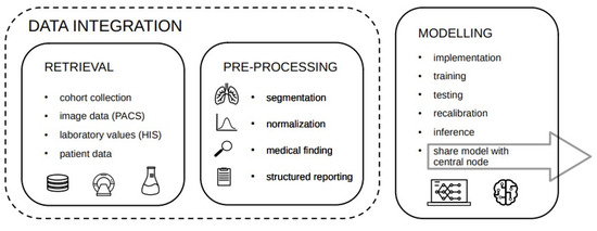
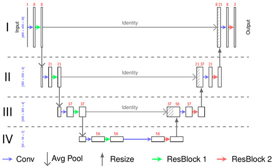
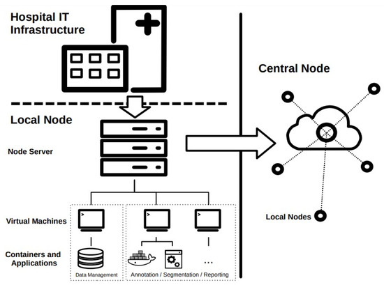
Challenges in Implementing the Local Node Infrastructure for a National Federated Machine Learning Network in Radiology
by
Paul-Philipp Jacobs, Constantin Ehrengut, Andreas Michael Bucher, Tobias Penzkofer, Mathias Lukas, Jens Kleesiek and Timm Denecke
Viewed by 870
Abstract
Data-driven machine learning in medical research and diagnostics needs large-scale datasets curated by clinical experts. The generation of large datasets can be challenging in terms of resource consumption and time effort, while generalizability and validation of the developed models significantly benefit from variety
[...] Read more.
Data-driven machine learning in medical research and diagnostics needs large-scale datasets curated by clinical experts. The generation of large datasets can be challenging in terms of resource consumption and time effort, while generalizability and validation of the developed models significantly benefit from variety in data sources. Training algorithms on smaller decentralized datasets through federated learning can reduce effort, but require the implementation of a specific and ambitious infrastructure to share data, algorithms and computing time. Additionally, it offers the opportunity of maintaining and keeping the data locally. Thus, data safety issues can be avoided because patient data must not be shared. Machine learning models are trained on local data by sharing the model and through an established network. In addition to commercial applications, there are also numerous academic and customized implementations of network infrastructures available. The configuration of these networks primarily differs, yet adheres to a standard framework composed of fundamental components. In this technical note, we propose basic infrastructure requirements for data governance, data science workflows, and local node set-up, and report on the advantages and experienced pitfalls in implementing the local infrastructure with the German Radiological Cooperative Network initiative as the use case example. We show how the infrastructure can be built upon some base components to reflect the needs of a federated learning network and how they can be implemented considering both local and global network requirements. After analyzing the deployment process in different settings and scenarios, we recommend integrating the local node into an existing clinical IT infrastructure. This approach offers benefits in terms of maintenance and deployment effort compared to external integration in a separate environment (e.g., the radiology department). This proposed groundwork can be taken as an exemplary development guideline for future applications of federated learning networks in clinical and scientific environments.
Full article
►▼
Show Figures
Open AccessProject Report
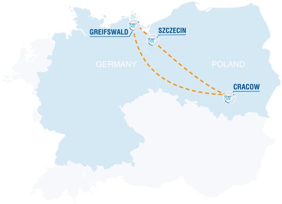
Pediatric Hematology and Oncology Center Integrated by Telemedicine: Experience, Challenges and First Results of a Cross Border Network
by
Tabea Troschke, Aleksandra Wieczorek, Konrad Kulinski, Tomasz Ociepa, Karolina Zielezinska, Holger N. Lode and Tomasz Urasinski
Cited by 2 | Viewed by 1259
Abstract
This article reports on the development, implementation and management of a German–Polish telemedicine network in the field of pediatric oncology and hematology in the Euroregion Pomerania. The achievements and challenges of joint medical case reviews involving patients and their care givers, as well
[...] Read more.
This article reports on the development, implementation and management of a German–Polish telemedicine network in the field of pediatric oncology and hematology in the Euroregion Pomerania. The achievements and challenges of joint medical case reviews involving patients and their care givers, as well as cross-border education activities for physicians, students and nursing staff, are presented. In addition to a progress report, the results of an evaluation of the participants and teachers, likewise the measurement of knowledge growth, are given.
Full article
►▼
Show Figures
Open AccessArticle
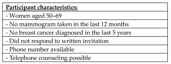
Reasons for Non-Attendance in the German National Mammography Screening Program: Which Barriers Can Be Overcome Using Telephone Counseling?—A Randomized Controlled Trial
by
Sebastian Fochler, Kerstin Weitmann, Martin Domin and Wolfgang Hoffmann
Viewed by 1276
Abstract
Introduction: Germany has established a national mammography screening program (MSP). Despite extensive awareness campaigns, the participation rate is only 54%, which is considerably below the European guidelines’ recommendation of at least 70%. Several reasons why women do not participate are already known. Telephone
[...] Read more.
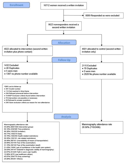
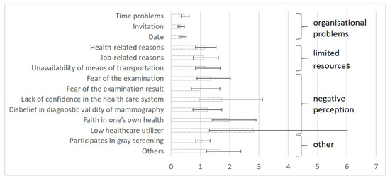
Introduction: Germany has established a national mammography screening program (MSP). Despite extensive awareness campaigns, the participation rate is only 54%, which is considerably below the European guidelines’ recommendation of at least 70%. Several reasons why women do not participate are already known. Telephone consultations along with invitation letters have improved the participation rate. Here, we analyzed the reasons for non-participation and offered barrier-specific counseling to examine which impediments can be overcome to improve participation. Study Design: In a randomized controlled trial, women who had not attended their proposed screening appointment in the MSP after a written invitation were contacted by telephone and asked why they did not attend. Barrier-specific counseling via telephone was then offered. Participation in the MSP was rechecked 3 months after counseling. Setting: 1772 women, aged 50–69 years, who had not scheduled a mammography screening after a written invitation were contacted by telephone and asked for their reasons for non-participation. Intervention: The reasons were recorded by the calling consultant and categorized either during the call or later based on their recorded statements. Afterward, the women received counseling specific to their statements and were given general information about the MSP. Main outcome measures: We categorized the reasons given, calculated their frequency, and analyzed the probabilities to which they could be successfully addressed in individual counseling. Participation rates were determined post-consultation according to the reason(s) indicated. Results: The data were analyzed in 2022. After exclusions, 1494 records were analyzed. Allowing for multiple reasons to be stated by every individual 3280 reasons for not attending were abstracted. The most frequent reason was participation in “gray screening” (51.5%), which included various breast cancer prevention measures outside the national MSP. Time problems (26.6%) and health reasons (17.3%) were also important. Counseling was most effective when women had not participated for scheduling reasons. Conclusion: Several reasons prevented women from participating in the MSP. Some reasons, such as time-related issues, could be overcome by telephone counseling, but others, like barriers resulting from fear of the examination procedure or its result, could not.
Full article
►▼
Show Figures
Open AccessArticle
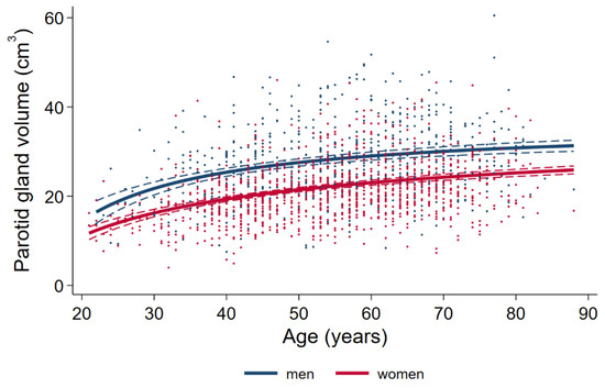
Population Based Average Parotid Gland Volume and Prevalence of Incidental Tumors in T1-MRI
by
Tina Brzoska, Till Ittermann, Friedrich Ihler, Carmela Koch, Markus Blaurock, Robin Bülow, Henry Völzke, Chia-Jung Busch and Achim Georg Beule
Cited by 1 | Viewed by 2596
Abstract
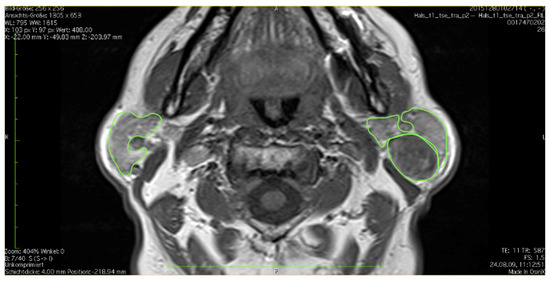
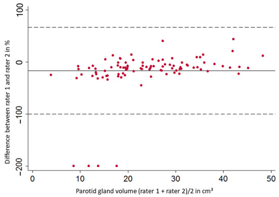
Representative epidemiologic data on the average volume of the parotid gland in a large population-based MRI survey is non-existent. Within the Study of Health in Pomerania (SHIP), we examined the parotid gland in 1725 non-contrast MRI-scans in T1 weighted sequence of axial layers.
[...] Read more.
Representative epidemiologic data on the average volume of the parotid gland in a large population-based MRI survey is non-existent. Within the Study of Health in Pomerania (SHIP), we examined the parotid gland in 1725 non-contrast MRI-scans in T1 weighted sequence of axial layers. Thus, a reliable standard operating procedure (Intraclass Correlation Coefficient > 0.8) could be established. In this study, we found an average, single sided parotid gland volume of 27.82 cm
3 (95% confidence interval (CI) 27.15 to 28.50) in male and 21.60 cm
3 (95% CI 21.16 to 22.05) in female subjects. We observed positive associations for age, body mass index (BMI), as well as male sex with parotid gland size in a multivariate model. The prevalence of incidental tumors within the parotid gland regardless of dignity was 3.94% in the Northeast German population, slightly higher than assumed. Further epidemiologic investigations regarding primary salivary gland diseases are necessary.
Full article
►▼
Show Figures
Open AccessArticle
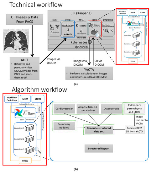
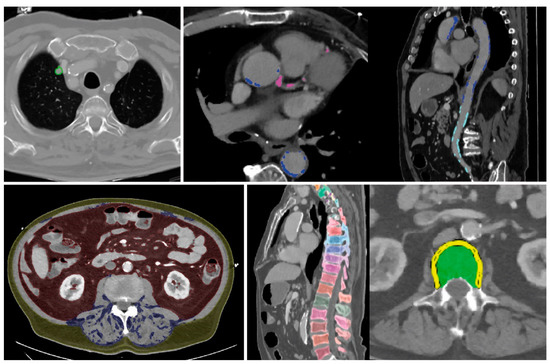
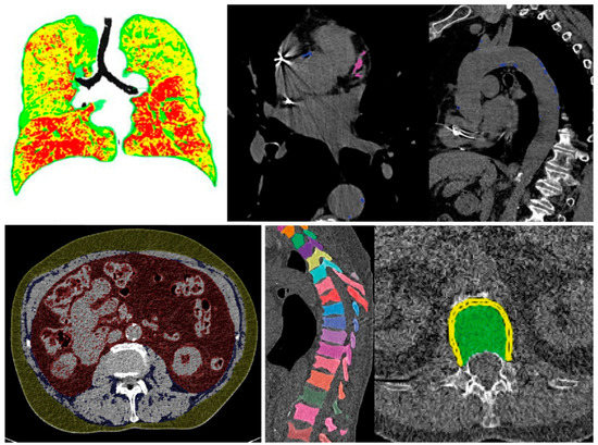
AI-Supported Comprehensive Detection and Quantification of Biomarkers of Subclinical Widespread Diseases at Chest CT for Preventive Medicine
by
Viktoria Palm, Tobias Norajitra, Oyunbileg von Stackelberg, Claus P. Heussel, Stephan Skornitzke, Oliver Weinheimer, Taisiya Kopytova, Andre Klein, Silvia D. Almeida, Michael Baumgartner, Dimitrios Bounias, Jonas Scherer, Klaus Kades, Hanno Gao, Paul Jäger, Marco Nolden, Elizabeth Tong, Kira Eckl, Johanna Nattenmüller, Tobias Nonnenmacher, Omar Naas, Julia Reuter, Arved Bischoff, Jonas Kroschke, Fabian Rengier, Kai Schlamp, Manuel Debic, Hans-Ulrich Kauczor, Klaus Maier-Hein and Mark O. Wielpützadd
Show full author list
remove
Hide full author list
| Viewed by 1981
Abstract
Automated image analysis plays an increasing role in radiology in detecting and quantifying image features outside of the perception of human eyes. Common AI-based approaches address a single medical problem, although patients often present with multiple interacting, frequently subclinical medical conditions. A holistic
[...] Read more.
Automated image analysis plays an increasing role in radiology in detecting and quantifying image features outside of the perception of human eyes. Common AI-based approaches address a single medical problem, although patients often present with multiple interacting, frequently subclinical medical conditions. A holistic imaging diagnostics tool based on artificial intelligence (AI) has the potential of providing an overview of multi-system comorbidities within a single workflow. An interdisciplinary, multicentric team of medical experts and computer scientists designed a pipeline, comprising AI-based tools for the automated detection, quantification and characterization of the most common pulmonary, metabolic, cardiovascular and musculoskeletal comorbidities in chest computed tomography (CT). To provide a comprehensive evaluation of each patient, a multidimensional workflow was established with algorithms operating synchronously on a decentralized Joined Imaging Platform (JIP). The results of each patient are transferred to a dedicated database and summarized as a structured report with reference to available reference values and annotated sample images of detected pathologies. Hence, this tool allows for the comprehensive, large-scale analysis of imaging-biomarkers of comorbidities in chest CT, first in science and then in clinical routine. Moreover, this tool accommodates the quantitative analysis and classification of each pathology, providing integral diagnostic and prognostic value, and subsequently leading to improved preventive patient care and further possibilities for future studies.
Full article
►▼
Show Figures
Open AccessArticle
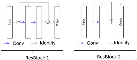
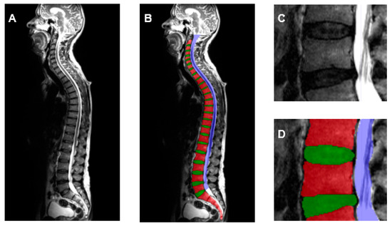
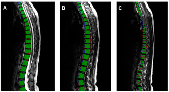
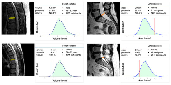
Application of a Deep Learning Approach to Analyze Large-Scale MRI Data of the Spine
by
Felix Streckenbach, Gundram Leifert, Thomas Beyer, Anita Mesanovic, Hanna Wäscher, Daniel Cantré, Sönke Langner, Marc-André Weber and Tobias Lindner
Viewed by 2064
Abstract
With its standardized MRI datasets of the entire spine, the German National Cohort (GNC) has the potential to deliver standardized biometric reference values for intervertebral discs (VD), vertebral bodies (VB) and spinal canal (SC). To handle such large-scale big data, artificial intelligence (AI)
[...] Read more.
With its standardized MRI datasets of the entire spine, the German National Cohort (GNC) has the potential to deliver standardized biometric reference values for intervertebral discs (VD), vertebral bodies (VB) and spinal canal (SC). To handle such large-scale big data, artificial intelligence (AI) tools are needed. In this manuscript, we will present an AI software tool to analyze spine MRI and generate normative standard values. 330 representative GNC MRI datasets were randomly selected in equal distribution regarding parameters of age, sex and height. By using a 3D U-Net, an AI algorithm was trained, validated and tested. Finally, the machine learning algorithm explored the full dataset (
n = 10,215). VB, VD and SC were successfully segmented and analyzed by using an AI-based algorithm. A software tool was developed to analyze spine-MRI and provide age, sex, and height-matched comparative biometric data. Using an AI algorithm, the reliable segmentation of MRI datasets of the entire spine from the GNC was possible and achieved an excellent agreement with manually segmented datasets. With the analysis of the total GNC MRI dataset with almost 30,000 subjects, it will be possible to generate real normative standard values in the future.
Full article
►▼
Show Figures
Open AccessProject Report
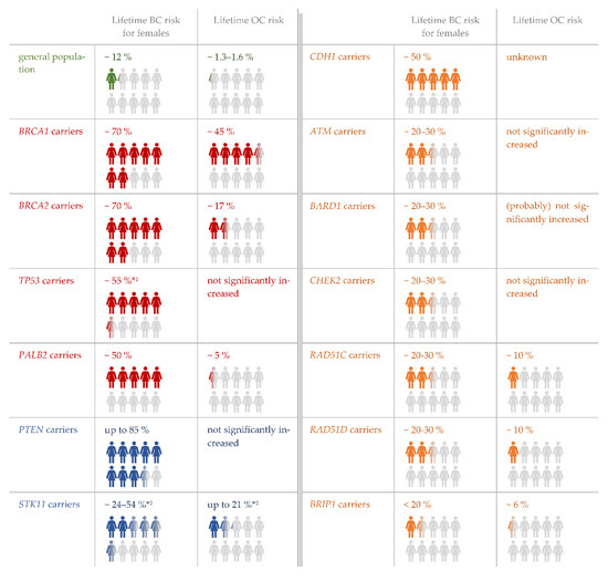
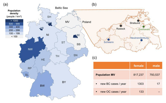
Hereditary Breast and Ovarian Cancer Service in Sparsely Populated Western Pomerania
by
Ute Felbor, Robin Bülow, Rita K. Schmutzler and Matthias Rath
Cited by 1 | Viewed by 1522
Abstract
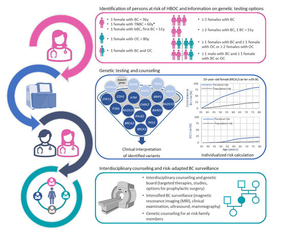
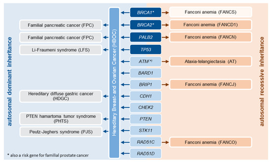
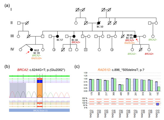
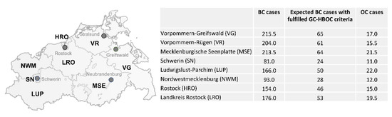
The German Consortium Hereditary Breast and Ovarian Cancer (GC-HBOC) consists of 23 academic centers striving to provide high-quality regional care for affected individuals and healthy at-risk family members. According to the standard operating procedures defined by the GC-HBOC, a Familial Breast and Ovarian
[...] Read more.
The German Consortium Hereditary Breast and Ovarian Cancer (GC-HBOC) consists of 23 academic centers striving to provide high-quality regional care for affected individuals and healthy at-risk family members. According to the standard operating procedures defined by the GC-HBOC, a Familial Breast and Ovarian Cancer Center was implemented at the University Medicine Greifswald over a four-year period from 2018 to 2021, despite the COVID-19 pandemic. Genetic analyses were performed in a total of 658 individuals, including 41 males, which paved the way to local annual risk-adapted breast cancer surveillance for 91 women and prophylactic surgery for 34 women in 2021. Our experience in the North Eastern part of Germany demonstrates that it is possible to establish a high-risk breast and ovarian cancer service even in a sparsely populated region. Major facilitators are the interdisciplinary collaboration of dedicated local experts, the support of the GC-HBOC, fruitful clinical and scientific cooperations and the use of technical improvements. As a blueprint, our project report may help to further expand the network of specialized and knowledge-generating care for HBOC families.
Full article
►▼
Show Figures
Open AccessArticle
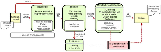
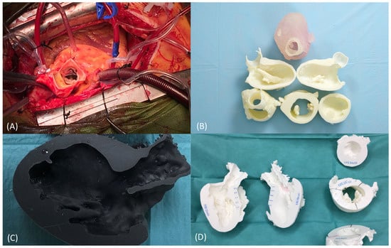
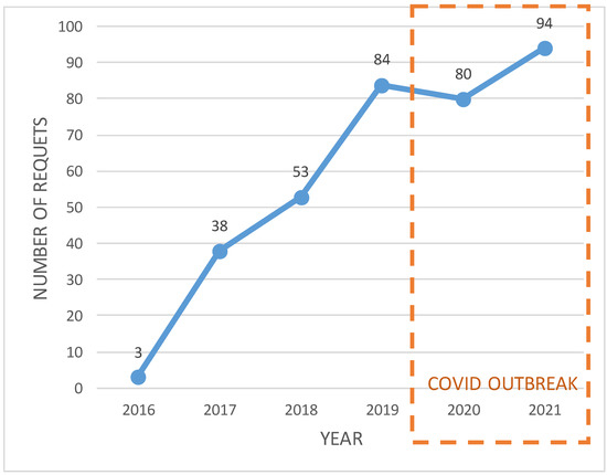
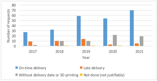
Implementation of an In-House 3D Manufacturing Unit in a Public Hospital’s Radiology Department
by
Ruben I. García, Ines Jauregui, Cristina del Amo, Ainhoa Gandiaga, Olivia Rodriguez, Leyre Margallo, Roberto Voces, Nerea Martin, Inés Gallego, Rikardo Minguez and Harkaitz Eguiraun
Cited by 2 | Viewed by 2483
Abstract
Objective: Three-dimensional printing has become a leading manufacturing technique in healthcare in recent years. Doubts in published studies regarding the methodological rigor and cost-effectiveness and stricter regulations have stopped the transfer of this technology in many healthcare organizations. The aim of this study
[...] Read more.
Objective: Three-dimensional printing has become a leading manufacturing technique in healthcare in recent years. Doubts in published studies regarding the methodological rigor and cost-effectiveness and stricter regulations have stopped the transfer of this technology in many healthcare organizations. The aim of this study was the evaluation and implementation of a 3D printing technology service in a radiology department. Methods: This work describes a methodology to implement a 3D printing service in a radiology department of a Spanish public hospital, considering leadership, training, workflow, clinical integration, quality processes and usability. Results: The results correspond to a 6-year period, during which we performed up to 352 cases, requested by 85 different clinicians. The training, quality control and processes required for the scaled implementation of an in-house 3D printing service are also reported. Conclusions: Despite the maturity of the technology and its impact on the clinic, it is necessary to establish new workflows to correctly implement them into the strategy of the health organization, adjusting it to the needs of clinicians and to their specific resources. Significance: This work allows hospitals to bridge the gap between research and 3D printing, setting up its transfer to clinical practice and using implementation methodology for decision support.
Full article
►▼
Show Figures
Open AccessReview
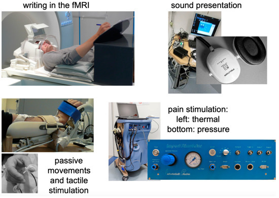
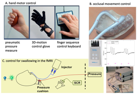
Functional MRI in Radiology—A Personal Review
by
Martin Lotze, Martin Domin, Sönke Langner and Thomas Platz
Viewed by 1778
Abstract
We, here, provide a personal review article on the development of a functional MRI in the radiology departments of two German university medicine units. Although the international community for human brain mapping has met since 1995, the researchers fascinated by human brain function
[...] Read more.
We, here, provide a personal review article on the development of a functional MRI in the radiology departments of two German university medicine units. Although the international community for human brain mapping has met since 1995, the researchers fascinated by human brain function are still young and innovative. However, the impact of functional magnetic resonance imaging (fMRI) on prognosis and treatment decisions is restricted, even though standardized methods have been developed. The tradeoff between the groundbreaking studies on brain function and the attempt to provide reliable biomarkers for clinical decisions is large. By describing some historical developments in the field of fMRI, from a personal view, the rise of this method in clinical neuroscience during the last 25 years might be understandable. We aim to provide some background for (a) the historical developments of fMRI, (b) the establishment of two research units for fMRI in the departments of radiology in Germany, and (c) a description of some contributions within the selected fields of systems neuroscience, clinical neurology, and behavioral psychology.
Full article
►▼
Show Figures
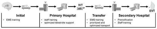
Open AccessArticle
Optimizing Time Management for Drip-and-Ship Stroke Patients Qualifying for Endovascular Therapy—A Single-Network Study
by
Kevin Hädrich, Pawel Krukowski, Jessica Barlinn, Matthias Gawlitza, Johannes C. Gerber, Volker Puetz, Jennifer Linn and Daniel P. O. Kaiser
Cited by 2 | Viewed by 1497
Abstract
BACKGROUND: We sought to identify factors for delayed drip-and-ship (DS) management in stroke patients transferred from primary hospitals to our comprehensive stroke center (CSC) for endovascular therapy (EVT). METHODS: We conducted a retrospective study of all patients transferred to our CSC for EVT
[...] Read more.
BACKGROUND: We sought to identify factors for delayed drip-and-ship (DS) management in stroke patients transferred from primary hospitals to our comprehensive stroke center (CSC) for endovascular therapy (EVT). METHODS: We conducted a retrospective study of all patients transferred to our CSC for EVT between 2016 and 2020. We analyzed emergency and hospital records to assess DS process times and factors predictive of delays. We dichotomized the admission period to 2016–2017 and 2018–2020 according to the main process optimization, including the introduction of a prenotification call. RESULTS: We included 869 DS patients (median age 76 years (IQR 65–82), NIHSS 16 (IQR 11–21), 278 min (IQR 243–335) from onset to EVT); 566 were transferred in 2018–2020. Admission in 2016–2017, during on-call, longer tranfer distance, and general anesthesia were factors independently associated with delayed onset to EVT time (F(5, 352) = 14.76,
p < 0.000). Other factors associated with delayed DS management were: transfer mode, primary hospital type, site of large-vessel occlusion, and intravenous thrombolysis. Total transfer time was faster for distances <50 km by ambulance and for distances >71 km by helicopter. CONCLUSION: Assessment of DS processes and times throughout the patient pathway allows identification of potentially modifiable factors for improvement of the very time-critical workflow for stroke patients.
Full article
►▼
Show Figures
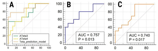
Open AccessArticle
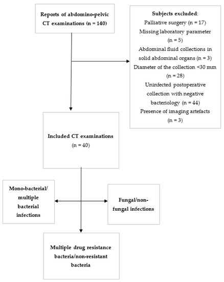
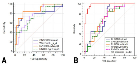
CT-Based Radiomic Analysis May Predict Bacteriological Features of Infected Intraperitoneal Fluid Collections after Gastric Cancer Surgery
by
Vlad Radu Puia, Roxana Adelina Lupean, Paul Andrei Ștefan, Alin Cornel Fetti, Dan Vălean, Florin Zaharie, Ioana Rusu, Lidia Ciobanu and Nadim Al-Hajjar
Cited by 3 | Viewed by 1498
Abstract
The ability of texture analysis (TA) features to discriminate between different types of infected fluid collections, as seen on computed tomography (CT) images, has never been investigated. The study comprised forty patients who had pathological post-operative fluid collections following gastric cancer surgery and
[...] Read more.
The ability of texture analysis (TA) features to discriminate between different types of infected fluid collections, as seen on computed tomography (CT) images, has never been investigated. The study comprised forty patients who had pathological post-operative fluid collections following gastric cancer surgery and underwent CT scans. Patients were separated into six groups based on advanced microbiological analysis of the fluid: mono bacterial (
n = 16)/multiple-bacterial (
n = 24)/fungal (
n = 14)/non-fungal (
n = 26) infection and drug susceptibility tests into: multiple drug-resistance bacteria (
n = 23) and non-resistant bacteria (
n = 17). Dedicated software was used to extract the collections’ TA parameters. The parameters obtained were used to compare fungal and non-fungal infections, mono-bacterial and multiple-bacterial infections, and multiresistant and non-resistant infections. Univariate and receiver operating characteristic analyses and the calculation of sensitivity (Se) and specificity (Sp) were used to identify the best-suited parameters for distinguishing between the selected groups. TA parameters were able to differentiate between fungal and non-fungal collections (ATeta3,
p = 0.02; 55% Se, 100% Sp), mono and multiple-bacterial (CN2D6AngScMom,
p = 0.03); 80% Se, 64.29% Sp) and between multiresistant and non-multiresistant collections (CN2D6Contrast,
p = 0.04; 100% Se, 50% Sp). CT-based TA can statistically differentiate between different types of infected fluid collections. However, it is unclear which of the fluids’ micro or macroscopic features are reflected by the texture parameters. In addition, this cohort is used as a training cohort for the imaging algorithm, with further validation cohorts being required to confirm the changes detected by the algorithm.
Full article
►▼
Show Figures
Open AccessArticle
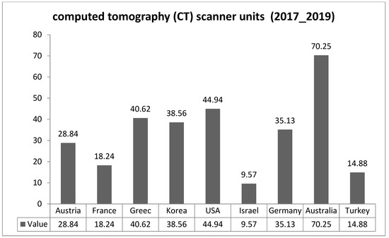
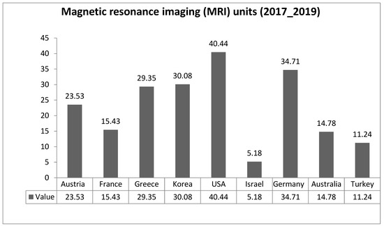
Assessment of Diagnostic Imaging Sector in Public Hospitals in Northern Jordan
by
Ammar A. Oglat
Cited by 1 | Viewed by 1888
Abstract
The most effective diagnostic methods in the medical field are diagnostic imaging techniques such as radiography, computed tomography (CT), magnetic resonance imaging (MRI), and nuclear medicine, which are used to visualize internal body to diagnose it, determine potential treatment, and evaluate and forecast
[...] Read more.
The most effective diagnostic methods in the medical field are diagnostic imaging techniques such as radiography, computed tomography (CT), magnetic resonance imaging (MRI), and nuclear medicine, which are used to visualize internal body to diagnose it, determine potential treatment, and evaluate and forecast care results. Therefore, the purpose of this research is to assess the diagnostic imaging sector, at three major public hospitals in the northern part of Jordan, according to regional and global requirements. The assessment approach was based on knowledge of the accessibility of diagnostic imaging equipment and its quality assurance and performance, the quantity and efficiency of radiological technologists, and the design of radiology units and medical imaging chambers in many aspects based on the use of two tools, a questionnaire and checklists, to accomplish a comprehensive evaluation. The response rate of radiological technologists was 66%. The assessment reveals a noticeable increase in the number of radiological technologists in general with high academic qualification level. Additionally, the number of diagnostic imaging equipment in Jordan revealed a large deficiency in the population–device balance, and through checklists that evaluated both CT and MRI units, it was revealed that the rate of following global requirements and occupational health and safety (OHS) standards was high. The basic supplies available in both the CT and MRI units alike were high, which indicates the high quality of healthcare provided in Jordan.
Full article
►▼
Show Figures
Open AccessArticle
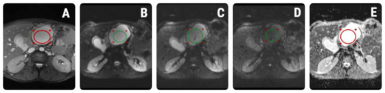
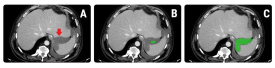
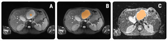
Quantitative MRI of Pancreatic Cystic Lesions: A New Diagnostic Approach
by
Paul Andrei Ștefan, Roxana Adelina Lupean, Andrei Lebovici, Csaba Csutak, Carmen Bianca Crivii, Iulian Opincariu and Cosmin Caraiani
Cited by 4 | Viewed by 1547
Abstract
The commonly used magnetic resonance (MRI) criteria can be insufficient for discriminating mucinous from non-mucinous pancreatic cystic lesions (PCLs). The histological differences between PCLs’ fluid composition may be reflected in MRI images, but cannot be assessed by visual evaluation alone. We investigate whether
[...] Read more.
The commonly used magnetic resonance (MRI) criteria can be insufficient for discriminating mucinous from non-mucinous pancreatic cystic lesions (PCLs). The histological differences between PCLs’ fluid composition may be reflected in MRI images, but cannot be assessed by visual evaluation alone. We investigate whether additional MRI quantitative parameters such as signal intensity measurements (SIMs) and radiomics texture analysis (TA) can aid the differentiation between mucinous and non-mucinous PCLs. Fifty-nine PCLs (mucinous,
n = 24; non-mucinous,
n = 35) are retrospectively included. The SIMs were performed by two radiologists on T2 and diffusion-weighted images (T2WI and DWI) and apparent diffusion coefficient (ADC) maps. A total of 550 radiomic features were extracted from the T2WI and ADC maps of every lesion. The SIMs and TA features were compared between entities using univariate, receiver-operating, and multivariate analysis. The SIM analysis showed no statistically significant differences between the two groups (
p = 0.69, 0.21–0.43, and 0.98 for T2, DWI, and ADC, respectively). Mucinous and non-mucinous PLCs were successfully discriminated by both T2-based (83.2–100% sensitivity and 69.3–96.2% specificity) and ADC-based (40–85% sensitivity and 60–96.67% specificity) radiomic features. SIMs cannot reliably discriminate between PCLs. Radiomics have the potential to augment the common MRI diagnosis of PLCs by providing quantitative and reproducible imaging features, but validation is required by further studies.
Full article
►▼
Show Figures
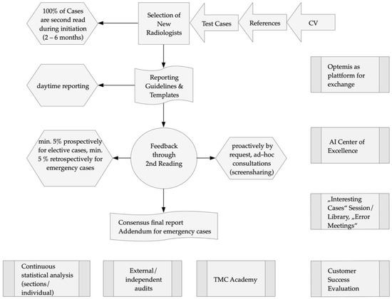
Open AccessProject Report
Quality Assurance of a Cross-Border and Sub-Specialized Teleradiology Service
by
Szabolcs Hetenyi, Leonie Goelz, Alexander Boehmcker and Carlos Schorlemmer
Cited by 3 | Viewed by 1924
Abstract
Background: The current literature discusses aspects of quality assurance (QA) and sub-specialization. However, the challenges of these topics in a teleradiology network have been less explored. In a project report, we aimed to review the development and enforcement of sub-specialized radiology at Telemedicine
[...] Read more.
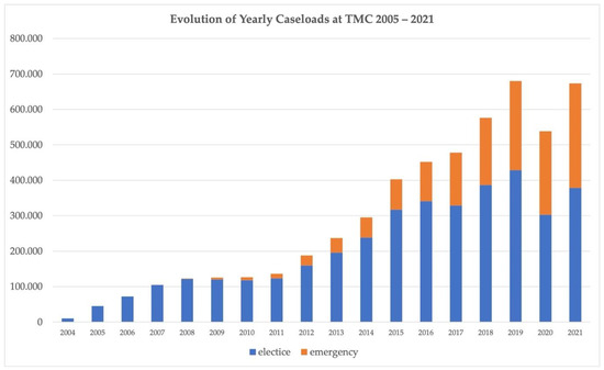
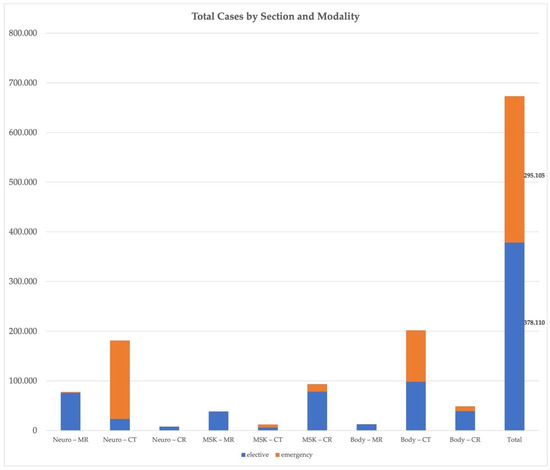
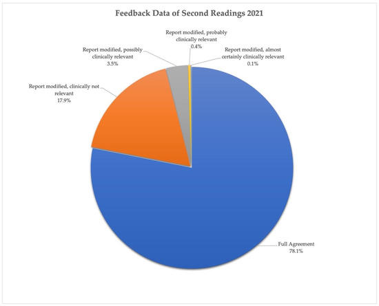
Background: The current literature discusses aspects of quality assurance (QA) and sub-specialization. However, the challenges of these topics in a teleradiology network have been less explored. In a project report, we aimed to review the development and enforcement of sub-specialized radiology at Telemedicine Clinic (TMC), one of the largest teleradiology providers in Europe, and to describe each step of its QA. Evaluation: The company-specific background was provided by the co-authors—current and former staff members of TMC. Detailed descriptions of the structures of sub-specialization and QA at TMC are provided. Exemplary quantitative evaluation of caseloads and disagreement rates of secondary reviews are illustrated. Description of Sub-specialization and Quality Assurance at TMC: Sub-specialization at TMC is divided into musculoskeletal radiology, neuroradiology, head and neck, a body, and an emergency section operating at local daytime in Europe and Australia. Quality assurance is based on a strict selection process of radiologists, specific reporting guidelines, feedback through the secondary reading of 100% of all radiology reports for new starters, and a minimum of 5% of radiology reports on a continuous basis for all other radiologists, knowledge sharing activities and ongoing training. The level of sub-specialization of each radiologist is monitored continuously on an individual basis in detail. After prospective secondary readings, the mean disagreement rate at TMC indicating at least possibly clinically relevant findings was 4% in 2021. Conclusion: With continuing and current developments in radiology in mind, the essential features of sub-specialization and innovative QA are relevant for further expansion of teleradiology services and for most radiology departments worldwide to respond to the increasing demand for value-based radiology.
Full article
►▼
Show Figures
Open AccessEditor’s ChoiceReview
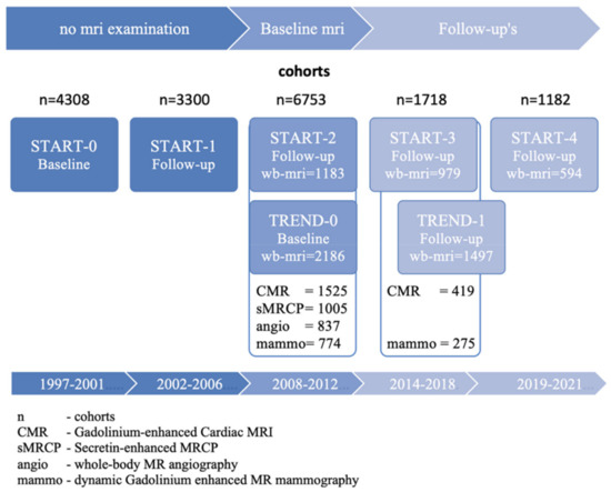
SHIP-MR and Radiology: 12 Years of Whole-Body Magnetic Resonance Imaging in a Single Center
by
Norbert Hosten, Robin Bülow, Henry Völzke, Martin Domin, Carsten Oliver Schmidt, Alexander Teumer, Till Ittermann, Matthias Nauck, Stephan Felix, Marcus Dörr, Marcello Ricardo Paulista Markus, Uwe Völker, Amro Daboul, Christian Schwahn, Birte Holtfreter, Torsten Mundt, Karl-Friedrich Krey, Stefan Kindler, Maria Mksoud, Stefanie Samietz, Reiner Biffar, Wolfgang Hoffmann, Thomas Kocher, Jean-Francois Chenot, Andreas Stahl, Frank Tost, Nele Friedrich, Stephanie Zylla, Anke Hannemann, Martin Lotze, Jens-Peter Kühn, Katrin Hegenscheid, Christian Rosenberg, Georgi Wassilew, Stefan Frenzel, Katharina Wittfeld, Hans J. Grabe and Marie-Luise Kromreyadd
Show full author list
remove
Hide full author list
| Viewed by 3844
Abstract
The Study of Health in Pomerania (SHIP), a population-based study from a rural state in northeastern Germany with a relatively poor life expectancy, supplemented its comprehensive examination program in 2008 with whole-body MR imaging at 1.5 T (SHIP-MR). We reviewed more than 100
[...] Read more.
The Study of Health in Pomerania (SHIP), a population-based study from a rural state in northeastern Germany with a relatively poor life expectancy, supplemented its comprehensive examination program in 2008 with whole-body MR imaging at 1.5 T (SHIP-MR). We reviewed more than 100 publications that used the SHIP-MR data and analyzed which sequences already produced fruitful scientific outputs and which manuscripts have been referenced frequently. Upon reviewing the publications about imaging sequences, those that used T1-weighted structured imaging of the brain and a gradient-echo sequence for R2* mapping obtained the highest scientific output; regarding specific body parts examined, most scientific publications focused on MR sequences involving the brain and the (upper) abdomen. We conclude that population-based MR imaging in cohort studies should define more precise goals when allocating imaging time. In addition, quality control measures might include recording the number and impact of published work, preferably on a bi-annual basis and starting 2 years after initiation of the study. Structured teaching courses may enhance the desired output in areas that appear underrepresented.
Full article
►▼
Show Figures
Open AccessProject Report
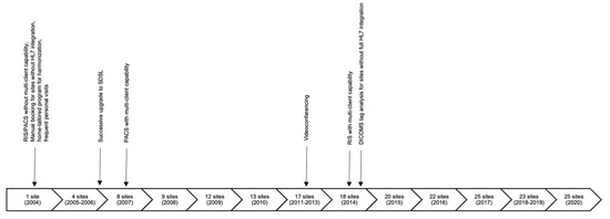
Obstacles and Solutions Driving the Development of a National Teleradiology Network
by
Leonie Goelz, Holger Arndt, Jens Hausmann, Christian Madeja and Sven Mutze
Cited by 4 | Viewed by 2310
Abstract
Background: Teleradiology has the potential to link medical experts and specialties despite geographical separation. In a project report about hospital-based teleradiology, the significance of technical and human factors during the implementation and growth of a teleradiology network are explored. Evaluation: The article identifies
[...] Read more.
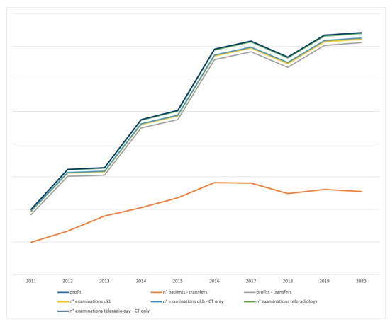
Background: Teleradiology has the potential to link medical experts and specialties despite geographical separation. In a project report about hospital-based teleradiology, the significance of technical and human factors during the implementation and growth of a teleradiology network are explored. Evaluation: The article identifies major obstacles during the implementation and growth of the teleradiology network of the Berlin Trauma Hospital (BG Unfallkrankenhaus Berlin) between 2004 and 2020 in semi-structured interviews with senior staff members. Quantitative analysis of examination numbers, patient numbers, and profits relates the efforts of the staff members to the monetary benefits and success of the network. Identification of qualitative and quantitative factors for success: Soft and hard facilitators and solutions driving the development of the national teleradiology network are identified. Obstacles were often solved by technical innovations, but the time span between required personal efforts, endurance, and flexibility of local and external team members. The article describes innovations driven by teleradiology and hints at the impact of teleradiology on modern medical care by relating the expansion of the teleradiology network to patient transfers and profits. Conclusion: In addition to technical improvements, interpersonal collaborations were key to the success of the teleradiology network of the Berlin Trauma Hospital and remained a unique feature and selling point of this teleradiology network.
Full article
►▼
Show Figures
Open AccessEditor’s ChoiceArticle
Are We Overdoing It? Changes in Diagnostic Imaging Workload during the Years 2010–2020 including the Impact of the SARS-CoV-2 Pandemic
by
Mateusz Winder, Aleksander Jerzy Owczarek, Jerzy Chudek, Joanna Pilch-Kowalczyk and Jan Baron
Cited by 32 | Viewed by 2998
Abstract
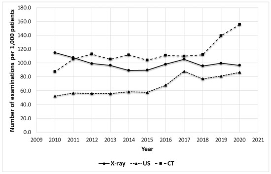
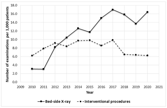
Since the 1990s, there has been a significant increase in the number of imaging examinations as well as a related increase in the healthcare expenditure and the exposure of the population to X-rays. This study aimed to analyze the workload trends in radiology
[...] Read more.
Since the 1990s, there has been a significant increase in the number of imaging examinations as well as a related increase in the healthcare expenditure and the exposure of the population to X-rays. This study aimed to analyze the workload trends in radiology during the last decade, including the impact of COVID-19 in a single university hospital in Poland and to identify possible solutions to the challenges that radiology could face in the future. We compared the annual amount of computed tomography (CT), radiography (X-ray), and ultrasound (US) examinations performed between the years 2010 and 2020 and analyzed the changes in the number of practicing radiologists in Poland. The mean number of patients treated in our hospital was 60,727 per year. During the last decade, the number of CT and US examinations nearly doubled (from 87.4 to 155.7 and from 52.1 to 86.5 per 1000 patients in 2010 and 2020 respectively), while X-ray examinations decreased from 115.1 to 96.9 per 1000 patients. The SARS-CoV-2 pandemic did not change the workload trends as more chest examinations were performed. AI, which contributed to the COVID-19 diagnosis, could aid radiologists in the future with the growing workload by increasing the efficiency of radiology departments as well as by potentially minimizing the related costs.
Full article
►▼
Show Figures


