


Gastroenterol. Insights 2023, 14(1), 110-120; https://doi.org/10.3390/gastroent14010008 - 26 Feb 2023
Viewed by 1554
Abstract
►
Show Figures
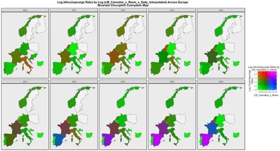
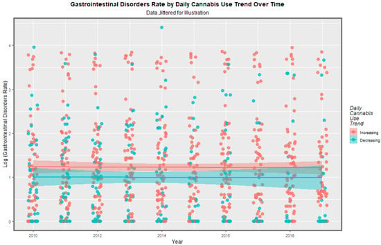
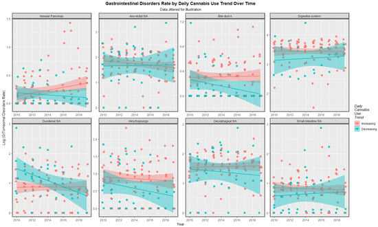
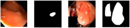
Serrated lesions in the colorectum include all epithelial neoplastic lesions, which show a sawtooth-like morphology in the epithelial crypts. Classification systems nosologically divide colon serrated polyps into three different categories, primarily emphasizing their micromorphological growth pattern and cytodifferentiation: (1) hyperplastic polyps, (2) sessile
[...] Read more.
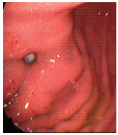
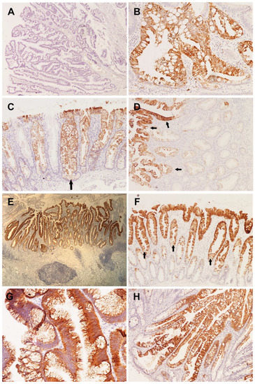
Serrated lesions in the colorectum include all epithelial neoplastic lesions, which show a sawtooth-like morphology in the epithelial crypts. Classification systems nosologically divide colon serrated polyps into three different categories, primarily emphasizing their micromorphological growth pattern and cytodifferentiation: (1) hyperplastic polyps, (2) sessile serrated adenomas/polyps and (3) traditional serrated adenomas. Overall, 109 patients with serrated lesions of the colon, who underwent endoscopic or surgical polypectomy/tumorectomy during one or multiple endoscopic or surgical interventions, over a four-year period, were analyzed. The average age of patients was 62.8 ± 11.6 years. The frequency of serrated lesions of the colon in male patients was 2.4 times higher than in females (70.6% vs. 29.4%). All sessile serrated lesions without dysplasia were positive for CK7 and statistically significant compared to other serrated lesions, if this positivity was present in the complete crypt (p = 0.005). CK20 positivity, which is limited to the upper half of the crypt, is a special feature of hyperplastic polyps compared to other serrated lesions, which is statistically significant (p = 0.0078). Whereas, CK20 positivity of complete crypts is a statistically significant feature of traditional serrated adenomas (p < 0.01). Differences in the expression pattern of cytokeratin 7 and 20 in different serrated lesions may indicate different pathways of colorectal carcinogenesis, and be diagnostically and prognostically useful.
Full article
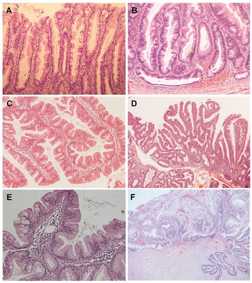
Figure 1


