Medical Optical Imaging
Share This Topical Collection
Editor
 Prof. Dr. Kwang Gi Kim
Prof. Dr. Kwang Gi Kim
E-Mail
Website
Collection Editor
1. Department of Biomedical Engineering, College of Medicine, Gachon University, Seongnam-si 13120, Republic of Korea
2. Department of Health Sciences and Technology, Gachon Advanced Institute for Health Sciences and Technology (GAIHST), Gachon University, Seongnam-si 13120, Republic of Korea
Interests: fluorescence optical imaging; biomarker; medical device; fluorescence device
Special Issues, Collections and Topics in MDPI journals
Topical Collection Information
Dear Colleagues,
Medical optical imaging includes a variety of techniques that use light to obtain images from inside the human body. Examples include optical microscopy, spectroscopy, endoscopy, scanning laser ophthalmoscopy, laser Doppler imaging, and optical coherence tomography. This Topical Collection aims to bring together scholars, professors, researchers, and administrators from around the world to use state-of-the-art technologies and concepts to significantly improve the field of medical optical imaging.
Prof. Dr. Kwang Gi Kim
Collection Editor
Manuscript Submission Information
Manuscripts should be submitted online at www.mdpi.com by registering and logging in to this website. Once you are registered, click here to go to the submission form. Manuscripts can be submitted until the deadline. All submissions that pass pre-check are peer-reviewed. Accepted papers will be published continuously in the journal (as soon as accepted) and will be listed together on the collection website. Research articles, review articles as well as short communications are invited. For planned papers, a title and short abstract (about 100 words) can be sent to the Editorial Office for announcement on this website.
Submitted manuscripts should not have been published previously, nor be under consideration for publication elsewhere (except conference proceedings papers). All manuscripts are thoroughly refereed through a single-blind peer-review process. A guide for authors and other relevant information for submission of manuscripts is available on the Instructions for Authors page. Diagnostics is an international peer-reviewed open access semimonthly journal published by MDPI.
Please visit the Instructions for Authors page before submitting a manuscript.
The Article Processing Charge (APC) for publication in this open access journal is 2600 CHF (Swiss Francs).
Submitted papers should be well formatted and use good English. Authors may use MDPI's
English editing service prior to publication or during author revisions.
Keywords
- medical optical imaging
- optical microscopy
- spectroscopy
- endoscopy
- scanning laser ophthalmoscopy
- laser Doppler imaging
- optical coherence tomography
Published Papers (5 papers)
Open AccessArticle
Joint Diagnostic Method of Tumor Tissue Based on Hyperspectral Spectral-Spatial Transfer Features
by
Jian Du, Chenglong Tao, Shuang Xue and Zhoufeng Zhang
Cited by 1 | Viewed by 958
Abstract
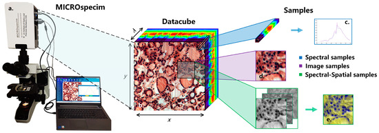
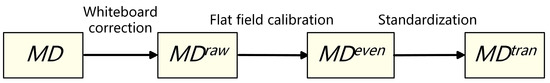
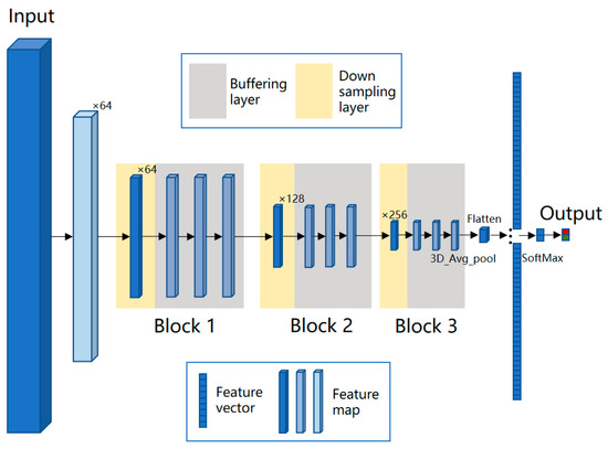
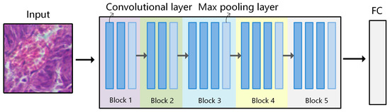
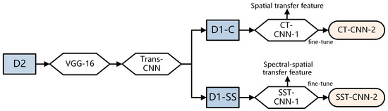
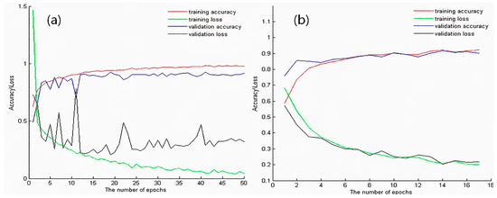
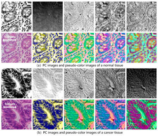
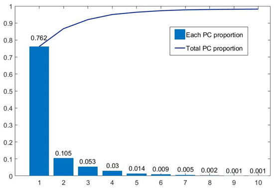
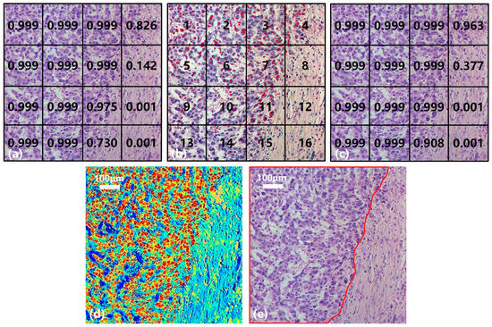
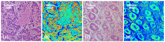
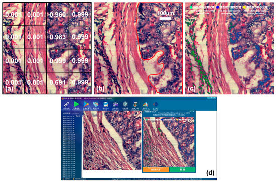
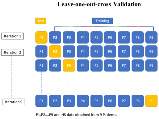
In order to improve the clinical application of hyperspectral technology in the pathological diagnosis of tumor tissue, a joint diagnostic method based on spectral-spatial transfer features was established by simulating the actual clinical diagnosis process and combining micro-hyperspectral imaging with large-scale pathological data.
[...] Read more.
In order to improve the clinical application of hyperspectral technology in the pathological diagnosis of tumor tissue, a joint diagnostic method based on spectral-spatial transfer features was established by simulating the actual clinical diagnosis process and combining micro-hyperspectral imaging with large-scale pathological data. In view of the limited sample volume of medical hyperspectral data, a multi-data transfer model pre-trained on conventional pathology datasets was applied to the classification task of micro-hyperspectral images, to explore the differences in spectral-spatial transfer features in the wavelength of 410–900 nm between tumor tissues and normal tissues. The experimental results show that the spectral-spatial transfer convolutional neural network (SST-CNN) achieved a classification accuracy of 95.46% for the gastric cancer dataset and 95.89% for the thyroid cancer dataset, thus outperforming models trained on single conventional digital pathology and single hyperspectral data. The joint diagnostic method established based on SST-CNN can complete the interpretation of a section of data in 3 min, thus providing a new technical solution for the rapid diagnosis of pathology. This study also explored problems involving the correlation between tumor tissues and typical spectral-spatial features, as well as the efficient transformation of conventional pathological and transfer spectral-spatial features, which solidified the theoretical research on hyperspectral pathological diagnosis.
Full article
►▼
Show Figures
Open AccessArticle
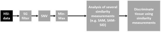
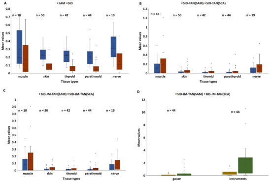
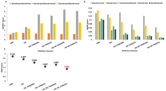
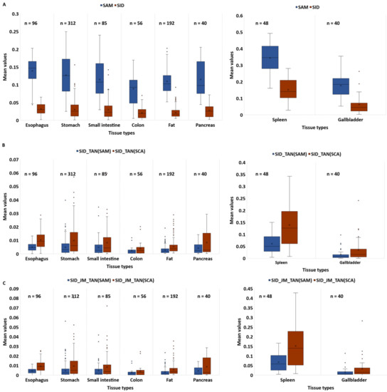
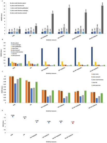
Spectral Similarity Measures for In Vivo Human Tissue Discrimination Based on Hyperspectral Imaging
by
Priya Pathak, Claire Chalopin, Laura Zick, Hannes Köhler, Annekatrin Pfahl, Nada Rayes, Ines Gockel, Thomas Neumuth, Andreas Melzer, Boris Jansen-Winkeln and Marianne Maktabi
Cited by 1 | Viewed by 1356
Abstract
Problem: Similarity measures are widely used as an approved method for spectral discrimination or identification with their applications in different areas of scientific research. Even though a range of works have been presented, only a few showed slightly promising results for human tissue,
[...] Read more.
Problem: Similarity measures are widely used as an approved method for spectral discrimination or identification with their applications in different areas of scientific research. Even though a range of works have been presented, only a few showed slightly promising results for human tissue, and these were mostly focused on pathological and non-pathological tissue classification. Methods: In this work, several spectral similarity measures on hyperspectral (HS) images of in vivo human tissue were evaluated for tissue discrimination purposes. Moreover, we introduced two new hybrid spectral measures, called SID-JM-TAN(SAM) and SID-JM-TAN(SCA). We analyzed spectral signatures obtained from 13 different human tissue types and two different materials (gauze, instruments), collected from HS images of 100 patients during surgeries. Results: The quantitative results showed the reliable performance of the different similarity measures and the proposed hybrid measures for tissue discrimination purposes. The latter produced higher discrimination values, up to 6.7 times more than the classical spectral similarity measures. Moreover, an application of the similarity measures was presented to support the annotations of the HS images. We showed that the automatic checking of tissue-annotated thyroid and colon tissues was successful in 73% and 60% of the total spectra, respectively. The hybrid measures showed the highest performance. Furthermore, the automatic labeling of wrongly annotated tissues was similar for all measures, with an accuracy of up to 90%. Conclusion: In future work, the proposed spectral similarity measures will be integrated with tools to support physicians in annotations and tissue labeling of HS images.
Full article
►▼
Show Figures
Open AccessArticle
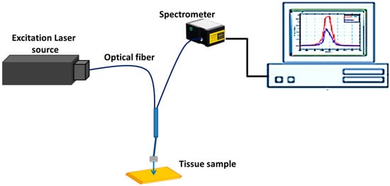
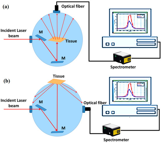
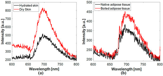
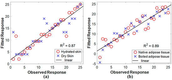
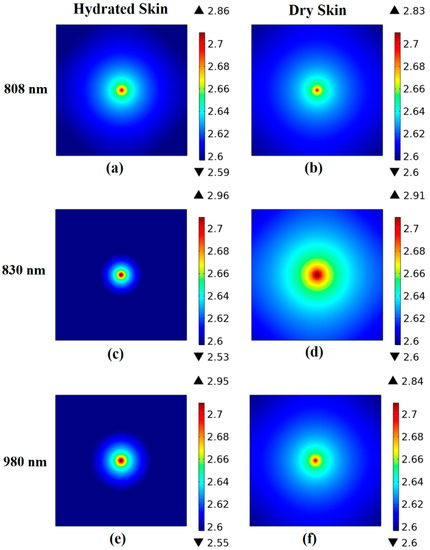
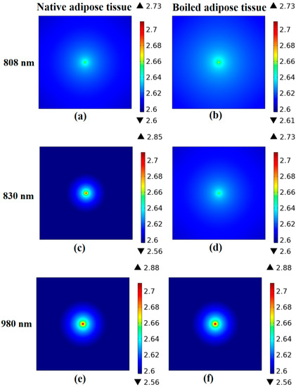
Optical Characterization of Biological Tissues Based on Fluorescence, Absorption, and Scattering Properties
by
Omnia Hamdy, Zienab Abdel-Salam and Mohamed Abdel-Harith
Cited by 13 | Viewed by 2093
Abstract
Optical diagnostics methods are significantly appealing in biological applications since they are non-destructive, safe, and minimally invasive. Laser-induced fluorescence is a promising optical spectrochemical analytical technique widely employed for tissue classification through molecular analysis of the studied samples after excitation with appropriate short-wavelength
[...] Read more.
Optical diagnostics methods are significantly appealing in biological applications since they are non-destructive, safe, and minimally invasive. Laser-induced fluorescence is a promising optical spectrochemical analytical technique widely employed for tissue classification through molecular analysis of the studied samples after excitation with appropriate short-wavelength laser light. On the other hand, diffuse optics techniques are used for tissue monitoring and differentiation based on their absorption and scattering characteristics in the red to the near-infrared spectra. Therefore, it is strongly foreseen to obtain promising results by combining these techniques. In the present work, tissues under different conditions (hydrated/dry skin and native/boiled adipose fat) were distinguished according to their fluorescence emission, absorption, and scattering properties. The selected tissues’ optical absorption and scattering parameters were determined via Kubelka–Munk mathematical model according to the experimental tissue reflectance and transmittance measurements. Such measurements were obtained using an optical configuration of integrating sphere and spectrometer at different laser wavelengths (808, 830, and 980 nm). Moreover, the diffusion equation was solved for the fluence rate at the sample surface using the finite element method. Furthermore, the accuracy of the obtained spectroscopic measurements was evaluated using partial least squares regression statistical analysis with 0.87 and 0.89 R-squared values for skin and adipose fat, respectively.
Full article
►▼
Show Figures
Open AccessArticle
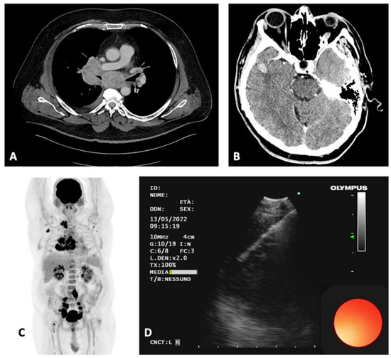
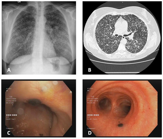
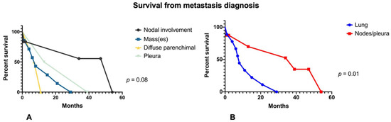
Diagnosis and Pattern Identification of Intrathoracic Malignant Melanoma Metastasis: A Retrospective Single Center Analysis
by
Matteo Fontana, Laura Rossi, Federica Ghinassi, Roberto Piro, Chiara Scelfo, Sofia Taddei, Eleonora Casalini, Patrizia Ruggiero, Chiara Pollorsi, Bianca Beghe’, Caterina Longo and Nicola Facciolongo
Viewed by 2022
Abstract
The lung is a frequent site of secondary malignancies. Melanoma is a malignant tumor originating from melanocytes, that accounts for the majority of death related to skin cancers. In advanced stages, it can also present with intrathoracic metastasis, particularly in the lungs, but
[...] Read more.
The lung is a frequent site of secondary malignancies. Melanoma is a malignant tumor originating from melanocytes, that accounts for the majority of death related to skin cancers. In advanced stages, it can also present with intrathoracic metastasis, particularly in the lungs, but infrequent intrathoracic manifestations are possible. A retrospective analysis of the cases referred to the pulmonary endoscopy unit of the hospital of Reggio Emilia in the last 10 years (since December 2012) was carried out, discovering 17 cases of melanoma metastasis with thoracic localizations, either with or without a diagnosis of primary melanoma. Four repetitive patterns of clinical-radiological presentation have been identified and described through the same number of paradigmatic clinical cases: nodal involvement (35%), lung mass(es) (41%), diffuse pulmonary involvement (12%), and pleural involvement (12%). These different presentations imply the use of different diagnostic techniques, with an overall high diagnostic yield (87.5%). Finally, a brief analysis of survival based on the pattern of presentation has been performed, finding no statistically significant differences between the four groups at metastasis diagnosis (
p-value = 0.06, median survival of respectively 54, 8, 9, and 26 months from metastasis diagnosis), while there is a significant difference considering patients with lung involvement versus nodal/pleural involvement (
p = 0.01).
Full article
►▼
Show Figures
Open AccessArticle
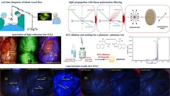
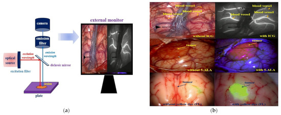
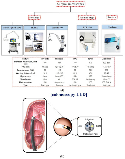
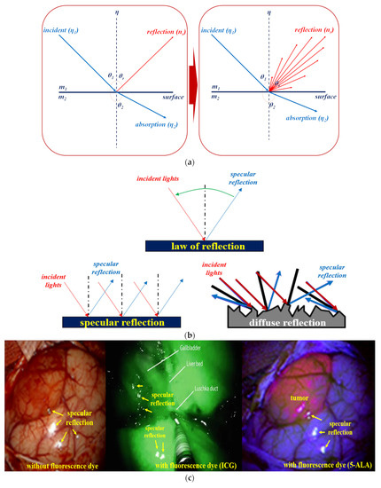
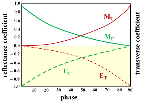
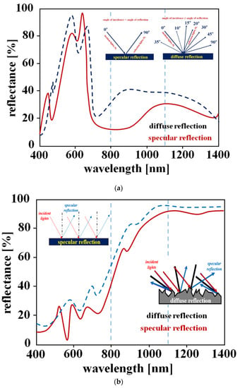
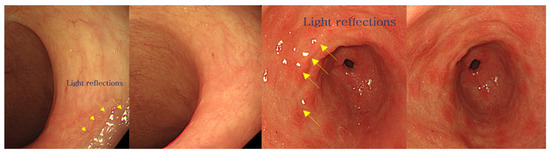
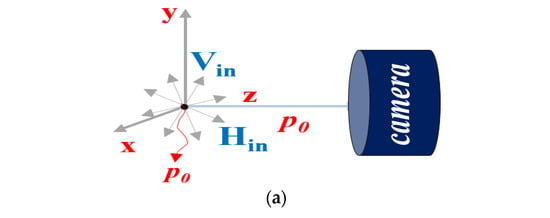
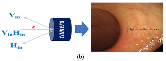
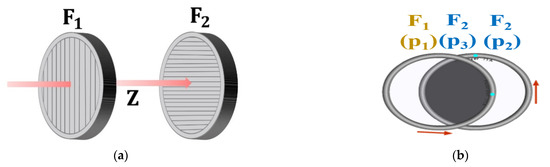
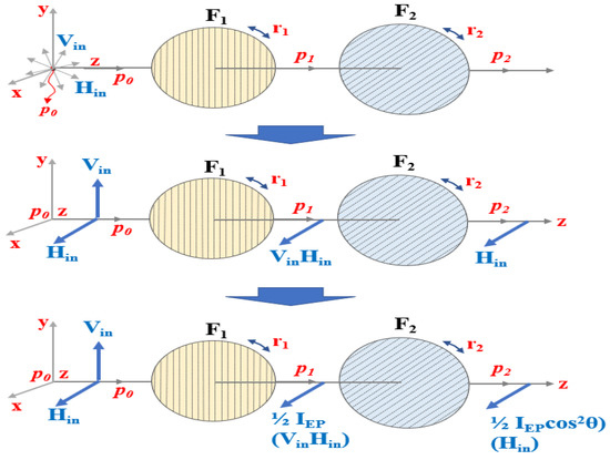
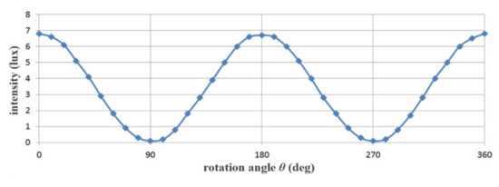
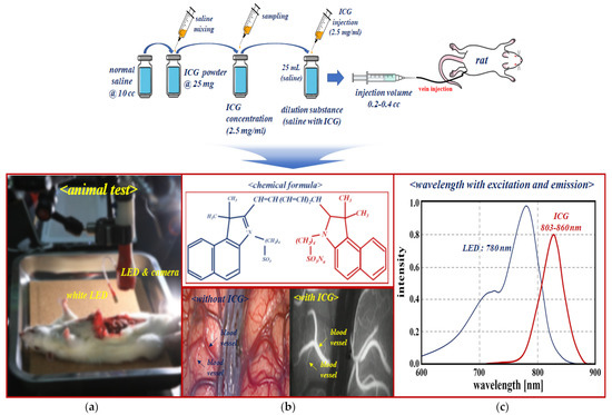
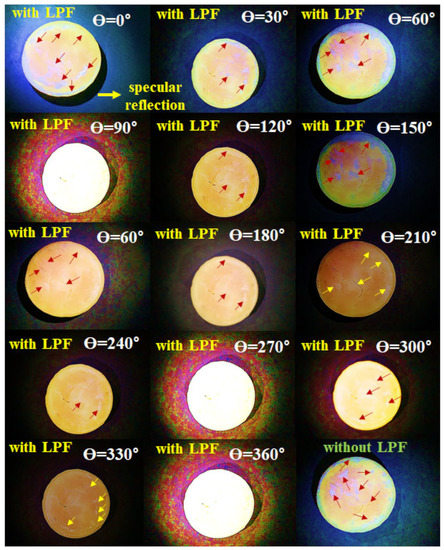
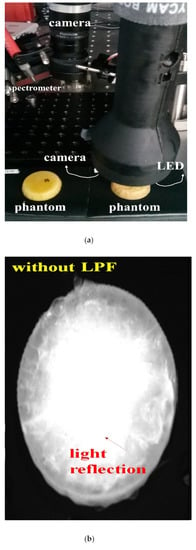
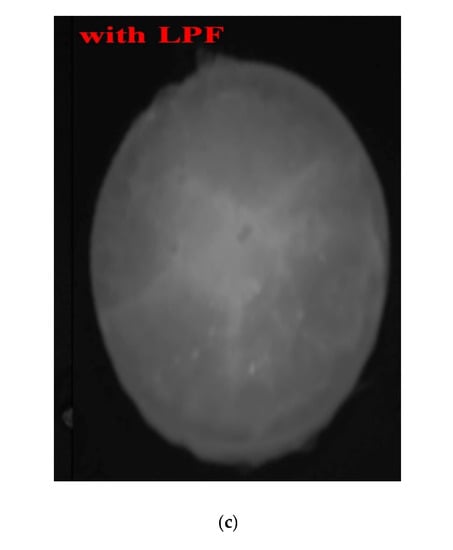
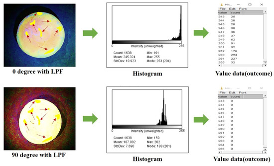
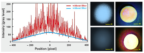
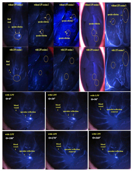
Reduction of Specular Reflection Based on Linear Polarization Control for Fluorescence-Induced Diagnostic Evaluation
by
Sangyun Lee, Kicheol Yoon, Jungmin Kim and Kwang Gi Kim
Viewed by 1916
Abstract
The primary goal of cancer surgery is to completely eliminate tumors. A real-time diagnostic method uses a fluorescence contrast agent and a surgical microscope to assess the status of tumor resection and the patient’s blood circulation. The biggest problem in imaging diagnostics using
[...] Read more.
The primary goal of cancer surgery is to completely eliminate tumors. A real-time diagnostic method uses a fluorescence contrast agent and a surgical microscope to assess the status of tumor resection and the patient’s blood circulation. The biggest problem in imaging diagnostics using a microscope is the specular reflection phenomenon. While observing a lesion, the observation field may be obstructed due to specular reflection, making it difficult to obtain accurate results during the diagnostic process. Herein we propose a method to reduce specular reflection during tumor diagnosis by introducing a linearly polarized filter for a surgical microscope system. The method of angular direction adjustment of the filter ensures that only the horizontally polarized light passes through it, thereby obstructing the specular reflection. As a result of removing specular reflection, clear images were obtained at 90° and 270°. This experiment was conducted using phantoms and animals. Our results prove that the proposed method can be applied to imaging cameras used in internal medicine, surgery, and radiology for diagnosis.
Full article
►▼
Show Figures


