Frontiers on Cancer Biomarkers

A topical collection in Diagnostics (ISSN 2075-4418). This collection belongs to the section "Pathology and Molecular Diagnostics".

Viewed by 2901

Editors


E-Mail Website
Collection Editor
1. Department of Immunochemistry Diagnostics, University Hospital Pilsen, Edvarda Benese 1128/13, 305 99 Pilsen, Czech Republic
2. Institute of Pharmacology and Toxicology, Charles University, Faculty of Medicine in Pilsen, alej Svobody 1655/76, 323 00 Pilsen, Czech Republic
Interests: tumor markers in cancer diagnostics and therapy control; hormones; growth factors; therapeutic drug monitoring
Institute of Chemistry, Slovak Academy of Sciences, Dubravska cesta 9, 845 38 Bratislava, Slovakia
Interests: biomarkers; cancer; diagnostics; glycans; lectins; biosensors; nanotechnology

Topical Collection Information

Dear Colleagues,

Cancer has probably accompanied humanity since its inception. The earliest cancerous growths in humans were found in Egyptian mummies dating back to ∼1500 BC. The first case of cancer in modern medicine was described in 1775 by a British surgeon by the name of Percivall Pott. At present, despite advanced treatment methods, early diagnosis is still a prerequisite for successful treatment.

Cancer biomarkers and advanced imaging techniques are the cornerstones of cancer diagnostics. The first cancer biomarker was reported in an 1848: the light chain of immunoglobulin that is present in the urine of myeloma patients. The modern era of monitoring malignant disease, however, began in the 1960s with the discovery of alpha-fetoprotein (AFP) and carcinoembryonic antigen (CEA); these discoveries were followed by the introduction of newly developed diagnostic techniques such as the radioimmunoassay. Subsequent progress was made in the 1980s when hybridoma technology enabled the development of cancer carbohydrate, prostate-specific, and other antigens that are still routinely used and are considered "classic" tumor markers. The past decade of biomarker development has been characterized by the completion of a number of genome-sequencing projects and the discovery of oncogenes and tumor-suppressor genes. Recent technologies are capable of performing parallel, not only serial analyses, and such new strategies represent a paradigm shift in the search for novel biomarkers.

In general, the current process in cancer biomarker development can be divided into five phases: preclinical studies; clinical assay development and validation; retrospective longitudinal studies; prospective screening; and randomized control trials.

This Issue will provide updates in the field of cancer biomarkers and introduce new techniques that are being used in their detection and development.

Prof. Dr. Radek Kučera
Dr. Jan Tkac
Collection Editors

Manuscript Submission Information

Manuscripts should be submitted online at www.mdpi.com by registering and logging in to this website. Once you are registered, click here to go to the submission form. Manuscripts can be submitted until the deadline. All submissions that pass pre-check are peer-reviewed. Accepted papers will be published continuously in the journal (as soon as accepted) and will be listed together on the collection website. Research articles, review articles as well as short communications are invited. For planned papers, a title and short abstract (about 100 words) can be sent to the Editorial Office for announcement on this website.

Submitted manuscripts should not have been published previously, nor be under consideration for publication elsewhere (except conference proceedings papers). All manuscripts are thoroughly refereed through a single-blind peer-review process. A guide for authors and other relevant information for submission of manuscripts is available on the Instructions for Authors page. Diagnostics is an international peer-reviewed open access semimonthly journal published by MDPI.

Please visit the Instructions for Authors page before submitting a manuscript. The Article Processing Charge (APC) for publication in this open access journal is 2600 CHF (Swiss Francs). Submitted papers should be well formatted and use good English. Authors may use MDPI's English editing service prior to publication or during author revisions.

Keywords

  • cancer
  • biomarkers
  • diagnostics
  • prognosis
  • recurrence
  • metastasis
  • therapy control
  • development
  • proteins
  • glycoproteins

Published Papers (2 papers)

2023

17 pages, 4351 KiB  
Article
Differentiation of Salivary Gland and Salivary Gland Tumor Tissue via Raman Imaging Combined with Multivariate Data Analysis
by Miriam C. Bassler, Mona Knoblich, Elena Gerhard-Hartmann, Ashutosh Mukherjee, Almoatazbellah Youssef, Rudolf Hagen, Lukas Haug, Miguel Goncalves, Agmal Scherzad, Manuel Stöth, Edwin Ostertag, Maria Steinke, Marc Brecht, Stephan Hackenberg and Till Jasper Meyer
Diagnostics 2024, 14(1), 92; https://doi.org/10.3390/diagnostics14010092 - 30 Dec 2023
Viewed by 844
Abstract
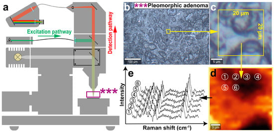
Salivary gland tumors (SGTs) are a relevant, highly diverse subgroup of head and neck tumors whose entity determination can be difficult. Confocal Raman imaging in combination with multivariate data analysis may possibly support their correct classification. For the analysis of the translational potential [...] Read more.
Salivary gland tumors (SGTs) are a relevant, highly diverse subgroup of head and neck tumors whose entity determination can be difficult. Confocal Raman imaging in combination with multivariate data analysis may possibly support their correct classification. For the analysis of the translational potential of Raman imaging in SGT determination, a multi-stage evaluation process is necessary. By measuring a sample set of Warthin tumor, pleomorphic adenoma and non-tumor salivary gland tissue, Raman data were obtained and a thorough Raman band analysis was performed. This evaluation revealed highly overlapping Raman patterns with only minor spectral differences. Consequently, a principal component analysis (PCA) was calculated and further combined with a discriminant analysis (DA) to enable the best possible distinction. The PCA-DA model was characterized by accuracy, sensitivity, selectivity and precision values above 90% and validated by predicting model-unknown Raman spectra, of which 93% were classified correctly. Thus, we state our PCA-DA to be suitable for parotid tumor and non-salivary salivary gland tissue discrimination and prediction. For evaluation of the translational potential, further validation steps are necessary. Full article
Show Figures

Figure 1

10 pages, 2720 KiB  
Article
The Stability of the Anti-Müllerian Hormone in Serum and Plasma Samples under Various Preanalytical Conditions
by Radana Vrzáková, Václav Šimánek, Ondřej Topolčan, Vladimír Vurm, David Slouka and Radek Kučera
Diagnostics 2023, 13(8), 1501; https://doi.org/10.3390/diagnostics13081501 - 21 Apr 2023
Cited by 3 | Viewed by 1074
Abstract
The anti-Müllerian hormone (AMH) is a glycoprotein that plays an important role in prenatal sex differentiation. It is used as a biomarker in polycystic ovary syndrome (PCOS) diagnostics, as well as for estimating an individual’s ovarian reserve and the ovarian response to hormonal [...] Read more.
The anti-Müllerian hormone (AMH) is a glycoprotein that plays an important role in prenatal sex differentiation. It is used as a biomarker in polycystic ovary syndrome (PCOS) diagnostics, as well as for estimating an individual’s ovarian reserve and the ovarian response to hormonal stimulation during in vitro fertilization (IVF). The aim of this study was to test the stability of AMH during various preanalytical conditions that are in accordance with the ISBER (International Society for Biological and Environmental Repositories) protocol. Plasma and serum samples were taken from each of the 26 participants. The samples were then processed according to the ISBER protocol. AMH levels were measured in all the samples simultaneously using the chemiluminescent kit ACCESS AMH in a UniCel® DxI 800 Immunoassay System (Beckman Coulter, Brea, CA, USA). The study proved that AMH retains a relatively high degree of stability during repeated freezing and thawing in serum. AMH was shown to be less stable in plasma samples. Room temperature proved to be the least suitable condition for the storage of samples before performing the biomarker analysis. During the testing of storage stability at 5–7 °C, the values decreased over time for all the plasma samples but remained stable in the serum samples. We proved that AMH is highly stable under various stress conditions. The anti-Müllerian hormone retained the greatest stability in the serum samples. Full article
Show Figures

Figure 1

Back to TopTop