


Cells 2023, 12(18), 2249; https://doi.org/10.3390/cells12182249 - 11 Sep 2023
Cited by 2 | Viewed by 1023
Abstract
►
Show Figures
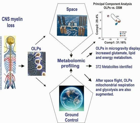
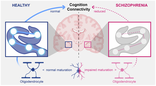
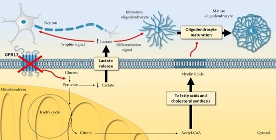
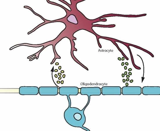
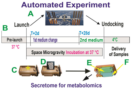
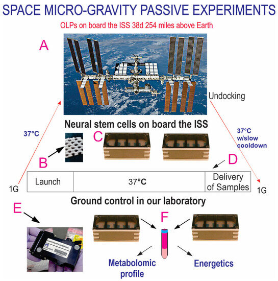
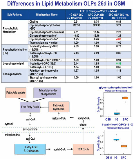
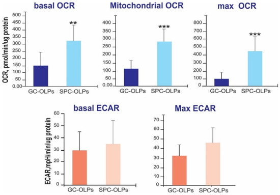
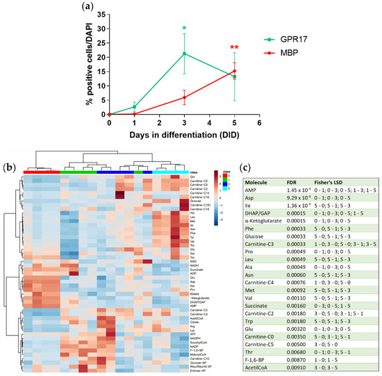
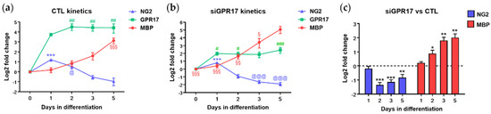
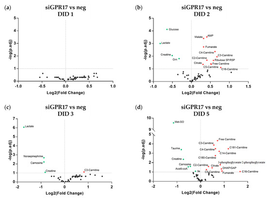
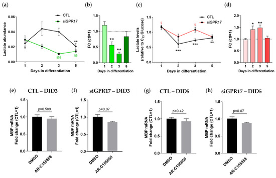
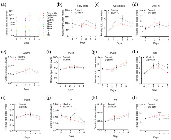
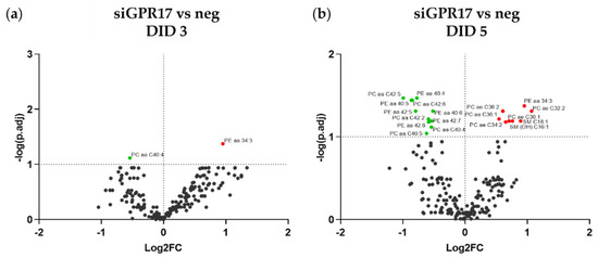
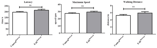
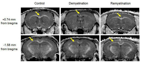
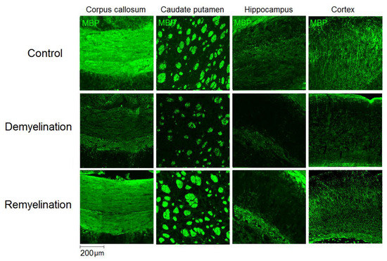
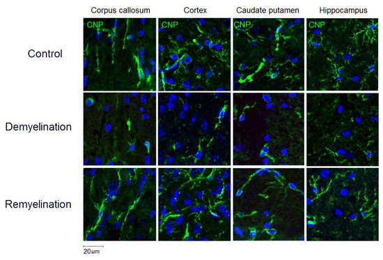
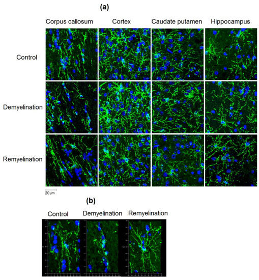
Intracranial hypertension (ICP) and visual impairment intracranial pressure (VIIP) are some of the sequels of long-term space missions. Here we sought to determine how space microgravity (µG) impacts the metabolomics profile of oligodendrocyte progenitors (OLPs), the myelin-forming cells in the central nervous system.
[...] Read more.
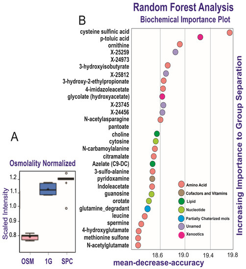
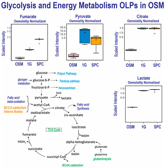
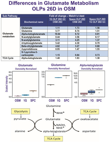
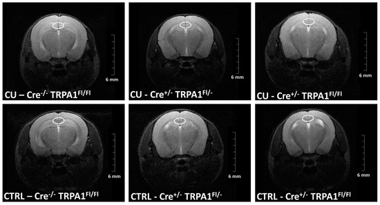
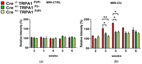
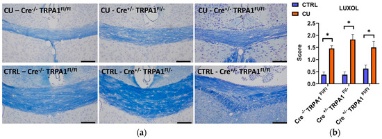
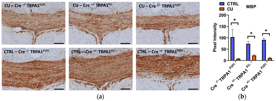
Intracranial hypertension (ICP) and visual impairment intracranial pressure (VIIP) are some of the sequels of long-term space missions. Here we sought to determine how space microgravity (µG) impacts the metabolomics profile of oligodendrocyte progenitors (OLPs), the myelin-forming cells in the central nervous system. We report increased glutamate and energy metabolism while the OLPs were in space for 26 days. We also show that after space flight, OLPs (SPC OLPs) display significantly increased mitochondrial respiration and glycolysis. These data are in agreement with our previous work using simulated microgravity. In addition, our global metabolomics approach allowed for the discovery of endogenous metabolites secreted by OLPs while in space that are significantly modulated by microgravity. Our results provide, for the first time, relevant information about the energetic state of OLPs while in space and after space flight. The functional and molecular relevance of these specific pathways are promising targets for therapeutic intervention for humans in long-term space missions to the moon, Mars and beyond.
Full article
Graphical abstract

