


Cells 2021, 10(12), 3331; https://doi.org/10.3390/cells10123331 - 27 Nov 2021
Cited by 6 | Viewed by 3870
Abstract
►
Show Figures
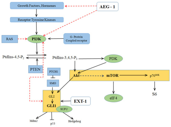
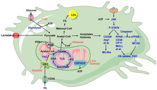
The majority of cardiovascular deaths are associated with acute coronary syndrome, especially ST-elevation myocardial infarction. Therapeutic reperfusion alone can contribute up to 40 percent of total infarct size following coronary artery occlusion, which is called ischemia-reperfusion injury (IRI). Its size depends on many
[...] Read more.
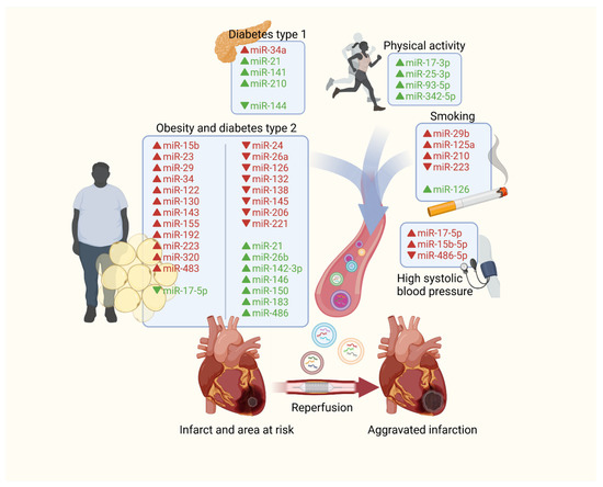
The majority of cardiovascular deaths are associated with acute coronary syndrome, especially ST-elevation myocardial infarction. Therapeutic reperfusion alone can contribute up to 40 percent of total infarct size following coronary artery occlusion, which is called ischemia-reperfusion injury (IRI). Its size depends on many factors, including the main risk factors of cardiovascular mortality, such as age, sex, systolic blood pressure, smoking, and total cholesterol level as well as obesity, diabetes, and physical effort. Extracellular vesicles (EVs) are membrane-coated particles released by every type of cell, which can carry content that affects the functioning of other tissues. Their role is essential in the communication between healthy and dysfunctional cells. In this article, data on the variability of the content of EVs in patients with the most prevalent cardiovascular risk factors is presented, and their influence on IRI is discussed.
Full article
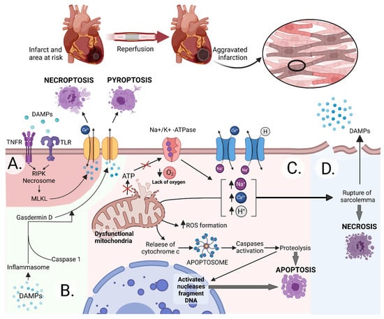
Figure 1


