


Cells 2024, 13(6), 502; https://doi.org/10.3390/cells13060502 - 13 Mar 2024
Viewed by 761
Abstract
►
Show Figures
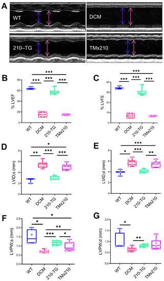
Heart failure with preserved ejection fraction (HFpEF) is associated with exercise intolerance due to alterations in the skeletal muscle (SKM). Leucine supplementation is known to alter the anabolic/catabolic balance and to improve mitochondrial function. Thus, we investigated the effect of leucine supplementation in
[...] Read more.
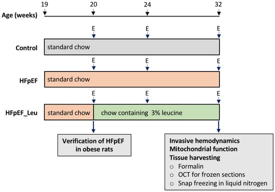
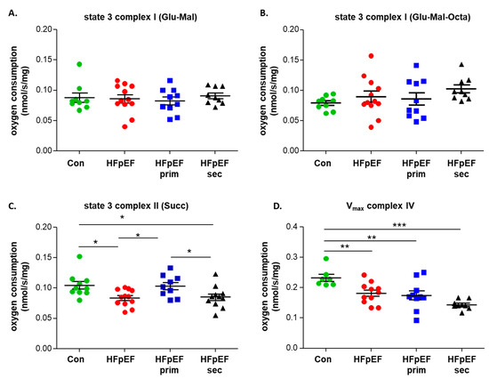
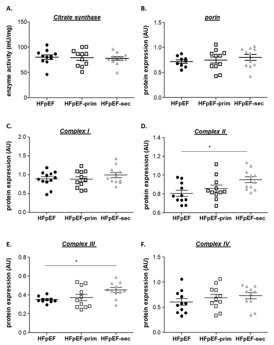
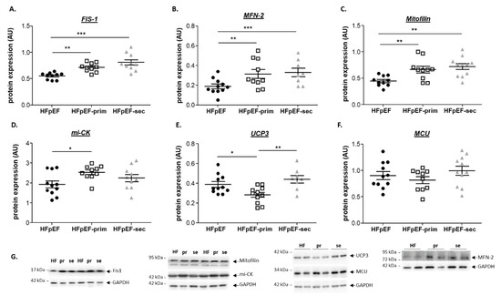
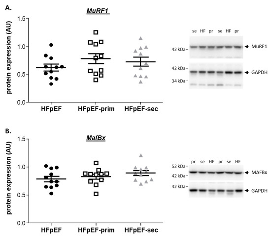
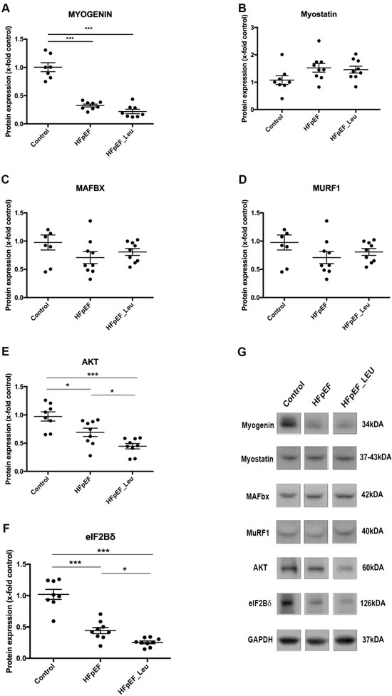
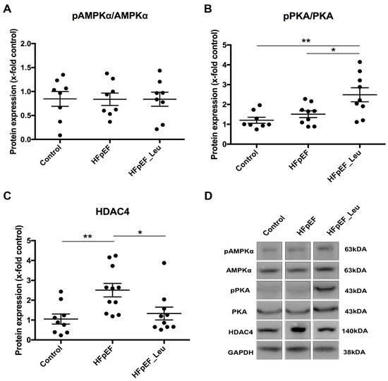
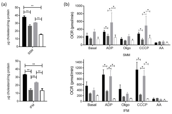
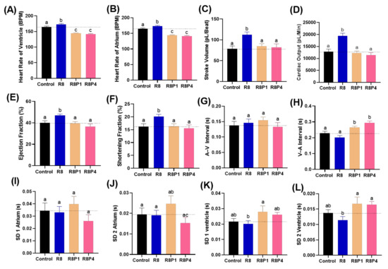
Heart failure with preserved ejection fraction (HFpEF) is associated with exercise intolerance due to alterations in the skeletal muscle (SKM). Leucine supplementation is known to alter the anabolic/catabolic balance and to improve mitochondrial function. Thus, we investigated the effect of leucine supplementation in both a primary and a secondary prevention approach on SKM function and factors modulating muscle function in an established HFpEF rat model. Female ZSF1 obese rats were randomized to an untreated, a primary prevention, and a secondary prevention group. For primary prevention, leucine supplementation was started before the onset of HFpEF (8 weeks of age) and for secondary prevention, leucine supplementation was started after the onset of HFpEF (20 weeks of age). SKM function was assessed at an age of 32 weeks, and SKM tissue was collected for the assessment of mitochondrial function and histological and molecular analyses. Leucine supplementation prevented the development of SKM dysfunction whereas it could not reverse it. In the primary prevention group, mitochondrial function improved and higher expressions of mitofilin, Mfn-2, Fis1, and miCK were evident in SKM. The expression of UCP3 was reduced whereas the mitochondrial content and markers for catabolism (MuRF1, MAFBx), muscle cross-sectional area, and SKM mass did not change. Our data show that leucine supplementation prevented the development of skeletal muscle dysfunction in a rat model of HFpEF, which may be mediated by improving mitochondrial function through modulating energy transfer.
Full article
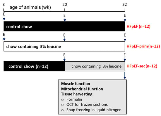
Figure 1


