


Cells 2023, 12(15), 1987; https://doi.org/10.3390/cells12151987 - 02 Aug 2023
Viewed by 1342
Abstract
►
Show Figures
Endothelin-1 (ET-1) overactivity has been implicated as a factor contributing to glaucomatous neuropathy, and it has been utilized in animal models of retinal ischemia. The functional effects of long-term ET-1 exposure and possible compensatory mechanisms have, however, not been investigated. This was therefore
[...] Read more.
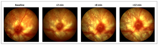
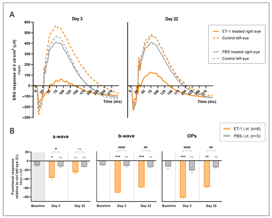
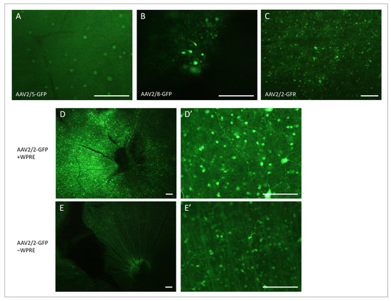
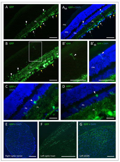
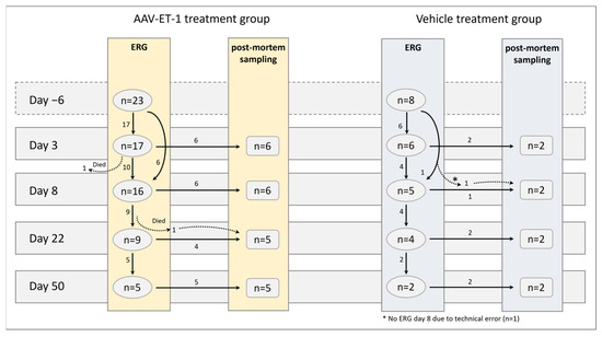
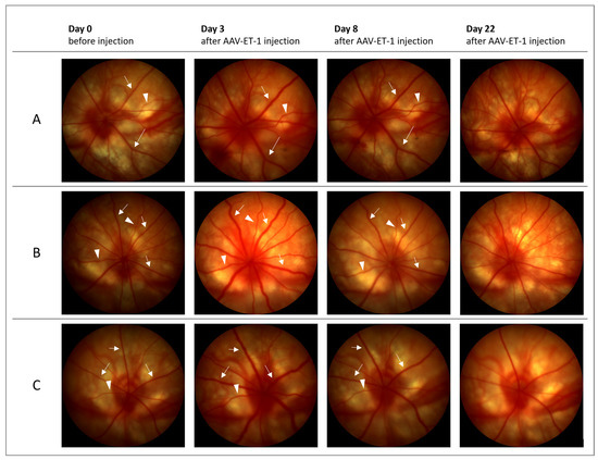
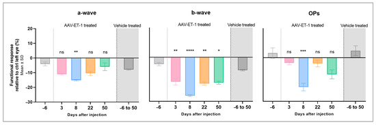
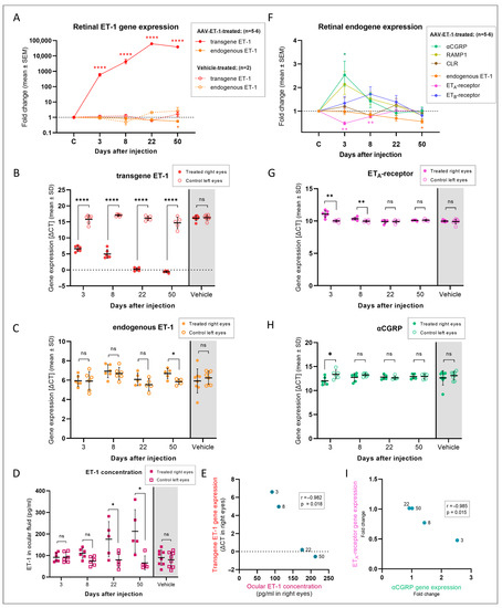
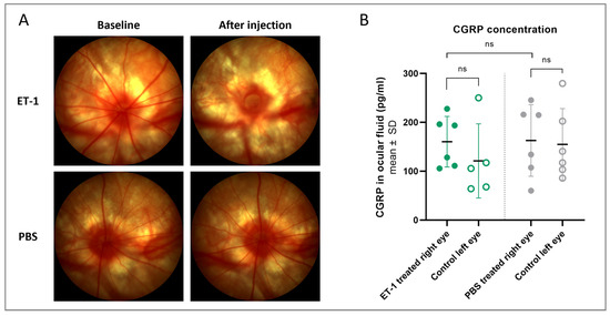
Endothelin-1 (ET-1) overactivity has been implicated as a factor contributing to glaucomatous neuropathy, and it has been utilized in animal models of retinal ischemia. The functional effects of long-term ET-1 exposure and possible compensatory mechanisms have, however, not been investigated. This was therefore the purpose of our study. ET-1 was delivered into rat eyes via a single intravitreal injection of 500 µM or via transgene delivery using an adeno-associated viral (AAV) vector. Retinal function was assessed using electroretinography (ERG) and the retinal expression of potentially compensatory genes was evaluated by means of qRT-PCR. Acute ET-1 delivery led to vasoconstriction and a significant reduction in the ERG response. AAV–ET-1 resulted in substantial transgene expression and ERG results similar to the acute ET-1 injections and comparable to other models of retinal ischemia. Compensatory changes were observed, including an increase in calcitonin gene-related peptide (CGRP) gene expression, which may both counterbalance the vasoconstrictive effects of ET-1 and provide neuroprotection. This chronic ET-1 ischemia model might be especially relevant to glaucoma research, mimicking the mild and repeated ischemic events in patients with long-term vascular dysfunction. The compensatory mechanisms, and particularly the role of vasodilatory CGRP in mitigating the retinal damage, warrant further investigation with the aim of evaluating new therapeutic strategies.
Full article
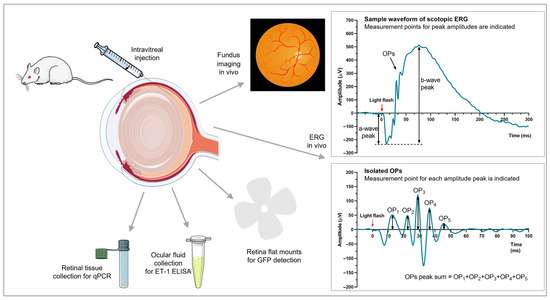
Figure 1

