The Pathogenesis of Neurological Disorders

A topical collection in Cells (ISSN 2073-4409). This collection belongs to the section "Cells of the Nervous System".

Viewed by 31167

Editor


E-Mail Website
Collection Editor
Department of Neurology, College of Medicine and Department of Physical Therapy, College of Health Professions, University of Tennessee Health Science Center, Memphis, TN, USA
Interests: Alzheimer’s disease; Parkinson’s disease; stroke; DNA damage; neuroinflammation; oxidative stress and antioxidants; regulation of motor function; gut microbiota in neurological diseases; long noncoding RNAs and CNS in regulation of muscle weakness
Special Issues, Collections and Topics in MDPI journals

Topical Collection Information

Dear Colleagues,

Neurological disorders are the leading cause of disability and second major cause of death worldwide. Currently, there are no therapeutic interventions that prevent or slow the progression of neurological disorders. The onset of neurological diseases mainly occurs in mid- to late life, so the prevalence is expected to increase as the population ages. Knowledge of the precise molecular and cellular mechanisms underlying neurodegeneration remains incomplete, and these gaps in our knowledge of fundamental neurobiology are a major barrier to therapeutic discovery. Thus, there is an unmet need to develop new and more effective therapeutic strategies that provide a higher quality of life for individuals affected with neurological disorders, which will only be achieved with a better understanding of the disease mechanisms.

During the past several decades, a growing body of research on several aspects has increased our understanding of neurological disorders. The aggregation and deposition of specific proteins is hypothesized to underlie several neurological disorders. However, the mechanistic connection between the process of protein aggregation and neuronal loss is not yet fully understood. Our knowledge of the role of oxidative stress, neuroinflammation, and neuro-immune interactions in the pathogenesis of neurological disorders has increased over the past several decades, but therapeutic and diagnostic interventions using this knowledge have not been widely implemented. For decades, the pathogenesis of neurlogical disorders has mostly been studied in the context of the brain; however, the contribution of peripheral influences in the development and function is largely unexplored. Alteration in gut microbiome composition, often referred to as gut dysbiosis, has recently been suggested to contribute to the pathogenesis of several neurological disorders. Similarly, alterations in the expressions of noncoding, RNAs especially microRNAs, have been extensively studied in neurological disorder; however, the functional role of lncRNAs in neurological disorders is still not understood well.

This Topical Collection shall highlight the global research efforts aimed at exploring causes, identifying disease biomarkers, and strategies/therapies that can attenuate disease symptoms and reduce the social and economic impact of neurological disorders. For this Topical Collection, we invite articles from researchers addressing their findings on novel mechanisms involving neuroinflammation, oxidative stress, gut–brain interaction, and lncRNAs in neurological disorders. Additionally, we hope to attract studies aimed at discovering disease biomarkers for early diagnosis and novel therapeutic approaches or strategies to prevent or strop the progression of neurological disorders.

Dr. Mohammad Moshahid Khan
Collection Editor

Manuscript Submission Information

Manuscripts should be submitted online at www.mdpi.com by registering and logging in to this website. Once you are registered, click here to go to the submission form. Manuscripts can be submitted until the deadline. All submissions that pass pre-check are peer-reviewed. Accepted papers will be published continuously in the journal (as soon as accepted) and will be listed together on the collection website. Research articles, review articles as well as short communications are invited. For planned papers, a title and short abstract (about 100 words) can be sent to the Editorial Office for announcement on this website.

Submitted manuscripts should not have been published previously, nor be under consideration for publication elsewhere (except conference proceedings papers). All manuscripts are thoroughly refereed through a single-blind peer-review process. A guide for authors and other relevant information for submission of manuscripts is available on the Instructions for Authors page. Cells is an international peer-reviewed open access semimonthly journal published by MDPI.

Please visit the Instructions for Authors page before submitting a manuscript. The Article Processing Charge (APC) for publication in this open access journal is 2700 CHF (Swiss Francs). Submitted papers should be well formatted and use good English. Authors may use MDPI's English editing service prior to publication or during author revisions.

Keywords

  • neurodegeneration
  • neuroinflammation
  • oxidative stress
  • lncRNAs
  • brain damage
  • gut–brain axis
  • neuroprotection

Published Papers (10 papers)

2023

Jump to: 2022, 2021, 2020

12 pages, 3749 KiB  
Article
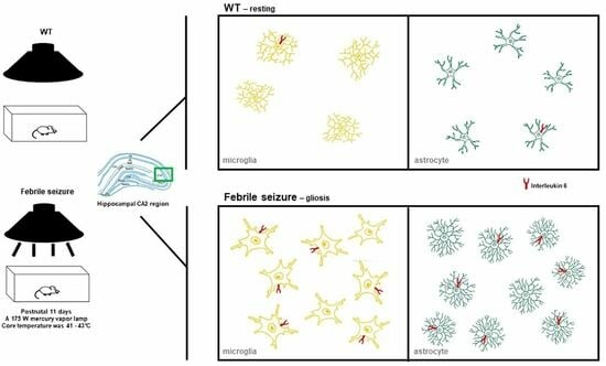
Febrile Seizure Causes Deficit in Social Novelty, Gliosis, and Proinflammatory Cytokine Response in the Hippocampal CA2 Region in Rats
by Yeon Hee Yu, Seong-Wook Kim, Hyuna Im, Yu Ran Lee, Gun Woo Kim, Seongho Ryu, Dae-Kyoon Park and Duk-Soo Kim
Cells 2023, 12(20), 2446; https://doi.org/10.3390/cells12202446 - 13 Oct 2023
Viewed by 847
Abstract
Febrile seizure (FS), which occurs as a response to fever, is the most common seizure that occurs in infants and young children. FS is usually accompanied by diverse neuropsychiatric symptoms, including impaired social behaviors; however, research on neuropsychiatric disorders and hippocampal inflammatory changes [...] Read more.
Febrile seizure (FS), which occurs as a response to fever, is the most common seizure that occurs in infants and young children. FS is usually accompanied by diverse neuropsychiatric symptoms, including impaired social behaviors; however, research on neuropsychiatric disorders and hippocampal inflammatory changes following febrile seizure occurrences is very limited. Here, we provide evidence linking FS occurrence with ASD pathogenesis in rats. We developed an FS juvenile rats model and found ASD-like abnormal behaviors including deficits in social novelty, repetitive behaviors, and hyperlocomotion. In addition, FS model juvenile rats showed enhanced levels of gliosis and inflammation in the hippocampal CA2 region and cerebellum. Furthermore, abnormal levels of social and repetitive behaviors persisted in adults FS model rats. These findings suggest that the inflammatory response triggered by febrile seizures in young children could potentially serve as a mediator of social cognitive impairments. Full article
Show Figures

Graphical abstract

32 pages, 5947 KiB  
Article
Impaired Insulin Signaling Alters Mediators of Hippocampal Synaptic Dynamics/Plasticity: A Possible Mechanism of Hyperglycemia-Induced Cognitive Impairment
by Mubeen A. Ansari, Aishah Al-Jarallah and Fawzi A. Babiker
Cells 2023, 12(13), 1728; https://doi.org/10.3390/cells12131728 - 27 Jun 2023
Viewed by 1156
Abstract
Alzheimer’s disease (AD) is a neurological condition that affects the elderly and is characterized by progressive and irreversible neurodegeneration in the cerebral cortex [...] Full article
Show Figures

Figure 1

2022

Jump to: 2023, 2021, 2020

20 pages, 1889 KiB  
Review
Role of Endogenous Lipopolysaccharides in Neurological Disorders
by Manjunath Kalyan, Ahmed Hediyal Tousif, Sharma Sonali, Chandrasekaran Vichitra, Tuladhar Sunanda, Sankar Simla Praveenraj, Bipul Ray, Vasavi Rakesh Gorantla, Wiramon Rungratanawanich, Arehally M. Mahalakshmi, M. Walid Qoronfleh, Tanya M. Monaghan, Byoung-Joon Song, Musthafa Mohamed Essa and Saravana Babu Chidambaram
Cells 2022, 11(24), 4038; https://doi.org/10.3390/cells11244038 - 14 Dec 2022
Cited by 22 | Viewed by 3929
Abstract
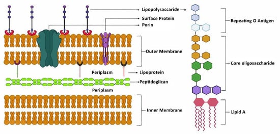
Lipopolysaccharide (LPS) is a cell-wall immunostimulatory endotoxin component of Gram-negative bacteria. A growing body of evidence reveals that alterations in the bacterial composition of the intestinal microbiota (gut dysbiosis) disrupt host immune homeostasis and the intestinal barrier function. Microbial dysbiosis leads to a [...] Read more.
Lipopolysaccharide (LPS) is a cell-wall immunostimulatory endotoxin component of Gram-negative bacteria. A growing body of evidence reveals that alterations in the bacterial composition of the intestinal microbiota (gut dysbiosis) disrupt host immune homeostasis and the intestinal barrier function. Microbial dysbiosis leads to a proinflammatory milieu and systemic endotoxemia, which contribute to the development of neurodegenerative diseases and metabolic disorders. Two important pathophysiological hallmarks of neurodegenerative diseases (NDDs) are oxidative/nitrative stress and inflammation, which can be initiated by elevated intestinal permeability, with increased abundance of pathobionts. These changes lead to excessive release of LPS and other bacterial products into blood, which in turn induce chronic systemic inflammation, which damages the blood–brain barrier (BBB). An impaired BBB allows the translocation of potentially harmful bacterial products, including LPS, and activated neutrophils/leucocytes into the brain, which results in neuroinflammation and apoptosis. Chronic neuroinflammation causes neuronal damage and synaptic loss, leading to memory impairment. LPS-induced inflammation causes inappropriate activation of microglia, astrocytes, and dendritic cells. Consequently, these alterations negatively affect mitochondrial function and lead to increases in oxidative/nitrative stress and neuronal senescence. These cellular changes in the brain give rise to specific clinical symptoms, such as impairment of locomotor function, muscle weakness, paralysis, learning deficits, and dementia. This review summarizes the contributing role of LPS in the development of neuroinflammation and neuronal cell death in various neurodegenerative diseases. Full article
Show Figures

Figure 1

11 pages, 1029 KiB  
Article
Ischemic Stroke Disrupts Sleep Homeostasis in Middle-Aged Mice
by Rishi Sharma, Abigail Chischolm, Meet Parikh, Adnan I. Qureshi, Pradeep Sahota and Mahesh M. Thakkar
Cells 2022, 11(18), 2818; https://doi.org/10.3390/cells11182818 - 09 Sep 2022
Cited by 3 | Viewed by 2193
Abstract
Sleep disturbances, including insomnia and excessive daytime sleepiness, are highly prevalent in patients with ischemic stroke (IS), which severely impacts recovery and rehabilitation efforts. However, how IS induces sleep disturbances is unclear. Three experiments were performed on middle-aged C57BL/6J mice, instrumented with sleep [...] Read more.
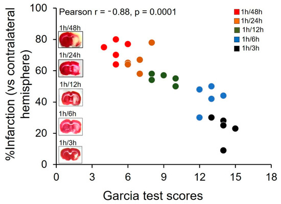
Sleep disturbances, including insomnia and excessive daytime sleepiness, are highly prevalent in patients with ischemic stroke (IS), which severely impacts recovery and rehabilitation efforts. However, how IS induces sleep disturbances is unclear. Three experiments were performed on middle-aged C57BL/6J mice, instrumented with sleep recording electrodes and/or subjected to 1 h of middle cerebral artery (MCAO; Stroke group) or sham (Sham group) occlusion to induce IS. After 48 h of reperfusion (a) experiment 1 verified sensorimotor deficit (using Garcia scale) and infarction (using TTC staining) in this mouse model; (b) experiment 2 examined the effects of IS on the quality (sleep latency and NREM delta power) and quantity (duration) of sleep; and (c) experiment 3 determined the effects of IS on sleep homeostasis using sleep deprivation (SD) and recovery sleep (RS) paradigm. Stroke mice display (a) a significant correlation between sensorimotor deficit and cerebral infarction; (b) insomnia-like symptoms (increased sleep latency, reduced NREM duration and delta power) during the light (inactive) period and daytime sleepiness-like symptoms during the dark (active) period mimicking sleep in IS patients; and (c) impairments in the markers of sleep pressure (during SD) and sleep dissipation (during RS). Our results suggest that IS disrupts sleep homeostasis to cause sleep disturbances. Full article
Show Figures

Figure 1

2021

Jump to: 2023, 2022, 2020

23 pages, 7206 KiB  
Article
The Parkinson’s Disease-Associated Protein DJ-1 Protects Dictyostelium Cells from AMPK-Dependent Outcomes of Oxidative Stress
by Suwei Chen, Sarah J. Annesley, Rasha A. F. Jasim and Paul R. Fisher
Cells 2021, 10(8), 1874; https://doi.org/10.3390/cells10081874 - 23 Jul 2021
Cited by 7 | Viewed by 2047
Abstract
Mitochondrial dysfunction has been implicated in the pathology of Parkinson’s disease (PD). In Dictyostelium discoideum, strains with mitochondrial dysfunction present consistent, AMPK-dependent phenotypes. This provides an opportunity to investigate if the loss of function of specific PD-associated genes produces cellular pathology by [...] Read more.
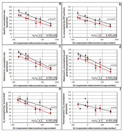
Mitochondrial dysfunction has been implicated in the pathology of Parkinson’s disease (PD). In Dictyostelium discoideum, strains with mitochondrial dysfunction present consistent, AMPK-dependent phenotypes. This provides an opportunity to investigate if the loss of function of specific PD-associated genes produces cellular pathology by causing mitochondrial dysfunction with AMPK-mediated consequences. DJ-1 is a PD-associated, cytosolic protein with a conserved oxidizable cysteine residue that is important for the protein’s ability to protect cells from the pathological consequences of oxidative stress. Dictyostelium DJ-1 (encoded by the gene deeJ) is located in the cytosol from where it indirectly inhibits mitochondrial respiration and also exerts a positive, nonmitochondrial role in endocytosis (particularly phagocytosis). Its loss in unstressed cells impairs endocytosis and causes correspondingly slower growth, while also stimulating mitochondrial respiration. We report here that oxidative stress in Dictyostelium cells inhibits mitochondrial respiration and impairs phagocytosis in an AMPK-dependent manner. This adds to the separate impairment of phagocytosis caused by DJ-1 knockdown. Oxidative stress also combines with DJ-1 loss in an AMPK-dependent manner to impair or exacerbate defects in phototaxis, morphogenesis and growth. It thereby phenocopies mitochondrial dysfunction. These results support a model in which the oxidized but not the reduced form of DJ-1 inhibits AMPK in the cytosol, thereby protecting cells from the adverse consequences of oxidative stress, mitochondrial dysfunction and the resulting AMPK hyperactivity. Full article
Show Figures

Figure 1

17 pages, 354 KiB  
Review
Genetic and Transcriptomic Biomarkers in Neurodegenerative Diseases: Current Situation and the Road Ahead
by Julie Lake, Catherine S. Storm, Mary B. Makarious and Sara Bandres-Ciga
Cells 2021, 10(5), 1030; https://doi.org/10.3390/cells10051030 - 27 Apr 2021
Cited by 12 | Viewed by 3181
Abstract
Neurodegenerative diseases are etiologically and clinically heterogeneous conditions, often reflecting a spectrum of disease rather than well-defined disorders. The underlying molecular complexity of these diseases has made the discovery and validation of useful biomarkers challenging. The search of characteristic genetic and transcriptomic indicators [...] Read more.
Neurodegenerative diseases are etiologically and clinically heterogeneous conditions, often reflecting a spectrum of disease rather than well-defined disorders. The underlying molecular complexity of these diseases has made the discovery and validation of useful biomarkers challenging. The search of characteristic genetic and transcriptomic indicators for preclinical disease diagnosis, prognosis, or subtyping is an area of ongoing effort and interest. The next generation of biomarker studies holds promise by implementing meaningful longitudinal and multi-modal approaches in large scale biobank and healthcare system scale datasets. This work will only be possible in an open science framework. This review summarizes the current state of genetic and transcriptomic biomarkers in Parkinson’s disease, Alzheimer’s disease, and amyotrophic lateral sclerosis, providing a comprehensive landscape of recent literature and future directions. Full article
23 pages, 7548 KiB  
Article
Traumatic Optic Neuropathy Is Associated with Visual Impairment, Neurodegeneration, and Endoplasmic Reticulum Stress in Adolescent Mice
by Shelby M. Hetzer, Fernanda Guilhaume-Correa, Dylan Day, Alicia Bedolla and Nathan K. Evanson
Cells 2021, 10(5), 996; https://doi.org/10.3390/cells10050996 - 23 Apr 2021
Cited by 11 | Viewed by 3320
Abstract
Traumatic brain injury (TBI) results in a number of impairments, often including visual symptoms. In some cases, visual impairments after head trauma are mediated by traumatic injury to the optic nerve, termed traumatic optic neuropathy (TON), which has few effective options for treatment. [...] Read more.
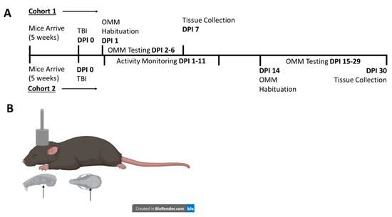
Traumatic brain injury (TBI) results in a number of impairments, often including visual symptoms. In some cases, visual impairments after head trauma are mediated by traumatic injury to the optic nerve, termed traumatic optic neuropathy (TON), which has few effective options for treatment. Using a murine closed-head weight-drop model of head trauma, we previously reported in adult mice that there is relatively selective injury to the optic tract and thalamic/brainstem projections of the visual system. In the current study, we performed blunt head trauma on adolescent C57BL/6 mice and investigated visual impairment in the primary visual system, now including the retina and using behavioral and histologic methods at new time points. After injury, mice displayed evidence of decreased optomotor responses illustrated by decreased optokinetic nystagmus. There did not appear to be a significant change in circadian locomotor behavior patterns, although there was an overall decrease in locomotor behavior in mice with head injury. There was evidence of axonal degeneration of optic nerve fibers with associated retinal ganglion cell death. There was also evidence of astrogliosis and microgliosis in major central targets of optic nerve projections. Further, there was elevated expression of endoplasmic reticulum (ER) stress markers in retinas of injured mice. Visual impairment, histologic markers of gliosis and neurodegeneration, and elevated ER stress marker expression persisted for at least 30 days after injury. The current results extend our previous findings in adult mice into adolescent mice, provide direct evidence of retinal ganglion cell injury after head trauma and suggest that axonal degeneration is associated with elevated ER stress in this model of TON. Full article
Show Figures

Figure 1

17 pages, 3151 KiB  
Article
Alterations in the Gut-Microbial-Inflammasome-Brain Axis in a Mouse Model of Alzheimer’s Disease
by Pradeep K. Shukla, David F. Delotterie, Jianfeng Xiao, Joseph F. Pierre, RadhaKrishna Rao, Michael P. McDonald and Mohammad Moshahid Khan
Cells 2021, 10(4), 779; https://doi.org/10.3390/cells10040779 - 01 Apr 2021
Cited by 47 | Viewed by 5933
Abstract
Alzheimer’s disease (AD), a progressive neurodegenerative disorder characterized by memory loss and cognitive decline, is a major cause of death and disability among the older population. Despite decades of scientific research, the underlying etiological triggers are unknown. Recent studies suggested that gut microbiota [...] Read more.
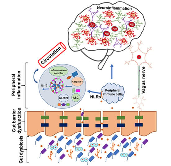
Alzheimer’s disease (AD), a progressive neurodegenerative disorder characterized by memory loss and cognitive decline, is a major cause of death and disability among the older population. Despite decades of scientific research, the underlying etiological triggers are unknown. Recent studies suggested that gut microbiota can influence AD progression; however, potential mechanisms linking the gut microbiota with AD pathogenesis remain obscure. In the present study, we provided a potential mechanistic link between dysbiotic gut microbiota and neuroinflammation associated with AD progression. Using a mouse model of AD, we discovered that unfavorable gut microbiota are correlated with abnormally elevated expression of gut NLRP3 and lead to peripheral inflammasome activation, which in turn exacerbates AD-associated neuroinflammation. To this end, we observe significantly altered gut microbiota compositions in young and old 5xFAD mice compared to age-matched non-transgenic mice. Moreover, 5xFAD mice demonstrated compromised gut barrier function as evident from the loss of tight junction and adherens junction proteins compared to non-transgenic mice. Concurrently, we observed increased expression of NLRP3 inflammasome and IL-1β production in the 5xFAD gut. Consistent with our hypothesis, increased gut–microbial–inflammasome activation is positively correlated with enhanced astrogliosis and microglial activation, along with higher expression of NLRP3 inflammasome and IL-1β production in the brains of 5xFAD mice. These data indicate that the elevated expression of gut–microbial–inflammasome components may be an important trigger for subsequent downstream activation of inflammatory and potentially cytotoxic mediators, and gastrointestinal NLRP3 may promote NLRP3 inflammasome-mediated neuroinflammation. Thus, modulation of the gut microbiota may be a potential strategy for the treatment of AD-related neurological disorders in genetically susceptible hosts. Full article
Show Figures

Graphical abstract

19 pages, 3085 KiB  
Article
Alteration of Neural Stem Cell Functions in Ataxia and Male Sterility Mice: A Possible Role of β-Tubulin Glutamylation in Neurodegeneration
by Abdullah Md. Sheikh, Shozo Yano, Shatera Tabassum, Koji Omura, Asuka Araki, Shingo Mitaki, Yoshie Ito, Shuai Huang and Atsushi Nagai
Cells 2021, 10(1), 155; https://doi.org/10.3390/cells10010155 - 14 Jan 2021
Cited by 6 | Viewed by 2754
Abstract
Ataxia and Male Sterility (AMS) is a mutant mouse strain that contains a missense mutation in the coding region of Nna1, a gene that encodes a deglutamylase. AMS mice exhibit early cerebellar Purkinje cell degeneration and an ataxic phenotype in an autosomal [...] Read more.
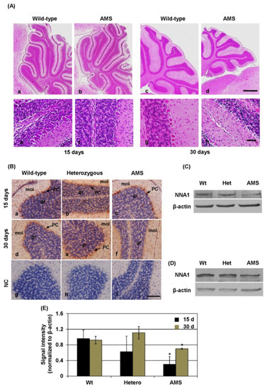
Ataxia and Male Sterility (AMS) is a mutant mouse strain that contains a missense mutation in the coding region of Nna1, a gene that encodes a deglutamylase. AMS mice exhibit early cerebellar Purkinje cell degeneration and an ataxic phenotype in an autosomal recessive manner. To understand the underlying mechanism, we generated neuronal stem cell (NSC) lines from wild-type (NMW7), Nna1 mutation heterozygous (NME), and Nna1 mutation homozygous (NMO1) mouse brains. The NNA1 levels were decreased, and the glutamylated tubulin levels were increased in NMO1 cultures as well as in the cerebellum of AMS mice at both 15 and 30 days of age. However, total β-tubulin protein levels were not altered in the AMS cerebellum. In NMO1 neurosphere cultures, β-tubulin protein levels were increased without changes at the transcriptional level. NMO1 grew faster than other NSC lines, and some of the neurospheres were attached to the plate after 3 days. Immunostaining revealed that SOX2 and nestin levels were decreased in NMO1 neurospheres and that the neuronal differentiation potentials were reduced in NMO1 cells compared to NME or NMW7 cells. These results demonstrate that the AMS mutation decreased the NNA1 levels and increased glutamylation in the cerebellum of AMS mice. The observed changes in glutamylation might alter NSC properties and the neuron maturation process, leading to Purkinje cell death in AMS mice. Full article
Show Figures

Figure 1

2020

Jump to: 2023, 2022, 2021

22 pages, 3627 KiB  
Article
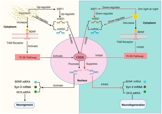
Dim Light at Night Induced Neurodegeneration and Ameliorative Effect of Curcumin
by Dhondup Namgyal, Kumari Chandan, Armiya Sultan, Mehreen Aftab, Sher Ali, Rachna Mehta, Hamed A. El-Serehy, Fahad A. Al-Misned and Maryam Sarwat
Cells 2020, 9(9), 2093; https://doi.org/10.3390/cells9092093 - 13 Sep 2020
Cited by 22 | Viewed by 3631
Abstract
It is a well-known fact that following a proper routine light/dark or diurnal rhythm controls almost all biological processes. With the introduction of modern lighting and artificial illumination systems, continuous exposure to light at night may lead to the disruption of diurnal rhythm. [...] Read more.
It is a well-known fact that following a proper routine light/dark or diurnal rhythm controls almost all biological processes. With the introduction of modern lighting and artificial illumination systems, continuous exposure to light at night may lead to the disruption of diurnal rhythm. However, the effect of light during the night on brain anatomy, physiology, and human body functions is less explored and poorly understood. In this study, we have evaluated the effect of exposure to dim light (5 lux) at night (dLAN) on Swiss Albino mice over a duration of three consecutive weeks. Results have revealed that exposure to dLAN led to an impairment of cognitive and non-cognitive behaviour, oxidative stress–mediated elevation of lipid peroxidation, and reduction of superoxide dismutase and catalase activity. It also led to the downregulation of hippocampal proteins (BDNF, Synapsin II and DCX) at both protein and mRNA level. Additionally, there was downregulation of CREB and SIRT1 mRNAs and neurodegeneration-associated miRNA21a-5p and miRNA34a-5p. The pyramidal and cortical neurons started showing pyknotic and chromatolysis characteristics. However, a dose of curcumin administered to the mice positively modulated these parameters in our experimental animals. We proposed the modulatory role of curcumin in addressing the deleterious effects of dLAN. Full article
Show Figures

Figure 1

Back to TopTop