


Cells 2023, 12(4), 529; https://doi.org/10.3390/cells12040529 - 06 Feb 2023
Cited by 2 | Viewed by 1718
Abstract
►
Show Figures
Progenitor cells isolated from the fetal liver can provide a unique cell source to generate new healthy tissue mass. Almost 20 years ago, it was demonstrated that rat fetal liver cells repopulate the normal host liver environment via a mechanism akin to cell
[...] Read more.
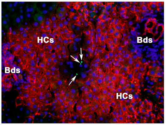
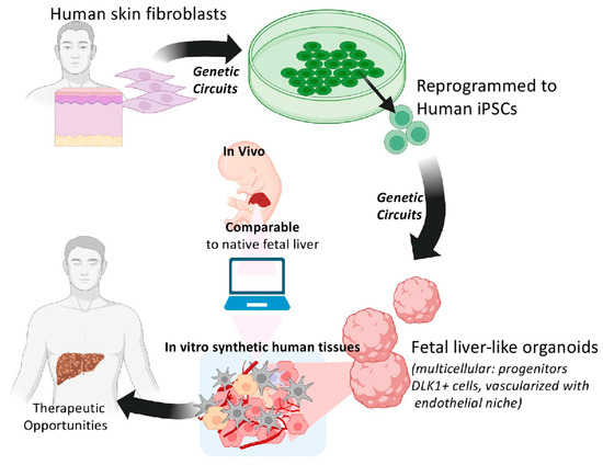
Progenitor cells isolated from the fetal liver can provide a unique cell source to generate new healthy tissue mass. Almost 20 years ago, it was demonstrated that rat fetal liver cells repopulate the normal host liver environment via a mechanism akin to cell competition. Activin A, which is produced by hepatocytes, was identified as an important player during cell competition. Because of reduced activin receptor expression, highly proliferative fetal liver stem/progenitor cells are resistant to activin A and therefore exhibit a growth advantage compared to hepatocytes. As a result, transplanted fetal liver cells are capable of repopulating normal livers. Important for cell-based therapies, hepatic stem/progenitor cells containing repopulation potential can be separated from fetal hematopoietic cells using the cell surface marker δ-like 1 (Dlk-1). In livers with advanced fibrosis, fetal epithelial stem/progenitor cells differentiate into functional hepatic cells and out-compete injured endogenous hepatocytes, which cause anti-fibrotic effects. Although fetal liver cells efficiently repopulate the liver, they will likely not be used for human cell transplantation. Thus, utilizing the underlying mechanism of repopulation and developed methods to produce similar growth-advantaged cells in vitro, e.g., human induced pluripotent stem cells (iPSCs), this approach has great potential for developing novel cell-based therapies in patients with liver disease. The present review gives a brief overview of the classic cell transplantation models and various cell sources studied as donor cell candidates. The advantages of fetal liver-derived stem/progenitor cells are discussed, as well as the mechanism of liver repopulation. Moreover, this article reviews the potential of in vitro developed synthetic human fetal livers from iPSCs and their therapeutic benefits.
Full article
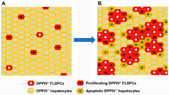
Figure 1