


Cells 2024, 13(4), 300; https://doi.org/10.3390/cells13040300 - 06 Feb 2024
Viewed by 952
Abstract
►
Show Figures
COVID-19, caused by severe acute respiratory syndrome coronavirus-2 (SARS-CoV-2), is characterized by a wide range of clinical symptoms and a poorly predictable disease course. Although in-depth transcriptomic investigations of peripheral blood samples from COVID-19 patients have been performed, the detailed molecular mechanisms underlying
[...] Read more.
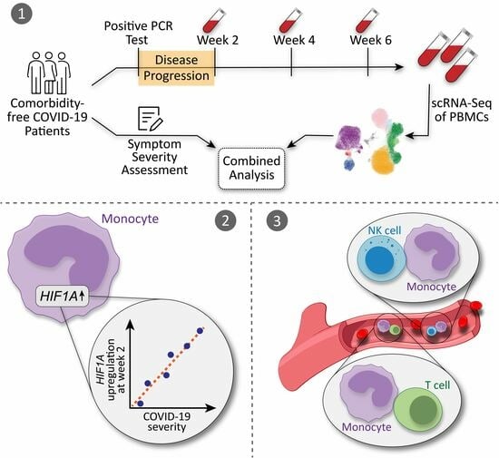
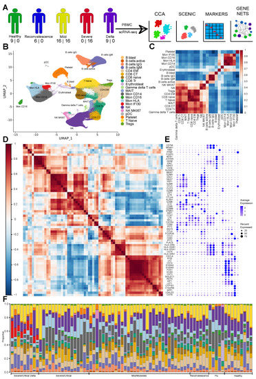
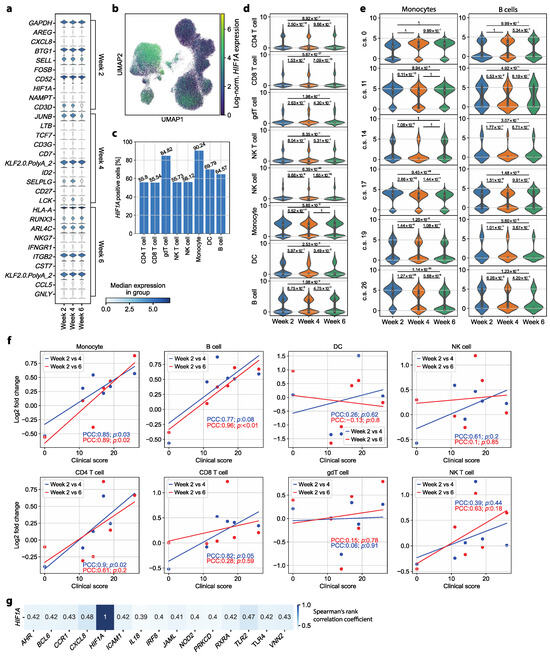
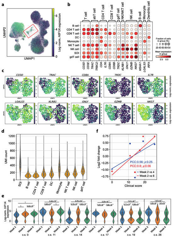
COVID-19, caused by severe acute respiratory syndrome coronavirus-2 (SARS-CoV-2), is characterized by a wide range of clinical symptoms and a poorly predictable disease course. Although in-depth transcriptomic investigations of peripheral blood samples from COVID-19 patients have been performed, the detailed molecular mechanisms underlying an asymptomatic, mild or severe disease course, particularly in patients without relevant comorbidities, remain poorly understood. While previous studies have mainly focused on the cellular and molecular dissection of ongoing COVID-19, we set out to characterize transcriptomic immune cell dysregulation at the single-cell level at different time points in patients without comorbidities after disease resolution to identify signatures of different disease severities in convalescence. With single-cell RNA sequencing, we reveal a role for hypoxia-inducible factor 1-alpha (HIF1A) as a severity-sensitive long-term immunological scar in circulating monocytes of convalescent COVID-19 patients. Additionally, we show that circulating complexes formed by monocytes with either T cells or NK cells represent a characteristic cellular marker in convalescent COVID-19 patients irrespective of their preceding symptom severity. Together, these results provide cellular and molecular correlates of recovery from COVID-19 and could help in immune monitoring and in the design of new treatment strategies.
Full article
Graphical abstract

