


Cells 2023, 12(4), 609; https://doi.org/10.3390/cells12040609 - 13 Feb 2023
Cited by 3 | Viewed by 2061
Abstract
►
Show Figures
Fibroblast growth factor 23 (FGF23) is a phosphaturic hormone produced mainly in osteocytes. In chronic kidney disease (CKD) FGF23 levels increase due to higher production, but also as the result of impaired cleavage and reduced excretion from the body. FGF23 has a significant
[...] Read more.
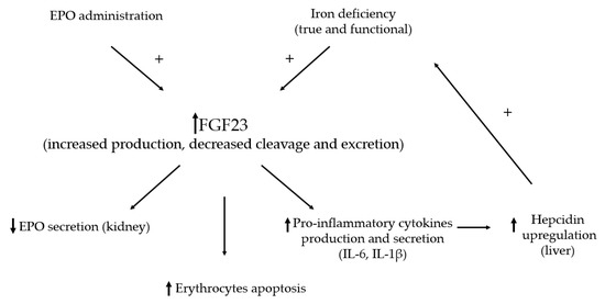
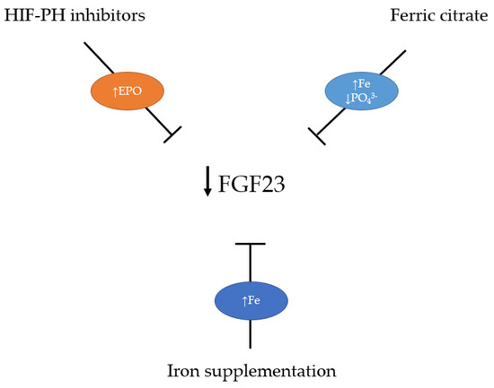
Fibroblast growth factor 23 (FGF23) is a phosphaturic hormone produced mainly in osteocytes. In chronic kidney disease (CKD) FGF23 levels increase due to higher production, but also as the result of impaired cleavage and reduced excretion from the body. FGF23 has a significant role in disturbed bone and mineral metabolism in CKD, which leads to a higher cardiovascular risk and mortality in these patients. Current research has emphasized the expression of FGF23 in cardiac myocytes, fibroblasts, and endothelial cells, and in addition to the effects on the kidney, its primary role is in cardiac remodeling in CKD patients. Recent discoveries found a significant link between increased FGF23 levels and anemia development in CKD. This review describes the FGF23 role in cardiac hypertrophy and anemia in the setting of CKD and discusses the best therapeutical approach for lowering FGF23 levels.
Full article
Figure 1


