


Cells 2023, 12(12), 1599; https://doi.org/10.3390/cells12121599 - 10 Jun 2023
Cited by 7 | Viewed by 2465
Abstract
►
Show Figures
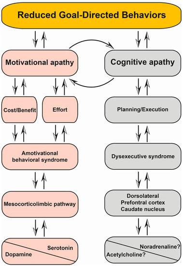
Apathy is commonly defined as a loss of motivation leading to a reduction in goal-directed behaviors. This multidimensional syndrome, which includes cognitive, emotional and behavioral components, is one of the most prevalent neuropsychiatric features of Parkinson’s disease (PD). It has been established that
[...] Read more.
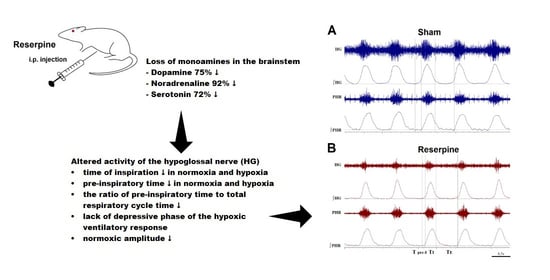
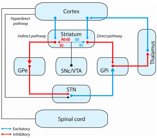
Apathy is commonly defined as a loss of motivation leading to a reduction in goal-directed behaviors. This multidimensional syndrome, which includes cognitive, emotional and behavioral components, is one of the most prevalent neuropsychiatric features of Parkinson’s disease (PD). It has been established that the prevalence of apathy increases as PD progresses. However, the pathophysiology and anatomic substrate of this syndrome remain unclear. Apathy seems to be underpinned by impaired anatomical structures that link the prefrontal cortex with the limbic system. It can be encountered in the prodromal stage of the disease and in fluctuating PD patients receiving bilateral chronic subthalamic nucleus stimulation. In these stages, apathy may be considered as a disorder of motivation that embodies amotivational behavioral syndrome, is underpinned by combined dopaminergic and serotonergic denervation and is dopa-responsive. In contrast, in advanced PD patients, apathy may be considered as cognitive apathy that announces cognitive decline and PD dementia, is underpinned by diffuse neurotransmitter system dysfunction and Lewy pathology spreading and is no longer dopa-responsive. In this review, we discuss the clinical patterns of apathy and their treatment, the neurobiological basis of apathy, the potential role of the anatomical structures involved and the pathways in motivational and cognitive apathy.
Full article
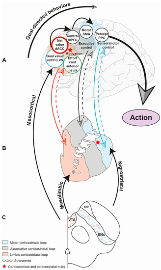
Figure 1


