


Cells 2024, 13(8), 690; https://doi.org/10.3390/cells13080690 - 16 Apr 2024
Viewed by 374
Abstract
►
Show Figures
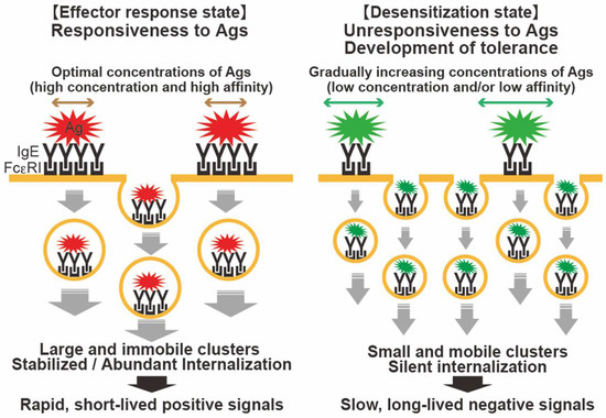
The significant role of mast cells in the development of allergic and inflammatory diseases is well-established. Among the various mechanisms of mast cell activation, the interaction of antigens/allergens with IgE and the subsequent binding of this complex to the high-affinity IgE receptor FcεRI
[...] Read more.
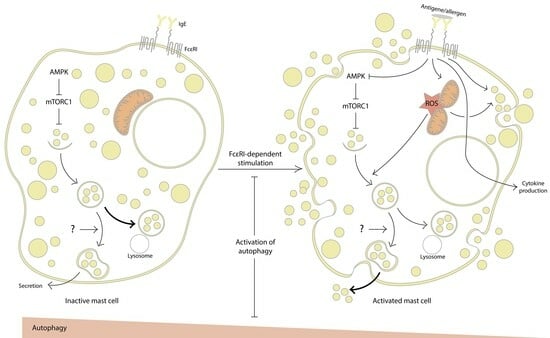
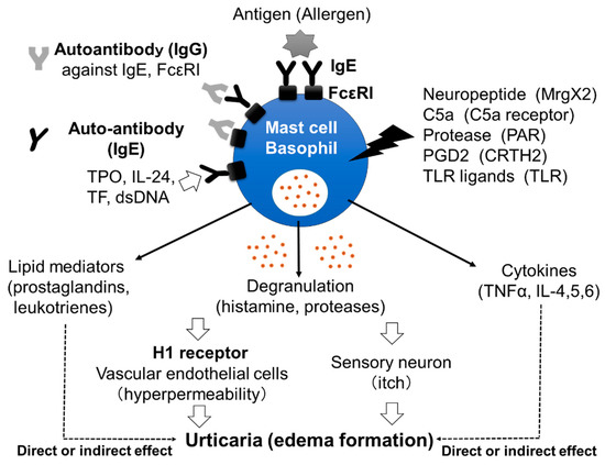
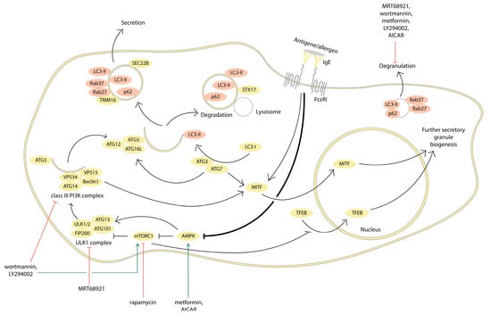
The significant role of mast cells in the development of allergic and inflammatory diseases is well-established. Among the various mechanisms of mast cell activation, the interaction of antigens/allergens with IgE and the subsequent binding of this complex to the high-affinity IgE receptor FcεRI stand out as the most studied and fundamental pathways. This activation process leads to the rapid exocytosis of granules containing preformed mediators, followed by the production of newly synthesized mediators, including a diverse array of cytokines, chemokines, arachidonic acid metabolites, and more. While conventional approaches to allergy control primarily focus on allergen avoidance and the use of antihistamines (despite their associated side effects), there is increasing interest in exploring novel methods to modulate mast cell activity in modern medicine. Recent evidence suggests a role for autophagy in mast cell activation, offering potential avenues for utilizing low-molecular-weight autophagy regulators in the treatment of allergic diseases. More specifically, mitochondria, which play an important role in the regulation of autophagy as well as mast cell activation, emerge as promising targets for drug development. This review examines the existing literature regarding the involvement of the molecular machinery associated with autophagy in FcεRI-dependent mast cell activation.
Full article
Graphical abstract

