


Cells 2023, 12(13), 1766; https://doi.org/10.3390/cells12131766 - 03 Jul 2023
Cited by 11 | Viewed by 3134
Abstract
►
Show Figures
Inflammasome complexes and their integral receptor proteins have essential roles in regulating the innate immune response and inflammation at the post-translational level. Yet despite their protective role, aberrant activation of inflammasome proteins and gain of function mutations in inflammasome component genes seem to
[...] Read more.
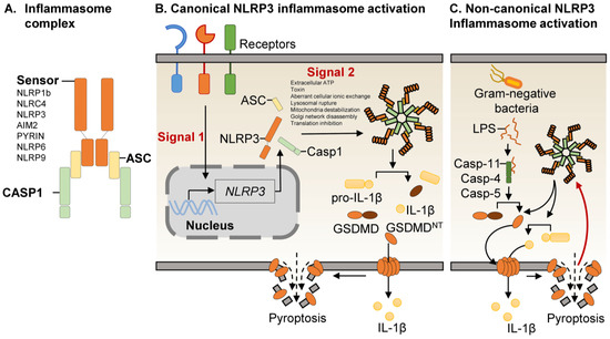
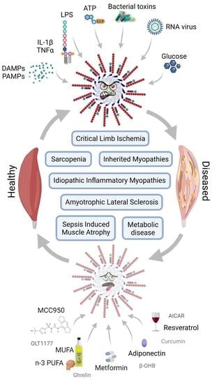
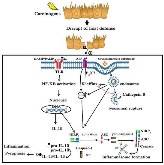
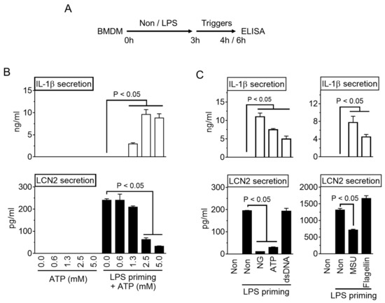
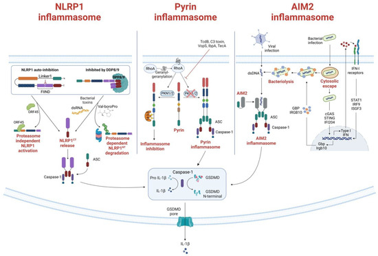
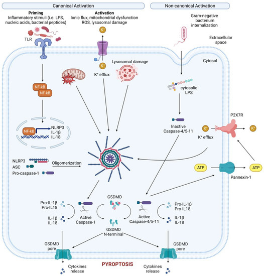
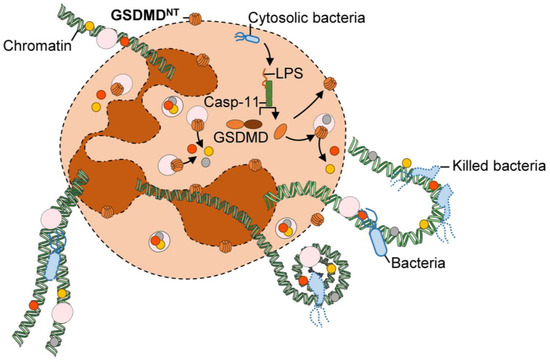
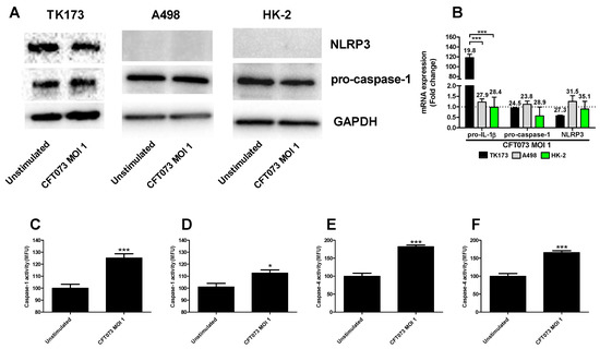
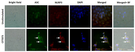
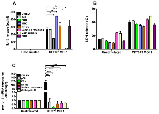
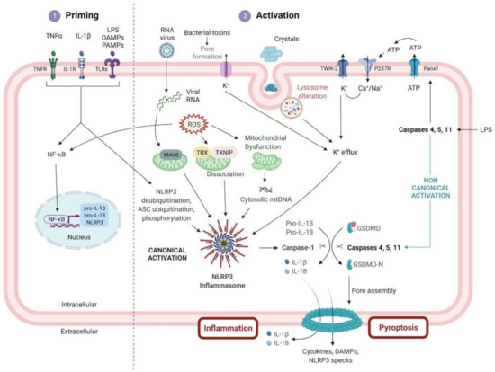
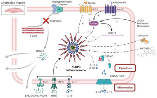
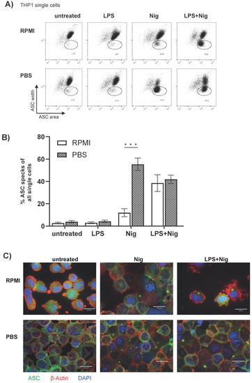
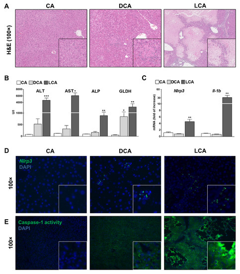
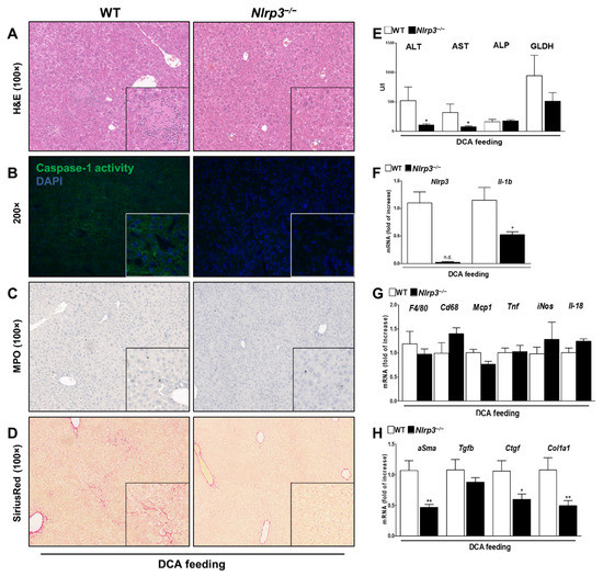
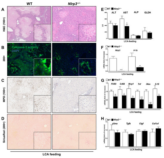
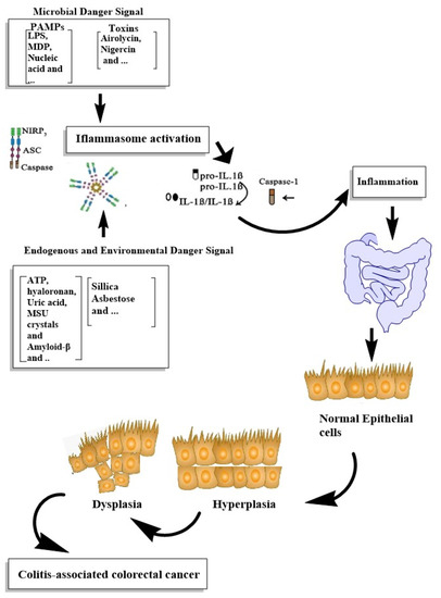
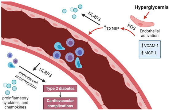
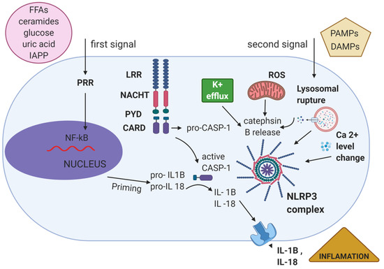
Inflammasome complexes and their integral receptor proteins have essential roles in regulating the innate immune response and inflammation at the post-translational level. Yet despite their protective role, aberrant activation of inflammasome proteins and gain of function mutations in inflammasome component genes seem to contribute to the development and progression of human autoimmune and autoinflammatory diseases. In the past decade, our understanding of inflammasome biology and activation mechanisms has greatly progressed. We therefore provide an up-to-date overview of the various inflammasomes and their known mechanisms of action. In addition, we highlight the involvement of various inflammasomes and their pathogenic mechanisms in common autoinflammatory, autoimmune and neurodegenerative diseases, including atherosclerosis, rheumatoid arthritis, systemic lupus erythematosus, inflammatory bowel disease, Alzheimer’s disease, Parkinson’s disease, and multiple sclerosis. We conclude by speculating on the future avenues of research needed to better understand the roles of inflammasomes in health and disease.
Full article
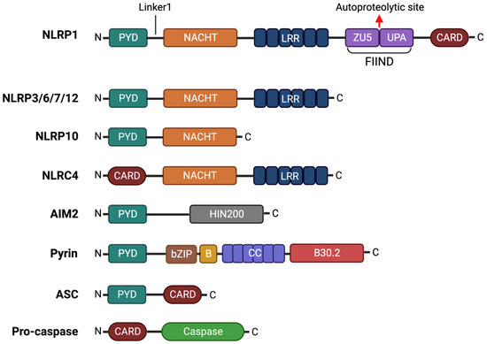
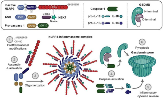
Figure 1
