


Cells 2022, 11(3), 436; https://doi.org/10.3390/cells11030436 - 27 Jan 2022
Cited by 19 | Viewed by 4537
Abstract
►
Show Figures
The hallmarks of Alzheimer’s disease (AD) pathology are senile plaques containing amyloid-beta (Aβ) and neurofibrillary tangles containing hyperphosphorylated tau. Additional pathologies often co-exist, whereas multiple pathogenic mechanisms are involved in AD, especially synaptic degeneration, which necessitate the need for synaptic integrity-related biomarkers alongside
[...] Read more.
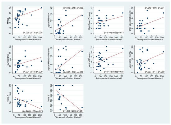
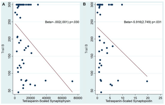
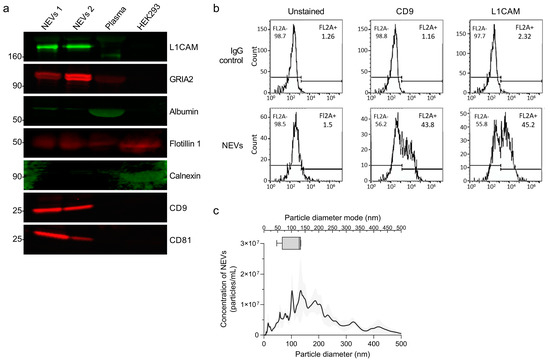
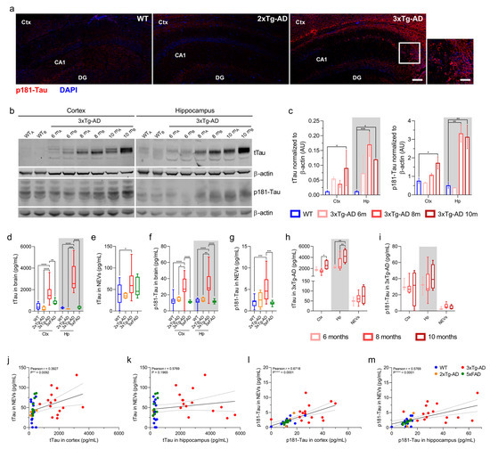
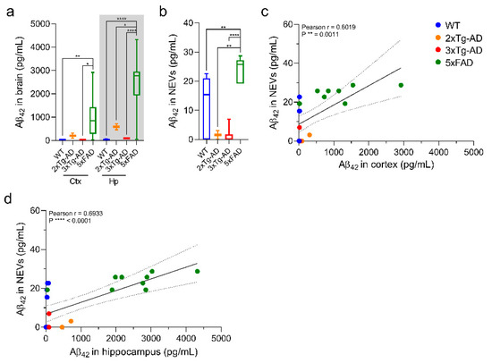
The hallmarks of Alzheimer’s disease (AD) pathology are senile plaques containing amyloid-beta (Aβ) and neurofibrillary tangles containing hyperphosphorylated tau. Additional pathologies often co-exist, whereas multiple pathogenic mechanisms are involved in AD, especially synaptic degeneration, which necessitate the need for synaptic integrity-related biomarkers alongside Aβ- and tau-related biomarkers. Plasma neuron-derived Extracellular Vesicles EVs (NDEVs) provide biomarkers related to Aβ and tau and synaptic degeneration. Here, to further establish the latter as a “liquid biopsy” for AD, we examined their relationship with ante-mortem cognition in pathologically-confirmed AD cases. We immunoprecipitated NDEVs by targeting neuronal marker L1CAM from ante-mortem plasma samples from 61 autopsy-confirmed cases of pure AD or AD with additional pathologies and measured Aβ42, p181-Tau, total Tau, synaptophysin, synaptopodin and three canonical EV markers, CD63, CD81 and CD9. Higher NDEV Aβ42 levels were consistently associated with better cognitive status, memory, fluency, working memory and executive function. Higher levels of NDEV synaptic integrity-related biomarkers were associated with better performance on executive function tasks. Our findings motivate the hypothesis that releasing Aβ42-laden NDEVs may be an adaptive mechanism in AD.
Full article
Figure 1


