


Cells 2022, 11(19), 3062; https://doi.org/10.3390/cells11193062 - 29 Sep 2022
Cited by 2 | Viewed by 1463
Abstract
►
Show Figures
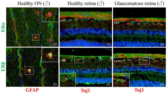
Deficiency of estradiol during the menopausal period is an important risk factor for neurodegenerative diseases, including various optic neuropathies. The aim of this study was to evaluate the impact of surgical menopause on the function and survival ratio of RGCs in the rat
[...] Read more.
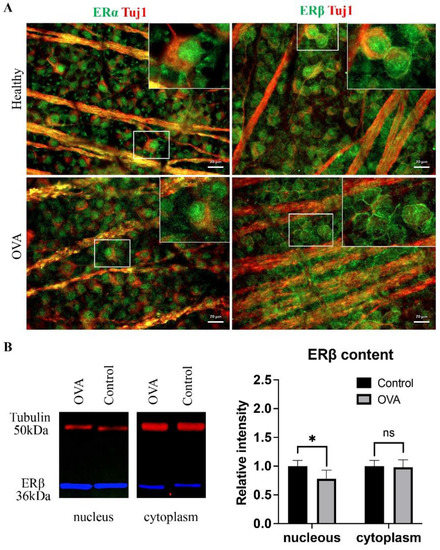
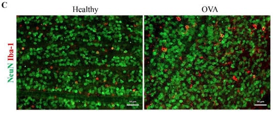
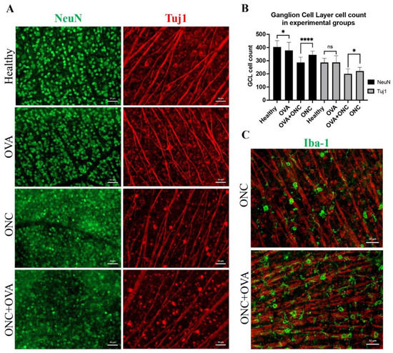
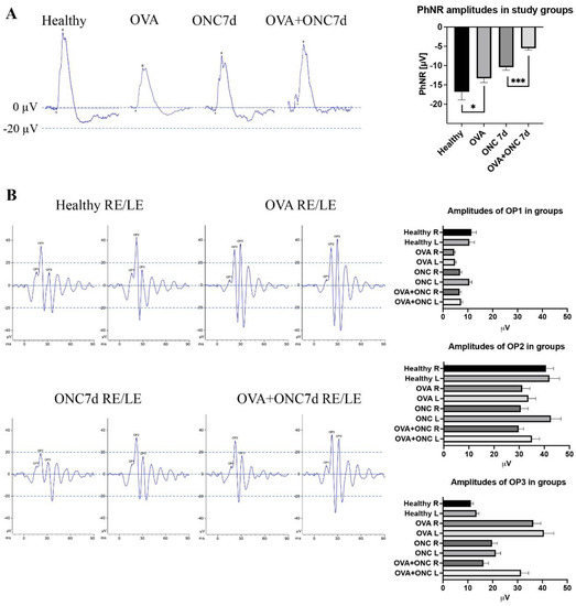
Deficiency of estradiol during the menopausal period is an important risk factor for neurodegenerative diseases, including various optic neuropathies. The aim of this study was to evaluate the impact of surgical menopause on the function and survival ratio of RGCs in the rat model of ONC (optic nerve crush). We used eight-week-old female Long Evans rats, divided into two main groups depending on the time between ovariectomy procedure (OVA) and euthanasia (two weeks vs. seven weeks), and subgroups—OVA, OVA + ONC, or ONC. Retinal function was assessed with electroretinography (ERG). RGC loss ratio was evaluated using immunolabelling and counting of RGCs. Seven weeks after OVA, the menopause morphologically affected interneurons but not RGC; however, when the ONC procedure was applied, RGCs appeared to be more susceptible to damage in case of deprivation of estrogens. In our analysis, PhNR (photopic negative responses) were severely diminished in the OVA + ONC group. A deprivation of estrogens in menopause results in accelerated retinal neurodegeneration that firstly involves retinal interneurons. The lack of estrogens increases the susceptibility of RGCs to insults.
Full article
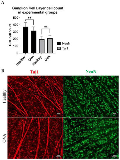
Figure 1
