


Cells 2022, 11(19), 2943; https://doi.org/10.3390/cells11192943 - 20 Sep 2022
Cited by 2 | Viewed by 2042
Abstract
►
Show Figures
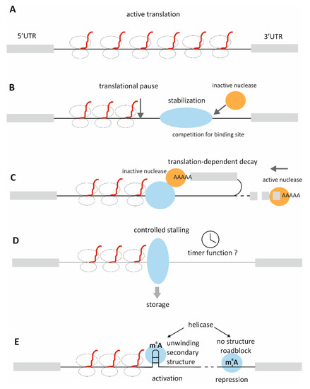
HAX1 is a human protein with no known homologues or structural domains. Mutations in the HAX1 gene cause severe congenital neutropenia through mechanisms that are poorly understood. Previous studies reported the RNA-binding capacity of HAX1, but the role of this binding in physiology
[...] Read more.
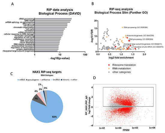
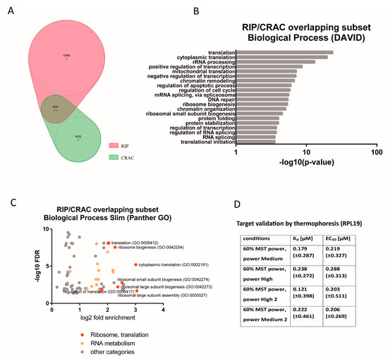
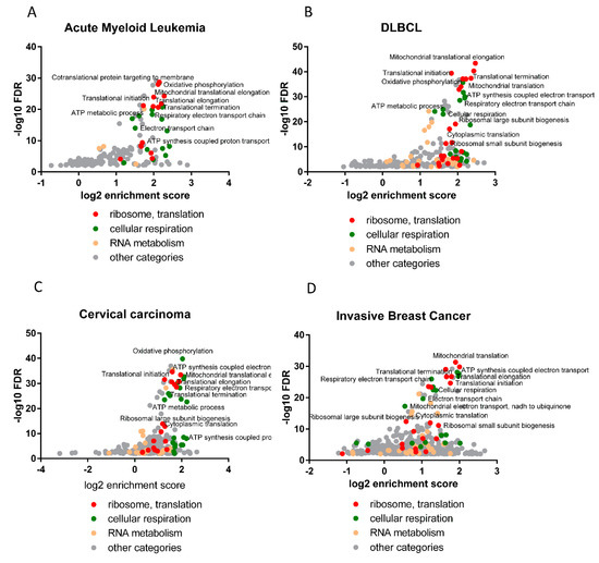
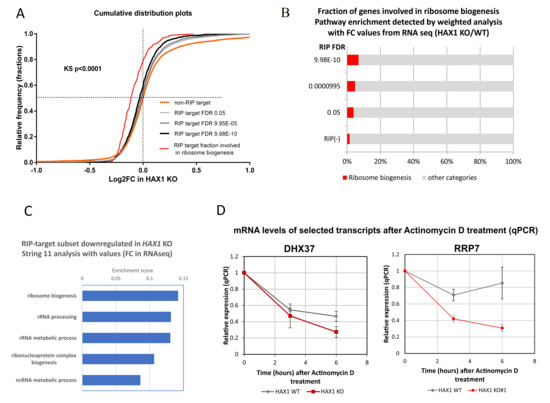
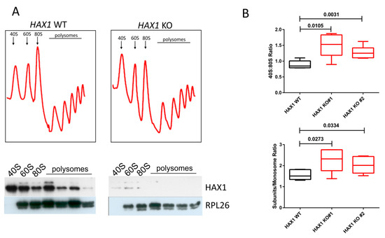
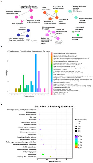
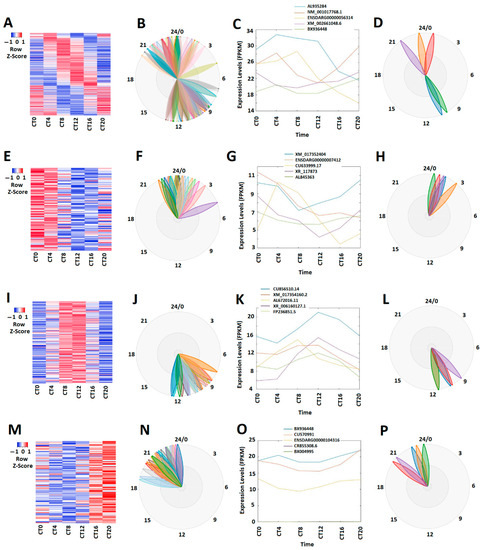
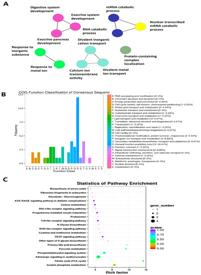
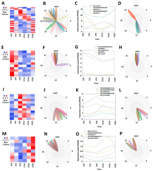
HAX1 is a human protein with no known homologues or structural domains. Mutations in the HAX1 gene cause severe congenital neutropenia through mechanisms that are poorly understood. Previous studies reported the RNA-binding capacity of HAX1, but the role of this binding in physiology and pathology remains unexplained. Here, we report the transcriptome-wide characterization of HAX1 RNA targets using RIP-seq and CRAC, indicating that HAX1 binds transcripts involved in translation, ribosome biogenesis, and rRNA processing. Using CRISPR knockouts, we find that HAX1 RNA targets partially overlap with transcripts downregulated in HAX1 KO, implying a role in mRNA stabilization. Gene ontology analysis demonstrated that genes differentially expressed in HAX1 KO (including genes involved in ribosome biogenesis and translation) are also enriched in a subset of genes whose expression correlates with HAX1 expression in four analyzed neoplasms. The functional connection to ribosome biogenesis was also demonstrated by gradient sedimentation ribosome profiles, which revealed differences in the small subunit:monosome ratio in HAX1 WT/KO. We speculate that changes in HAX1 expression may be important for the etiology of HAX1-linked diseases through dysregulation of translation.
Full article
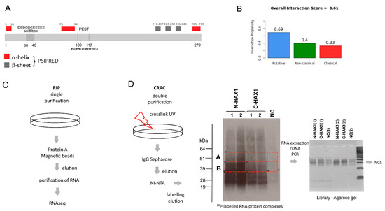
Figure 1
