


Cells 2023, 12(16), 2032; https://doi.org/10.3390/cells12162032 - 10 Aug 2023
Viewed by 893
Abstract
►
Show Figures
Thorase belongs to the AAA+ ATPase family, which plays a critical role in maintaining cellular homeostasis. Our previous work reported that Thorase was highly expressed in brain tissue, especially in the cerebellum. However, the roles of Thorase in the cerebellum have still not
[...] Read more.
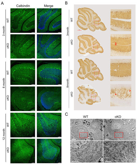
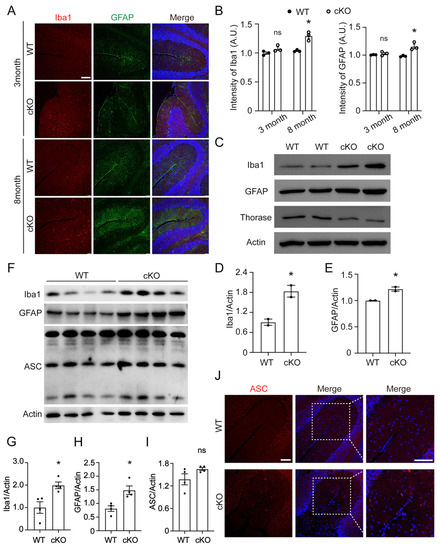
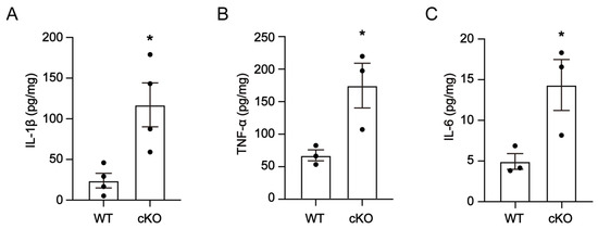
Thorase belongs to the AAA+ ATPase family, which plays a critical role in maintaining cellular homeostasis. Our previous work reported that Thorase was highly expressed in brain tissue, especially in the cerebellum. However, the roles of Thorase in the cerebellum have still not been characterized. In this study, we generated conditional knockout mice (cKO) with Thorase deletion in Purkinje cells. Thorase cKO mice exhibited cerebellar degenerative diseases-like behavior and significant impairment in motor coordination. Thorase deletion resulted in more Purkinje neuron apoptosis, leading to Purkinje cell loss in the cerebellum of Thorase cKO mice. We also found enhanced expression of the inflammatory protein ASC, IL-1β, IL-6 and TNF-α in the Thorase cKO cerebellum, which contributed to the pathogenesis of cerebellar degenerative disease. Our findings provide a better understanding of the role of Thorase in the cerebellum, which is a theoretical basis for Thorase as a therapeutic drug target for neurodegenerative diseases.
Full article
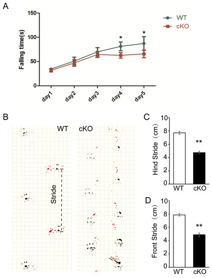
Figure 1