


Cells 2023, 12(23), 2709; https://doi.org/10.3390/cells12232709 - 26 Nov 2023
Viewed by 1379
Abstract
►
Show Figures
Ischemic thrombotic disease, characterized by the formation of obstructive blood clots within arteries or veins, is a condition associated with life-threatening events, such as stroke, myocardial infarction, deep vein thrombosis, and pulmonary embolism. The conventional therapeutic strategy relies on treatments with anticoagulants that
[...] Read more.
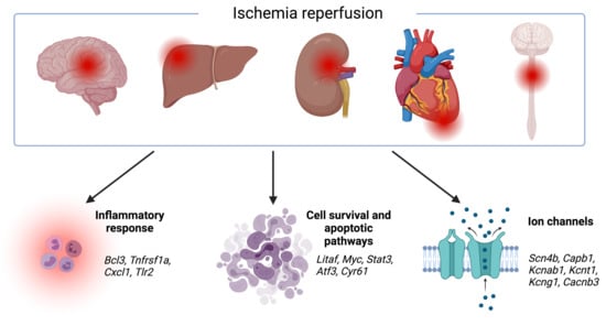
Ischemic thrombotic disease, characterized by the formation of obstructive blood clots within arteries or veins, is a condition associated with life-threatening events, such as stroke, myocardial infarction, deep vein thrombosis, and pulmonary embolism. The conventional therapeutic strategy relies on treatments with anticoagulants that unfortunately pose an inherent risk of bleeding complications. These anticoagulants primarily target clotting factors, often overlooking upstream events, including the release of neutrophil extracellular traps (NETs). Neutrophils are integral components of the innate immune system, traditionally known for their role in combating pathogens through NET formation. Emerging evidence has now revealed that NETs contribute to a prothrombotic milieu by promoting platelet activation, increasing thrombin generation, and providing a scaffold for clot formation. Additionally, NET components enhance clot stability and resistance to fibrinolysis. Clinical and preclinical studies have underscored the mechanistic involvement of NETs in the pathogenesis of thrombotic complications, since the clots obtained from patients and experimental models consistently exhibit the presence of NETs. Given these insights, the inhibition of NETs or NET formation is emerging as a promising therapeutic approach for ischemic thrombotic diseases. Recent investigations also implicate a role for the nucleotide-binding oligomerization domain (NOD)-like receptor family pyrin domain-containing 3 (NLRP3) inflammasome as a mediator of NETosis and thrombosis, suggesting that NLRP3 inhibition may also hold potential for mitigating thrombotic events. Therefore, future preclinical and clinical studies aimed at identifying and validating NLRP3 inhibition as a novel therapeutic intervention for thrombotic disorders are imperative.
Full article
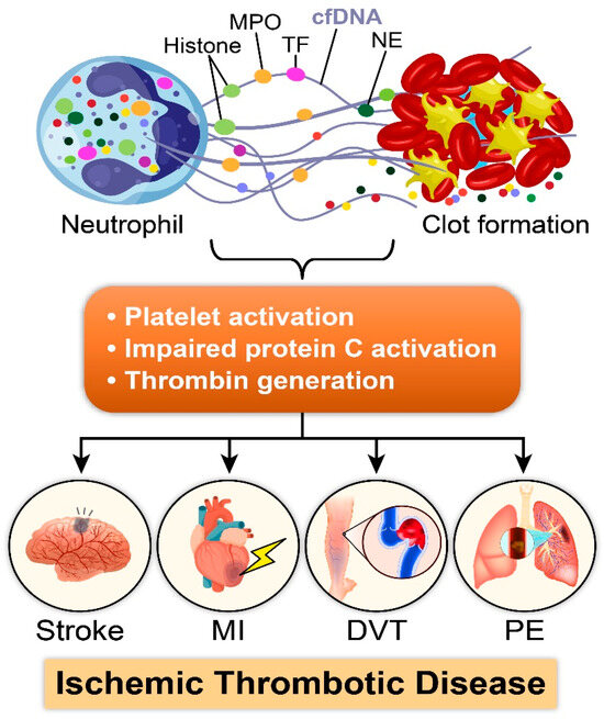
Figure 1