


Cells 2022, 11(15), 2299; https://doi.org/10.3390/cells11152299 - 26 Jul 2022
Cited by 2 | Viewed by 2911
Abstract
►
Show Figures
During vertebrate development, embryonic cells pass through a continuum of transitory pluripotent states that precede multi-lineage commitment and morphogenesis. Such states are referred to as “refractory/naïve” and “competent/formative” pluripotency. The molecular mechanisms maintaining refractory pluripotency or driving the transition to competent pluripotency, as
[...] Read more.
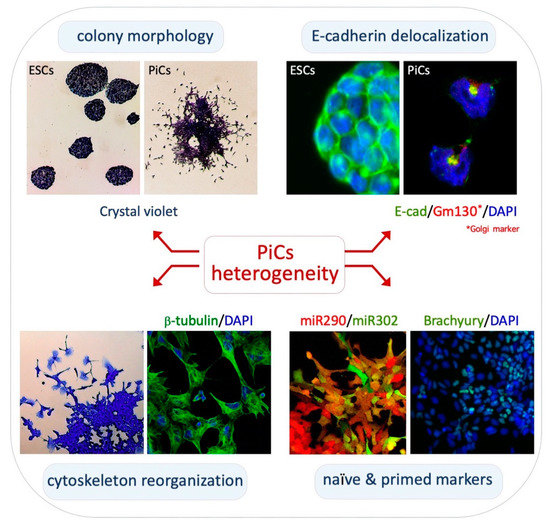
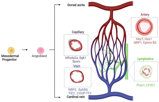
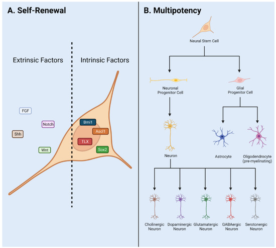
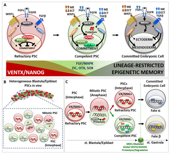
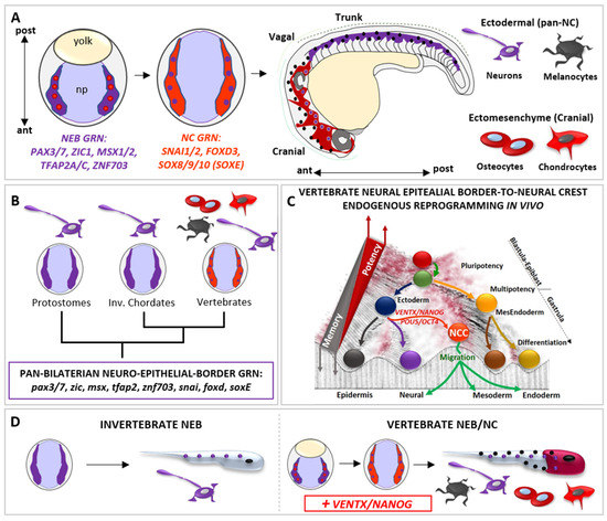
During vertebrate development, embryonic cells pass through a continuum of transitory pluripotent states that precede multi-lineage commitment and morphogenesis. Such states are referred to as “refractory/naïve” and “competent/formative” pluripotency. The molecular mechanisms maintaining refractory pluripotency or driving the transition to competent pluripotency, as well as the cues regulating multi-lineage commitment, are evolutionarily conserved. Vertebrate-specific “Developmental Potential Guardians” (vsDPGs; i.e., VENTX/NANOG, POU5/OCT4), together with MEK1 (MAP2K1), coordinate the pluripotency continuum, competence for multi-lineage commitment and morphogenesis in vivo. During neurulation, vsDPGs empower ectodermal cells of the neuro-epithelial border (NEB) with multipotency and ectomesenchyme potential through an “endogenous reprogramming” process, giving rise to the neural crest cells (NCCs). Furthermore, vsDPGs are expressed in undifferentiated-bipotent neuro-mesodermal progenitor cells (NMPs), which participate in posterior axis elongation and growth. Finally, vsDPGs are involved in carcinogenesis, whereby they confer selective advantage to cancer stem cells (CSCs) and therapeutic resistance. Intriguingly, the heterogenous distribution of vsDPGs in these cell types impact on cellular potential and features. Here, we summarize the findings about the role of vsDPGs during vertebrate development and their selective advantage in evolution. Our aim to present a holistic view regarding vsDPGs as facilitators of both cell plasticity/adaptability and morphological innovation/variation. Moreover, vsDPGs may also be at the heart of carcinogenesis by allowing malignant cells to escape from physiological constraints and surveillance mechanisms.
Full article

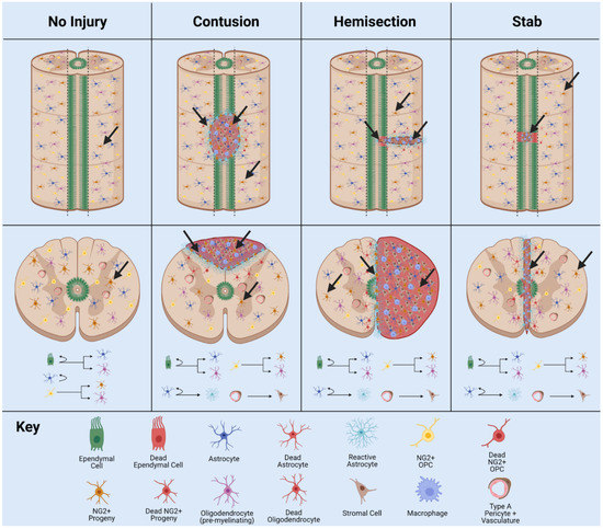
Figure 1