Magnetic Fields and Cells
A topical collection in Cells (ISSN 2073-4409). This collection belongs to the section "Cellular Biophysics".
Viewed by 49840
Share This Topical Collection
Editors
 Dr. Vitalii Zablotskii
Dr. Vitalii Zablotskii
E-Mail
Website
Collection Editor
Department of Optical and Biophysical Systems, Institute of Physics of the Czech Academy of Sciences, Prague, Czech Republic
Interests: magnetic field effects on living cells and organisms; targeted magnetic drug and cell delivery; magnetic nanostructures; magnetic phase transitions
Special Issues, Collections and Topics in MDPI journals
Dr. Xin Zhang
Dr. Xin Zhang
E-Mail
Website
Collection Editor
CAS Key Laboratory of High Magnetic Field and Ion Beam Physical Biology, High Magnetic Field
Laboratory, Hefei Institutes of Physical Science, Chinese Academy of Sciences, Hefei, China
Interests: cellular responses; the interplay between membrane trafficking and cell division; molecular mechanisms and potential biomedical applications of static magnetic fields; low-frequency magnetic fields
Special Issues, Collections and Topics in MDPI journals
Topical Collection Information
Dear Colleagues,
The remote and noninvasive control of living cells using magnetic fields (MFs) is at the top of the wish list of many researchers working in biology and medicine. The physical basis of the idea is that magnetic fields penetrate biological tissue practically without attenuation, but at specific sites in a cell, an MF is capable of triggering a wide spectrum of molecular processes, thereby drastically changing the whole-cell machinery and fate.
Knowledge of interaction mechanisms between living cells and MFs opens avenues for new research and MF applications in the following areas: magnetobiology, magnetogenetics, neuroscience, cell therapy, cell reprogramming, transcriptional responses to MFs, magnetic cryoconservation of cells and organs, cell magnetophoresis, membrane magnetoporation, biotechnologies using MFs, cell levitation and tissue engineering with MFs, magnetic carriers, magnetic nanorobots for applications in medicine, etc.
Therefore, precise knowledge of the specific cellular and molecular mechanisms behind magnetic field–cell interactions is a top priority for the scientific community.
In this Topical Collection of Cells, we invite both review papers discussing the current state of the art regarding the impact of magnetic fields on cells as well as research articles reporting new effects of MFs on the cell machinery, including reports describing novel technologies relevant to the study of biomagnetic effects at the cellular level.
Dr. Vitalii Zablotskii
Dr. Xin Zhang
Collection Editors
Manuscript Submission Information
Manuscripts should be submitted online at www.mdpi.com by registering and logging in to this website. Once you are registered, click here to go to the submission form. Manuscripts can be submitted until the deadline. All submissions that pass pre-check are peer-reviewed. Accepted papers will be published continuously in the journal (as soon as accepted) and will be listed together on the collection website. Research articles, review articles as well as short communications are invited. For planned papers, a title and short abstract (about 100 words) can be sent to the Editorial Office for announcement on this website.
Submitted manuscripts should not have been published previously, nor be under consideration for publication elsewhere (except conference proceedings papers). All manuscripts are thoroughly refereed through a single-blind peer-review process. A guide for authors and other relevant information for submission of manuscripts is available on the Instructions for Authors page. Cells is an international peer-reviewed open access semimonthly journal published by MDPI.
Please visit the Instructions for Authors page before submitting a manuscript.
The Article Processing Charge (APC) for publication in this open access journal is 2700 CHF (Swiss Francs).
Submitted papers should be well formatted and use good English. Authors may use MDPI's
English editing service prior to publication or during author revisions.
Keywords
- MF–cell interaction mechanisms
- magnetic field sensing
- effects of MFs on cell differentiation and proliferation
- molecular targets of MFs
- MFs in treating diseases
- magnetogenetics
- cytoskeleton, membrane, and ROS
- ion channel gating in MFs, Ca++ signaling
- radical pair mechanism
- magnetic control of neuronal activity
- plant cells in MFs
Published Papers (15 papers)
Open AccessArticle
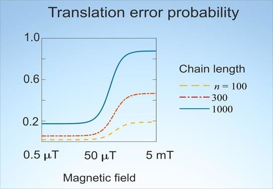
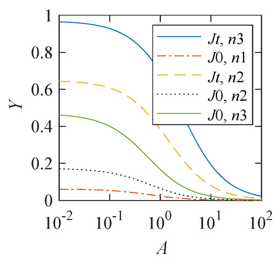
Statistical Amplification of the Effects of Weak Magnetic Fields in Cellular Translation
by
Vladimir N. Binhi
Cited by 3 | Viewed by 1595
Abstract
We assume that the enzymatic processes of recognition of amino acids and their addition to the synthesized molecule in cellular translation include the formation of intermediate pairs of radicals with spin-correlated electrons. The mathematical model presented describes the changes in the probability of
[...] Read more.
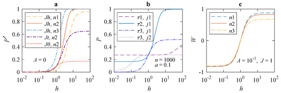
We assume that the enzymatic processes of recognition of amino acids and their addition to the synthesized molecule in cellular translation include the formation of intermediate pairs of radicals with spin-correlated electrons. The mathematical model presented describes the changes in the probability of incorrectly synthesized molecules in response to a change in the external weak magnetic field. A relatively high chance of errors has been shown to arise from the statistical enhancement of the low probability of local incorporation errors. This statistical mechanism does not require a long thermal relaxation time of electron spins of about 1
s—a conjecture often used to match theoretical models of magnetoreception with experiments. The statistical mechanism allows for experimental verification by testing the usual Radical Pair Mechanism properties. In addition, this mechanism localizes the site where magnetic effects originate, the ribosome, which makes it possible to verify it by biochemical methods. This mechanism predicts a random nature of the nonspecific effects caused by weak and hypomagnetic fields and agrees with the diversity of biological responses to a weak magnetic field.
Full article
►▼
Show Figures
Open AccessArticle
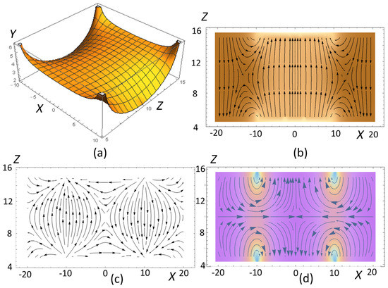
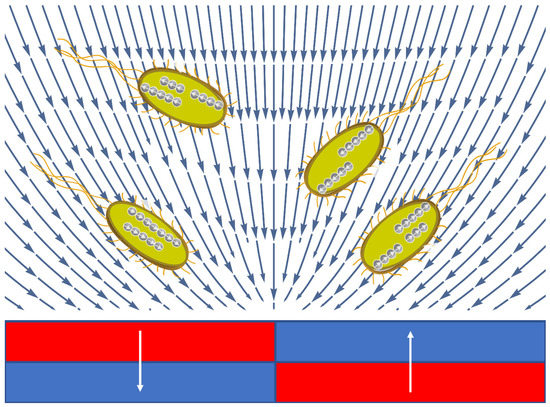
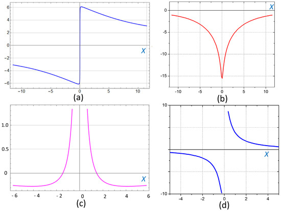
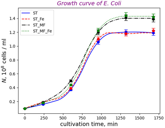
Gradient Magnetic Field Accelerates Division of E. coli Nissle 1917
by
Svitlana Gorobets, Oksana Gorobets, Iryna Sharai, Tatyana Polyakova and Vitalii Zablotskii
Cited by 1 | Viewed by 2040
Abstract
Cell-cycle progression is regulated by numerous intricate endogenous mechanisms, among which intracellular forces and protein motors are central players. Although it seems unlikely that it is possible to speed up this molecular machinery by applying tiny external forces to the cell, we show
[...] Read more.
Cell-cycle progression is regulated by numerous intricate endogenous mechanisms, among which intracellular forces and protein motors are central players. Although it seems unlikely that it is possible to speed up this molecular machinery by applying tiny external forces to the cell, we show that magnetic forcing of magnetosensitive bacteria reduces the duration of the mitotic phase. In such bacteria, the coupling of the cell cycle to the splitting of chains of biogenic magnetic nanoparticles (BMNs) provides a biological realization of such forcing. Using a static gradient magnetic field of a special spatial configuration, in probiotic bacteria
E. coli Nissle 1917, we shortened the duration of the mitotic phase and thereby accelerated cell division. Thus, focused magnetic gradient forces exerted on the BMN chains allowed us to intervene in the processes of division and growth of bacteria. The proposed magnetic-based cell division regulation strategy can improve the efficiency of microbial cell factories and medical applications of magnetosensitive bacteria.
Full article
►▼
Show Figures
Open AccessArticle
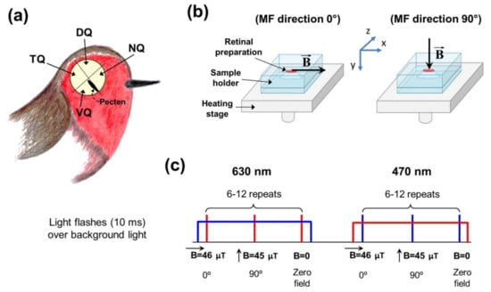
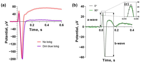
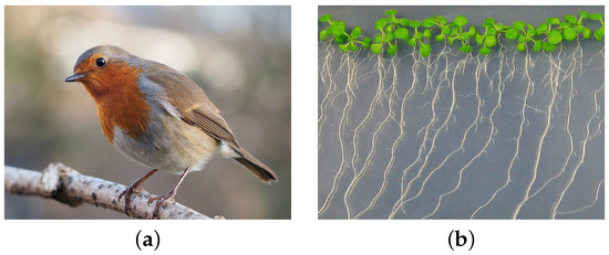
Magnetoreceptory Function of European Robin Retina: Electrophysiological and Morphological Non-Homogeneity
by
Alexander Yu. Rotov, Arsenii A. Goriachenkov, Roman V. Cherbunin, Michael L. Firsov, Nikita Chernetsov and Luba A. Astakhova
Cited by 2 | Viewed by 1750
Abstract
The avian magnetic compass allows orientation during migration and is shown to function properly under short-wavelength but not long-wavelength visible light. Therefore, the magnetoreceptive system is assumed to be light- and wavelength-dependent and localized in the retina of the eye. Putative candidates for
[...] Read more.
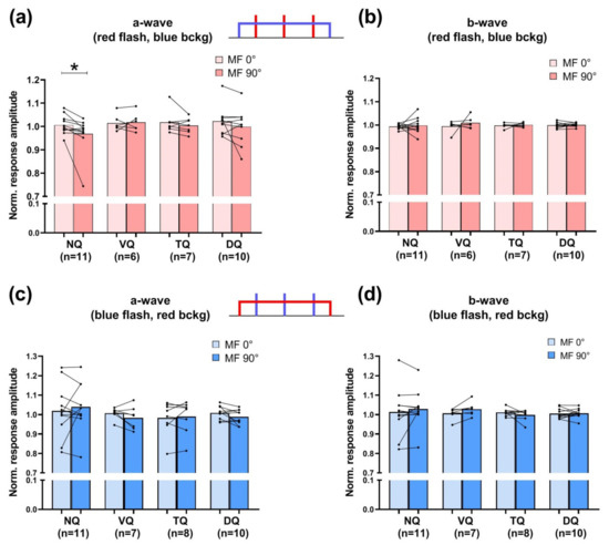
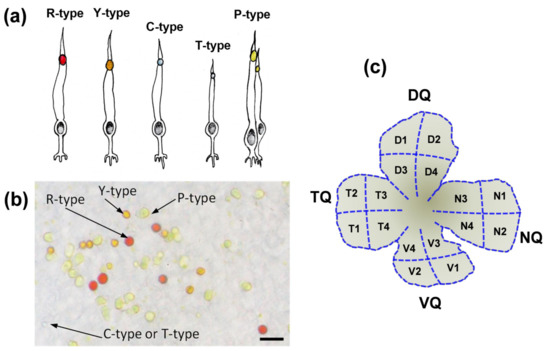
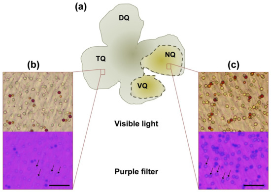
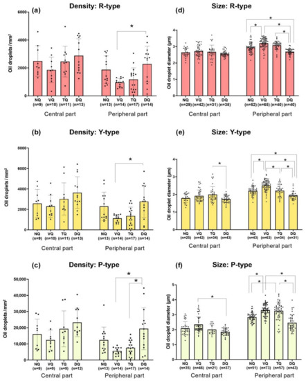
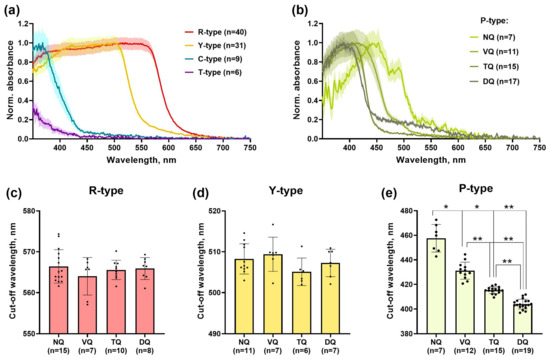
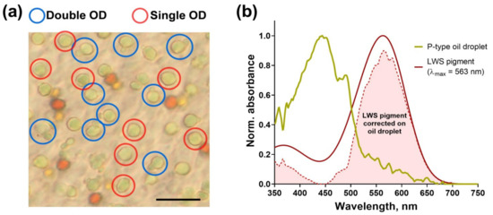
The avian magnetic compass allows orientation during migration and is shown to function properly under short-wavelength but not long-wavelength visible light. Therefore, the magnetoreceptive system is assumed to be light- and wavelength-dependent and localized in the retina of the eye. Putative candidates for the role of primary magnetosensory molecules are the cryptochromes that are known to be expressed in the avian retina and must be able to interact with phototransduction proteins. Previously, we reported that in migratory birds change in magnetic field direction induces significant effects on electroretinogram amplitude in response to blue flashes, and such an effect was observed only in the nasal quadrant of the retina. Here, we report new electroretinographic, microscopic and microspectrophotometric data on European robins, confirming the magnetosensitivity of the retinal nasal quadrant after applying the background illumination. We hypothesized that magnetoreceptive distinction of this region may be related to its morphology and analyzed the retinal distribution and optical properties of oil droplets, the filtering structures within cones. We found that the nasal quadrant contains double cones with the most intensely colorized oil droplets compared to the rest of the retina, which may be related to its magnetosensory function.
Full article
►▼
Show Figures
Open AccessArticle
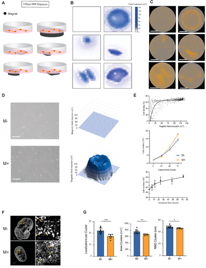
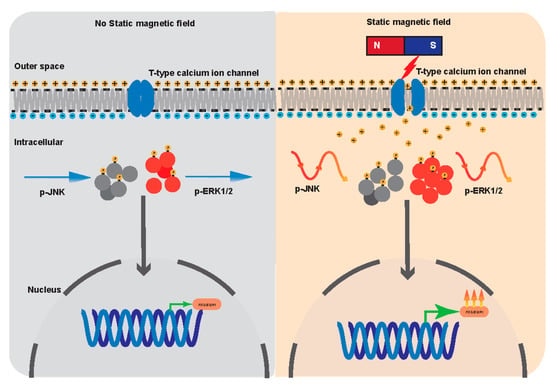
Static Magnetic Fields Regulate T-Type Calcium Ion Channels and Mediate Mesenchymal Stem Cells Proliferation
by
Haokaifeng Wu, Chuang Li, Muqaddas Masood, Zhen Zhang, Esther González-Almela, Alvaro Castells-Garcia, Gaoyang Zou, Xiaoduo Xu, Luqin Wang, Guoqing Zhao, Shengyong Yu, Ping Zhu, Bo Wang, Dajiang Qin and Jing Liu
Cited by 14 | Viewed by 3038
Abstract
The static magnetic fields (SMFs) impact on biological systems, induce a variety of biological responses, and have been applied to the clinical treatment of diseases. However, the underlying mechanisms remain largely unclear. In this report, by using human mesenchymal stem cells (MSCs) as
[...] Read more.
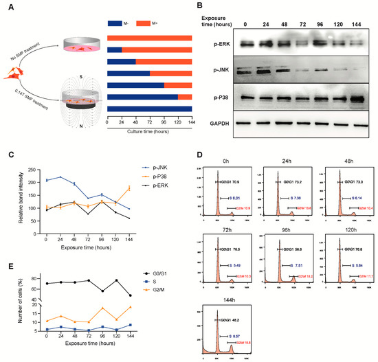
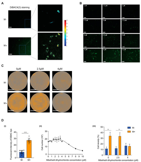
The static magnetic fields (SMFs) impact on biological systems, induce a variety of biological responses, and have been applied to the clinical treatment of diseases. However, the underlying mechanisms remain largely unclear. In this report, by using human mesenchymal stem cells (MSCs) as a model, we investigated the biological effect of SMFs at a molecular and cellular level. We showed that SMF exposure promotes MSC proliferation and activates the expression of transcriptional factors such as
FOS (Fos Proto-Oncogene, AP-1 Transcription Factor Subunit) and
EGR1 (Early Growth Response 1). In addition, the expression of signal-transduction proteins p-ERK1/2 and p-JNK oscillate periodically with SMF exposure time. Furthermore, we found that the inhibition of the T-type calcium ion channels negates the biological effects of SMFs on MSCs. Together, we revealed that the SMFs regulate T-type calcium ion channels and mediate MSC proliferation via the MAPK signaling pathways.
Full article
►▼
Show Figures
Open AccessArticle
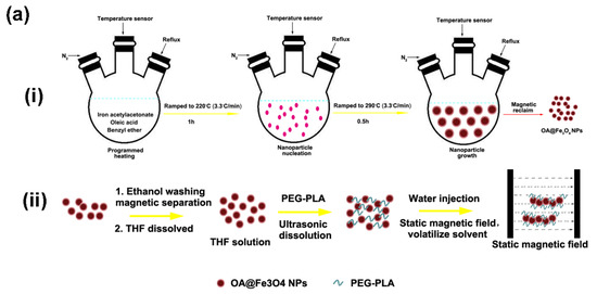
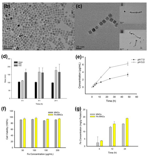
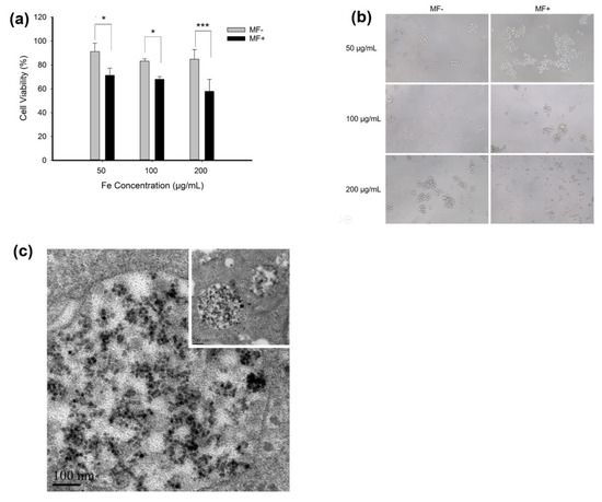
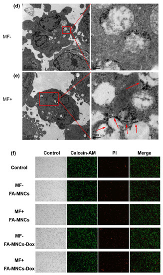
Some Preliminary Results to Eradicate Leukemic Cells in Extracorporeal Circulation by Actuating Doxorubicin-Loaded Nanochains of Fe3O4 Nanoparticles
by
Xiawen Zheng, Xiaoli Mai, Siyuan Bao, Peng Wang, Yu Hong, Yuexia Han, Jianfei Sun and Fei Xiong
Cited by 1 | Viewed by 2210
Abstract
Leukemia is a non-solid cancer which features the malignant proliferation of leukocytes. Excessive leukocytes of lesions in peripheral blood will infiltrate organs, resulting in intumescence and weakening treatment efficiency. In this study, we proposed a novel approach for targeted clearance of the leukocytes
[...] Read more.
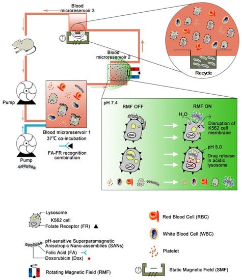
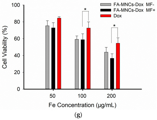
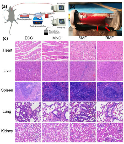
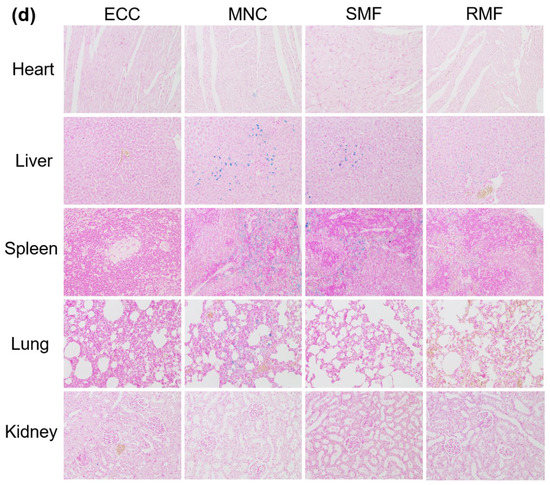
Leukemia is a non-solid cancer which features the malignant proliferation of leukocytes. Excessive leukocytes of lesions in peripheral blood will infiltrate organs, resulting in intumescence and weakening treatment efficiency. In this study, we proposed a novel approach for targeted clearance of the leukocytes in the peripheral blood ex vivo, which employed magnetic nanochains to selectively destroy the leukocytes of the lesions. The nanochains were doxorubicin-loaded nanochains of Fe
3O
4 nanoparticles which were fabricated by the solvent exchange method combined with magnetic field-directed self-assembly. Firstly, the nanochains were added into the peripheral blood during extracorporeal circulation and subjected to a rotational magnetic field for actuation. The leukocytes of the lesion were then conjugated by the nanochains via folic acid (FA) targeting. Finally, the rotational magnetic field actuated the nanochains to release the drugs and effectively damage the cytomembrane of the leukocytes. This strategy was conceptually shown in vitro (K562 cell line) and the method’s safety was evaluated in a rat model. The preliminary results demonstrate that the nanochains are biocompatible and suitable as drug carriers, showing direct lethal action to the leukemic cells combined with a rotational magnetic field. More importantly to note is that the nanochains can be effectively kept from entry into the body. We believe this extracorporeal circulation-based strategy by activating nanochains magnetically could serve as a potential method for leukemia treatment in the future.
Full article
►▼
Show Figures
Open AccessFeature PaperReview
Spatial Manipulation of Particles and Cells at Micro- and Nanoscale via Magnetic Forces
by
Larissa V. Panina, Anastasiya Gurevich, Anna Beklemisheva, Alexander Omelyanchik, Kateryna Levada and Valeria Rodionova
Cited by 11 | Viewed by 2862
Abstract
The importance of magnetic micro- and nanoparticles for applications in biomedical technology is widely recognised. Many of these applications, including tissue engineering, cell sorting, biosensors, drug delivery, and lab-on-chip devices, require remote manipulation of magnetic objects. High-gradient magnetic fields generated by micromagnets in
[...] Read more.
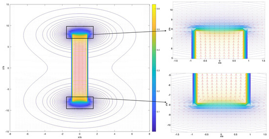
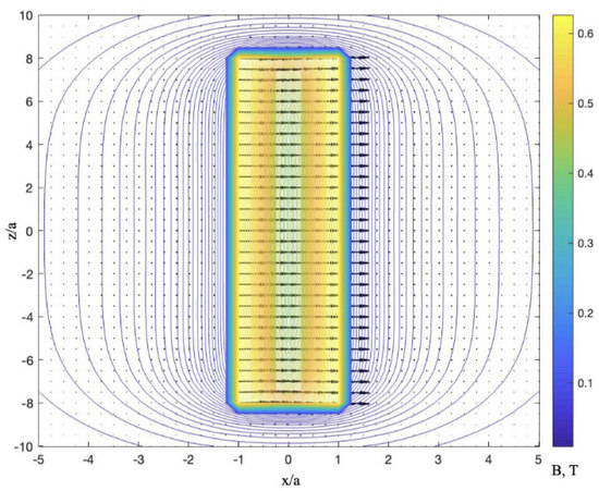
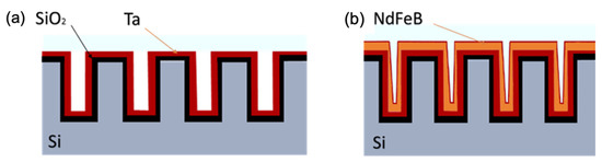
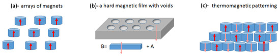
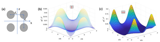
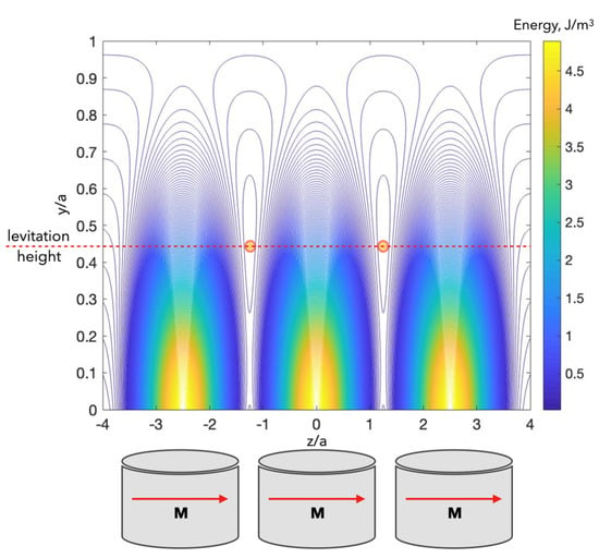
The importance of magnetic micro- and nanoparticles for applications in biomedical technology is widely recognised. Many of these applications, including tissue engineering, cell sorting, biosensors, drug delivery, and lab-on-chip devices, require remote manipulation of magnetic objects. High-gradient magnetic fields generated by micromagnets in the range of 10
3–10
5 T/m are sufficient for magnetic forces to overcome other forces caused by viscosity, gravity, and thermal fluctuations. In this paper, various magnetic systems capable of generating magnetic fields with required spatial gradients are analysed. Starting from simple systems of individual magnets and methods of field computation, more advanced magnetic microarrays obtained by lithography patterning of permanent magnets are introduced. More flexible field configurations can be formed with the use of soft magnetic materials magnetised by an external field, which allows control over both temporal and spatial field distributions. As an example, soft magnetic microwires are considered. A very attractive method of field generation is utilising tuneable domain configurations. In this review, we discuss the force requirements and constraints for different areas of application, emphasising the current challenges and how to overcome them.
Full article
►▼
Show Figures
Open AccessArticle
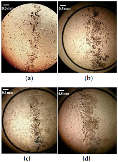
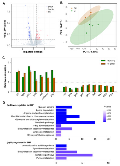
Static Magnetic Field Inhibits Growth of Escherichia coli Colonies via Restriction of Carbon Source Utilization
by
Haodong Li, Runnan Xie, Xiang Xu, Xingru Liao, Jiaxin Guo, Yanwen Fang, Zhicai Fang and Jirong Huang
Cited by 4 | Viewed by 2798
Abstract
Magnetobiological effects on growth and virulence have been widely reported in
Escherichia coli (
E. coli). However, published results are quite varied and sometimes conflicting because the underlying mechanism remains unknown. Here, we reported that the application of 250 mT static magnetic
[...] Read more.
Magnetobiological effects on growth and virulence have been widely reported in
Escherichia coli (
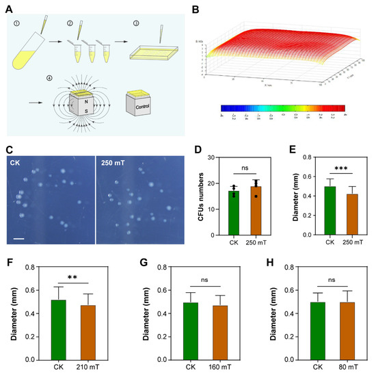
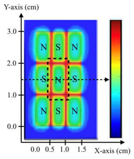
E. coli). However, published results are quite varied and sometimes conflicting because the underlying mechanism remains unknown. Here, we reported that the application of 250 mT static magnetic field (SMF) significantly reduces the diameter of
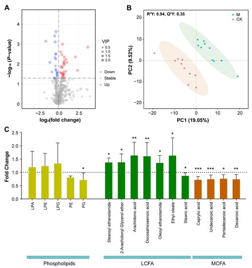
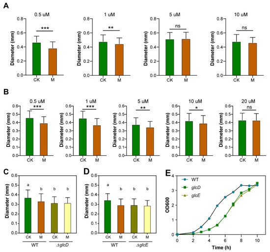
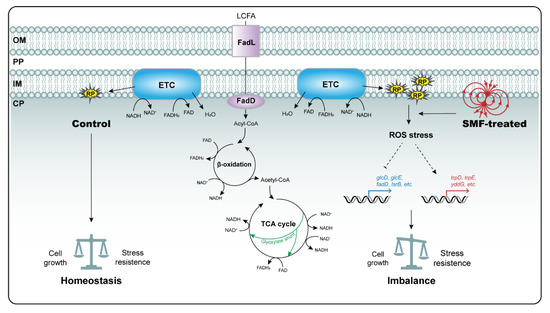
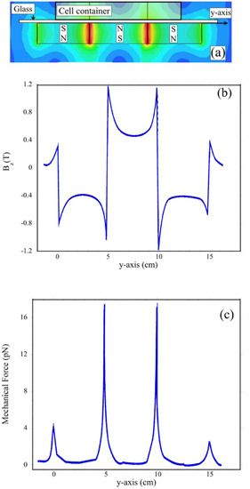
E. coli colony-forming units (CFUs) but has no impact on the number of CFUs. Transcriptomic analysis revealed that the inhibitory effect of SMF is attributed to differentially expressed genes (DEGs) primarily involved in carbon source utilization. Consistently, the addition of glycolate or glyoxylate to the culture media successfully restores the bacterial phenotype in SMF, and knockout mutants lacking glycolate oxidase are no longer sensitive to SMF. These results suggest that SMF treatment results in a decrease in glycolate oxidase activity. In addition, metabolomic assay showed that long-chain fatty acids (LCFA) accumulate while phosphatidylglycerol and middle-chain fatty acids decrease in the SMF-treated bacteria, suggesting that SMF inhibits LCFA degradation. Based on the published evidence together with ours derived from this study, we propose a model showing that free radicals generated by LCFA degradation are the primary target of SMF action, which triggers the bacterial oxidative stress response and ultimately leads to growth inhibition.
Full article
►▼
Show Figures
Open AccessArticle
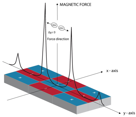
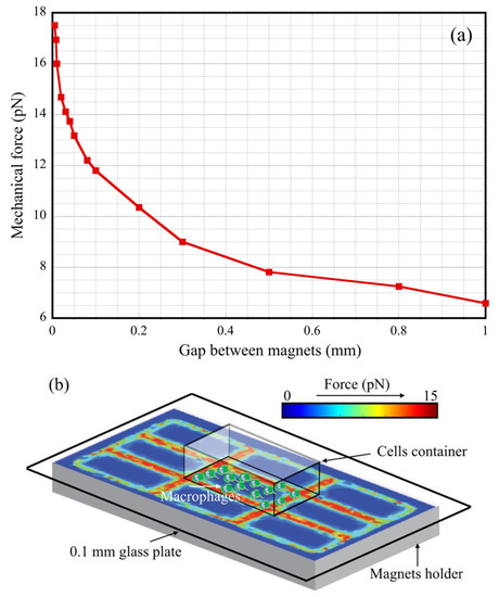
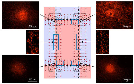
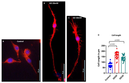
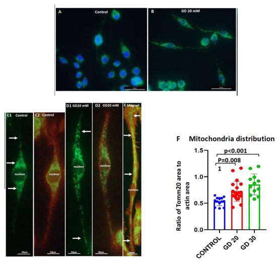
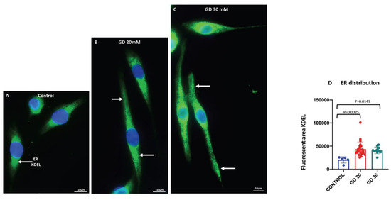
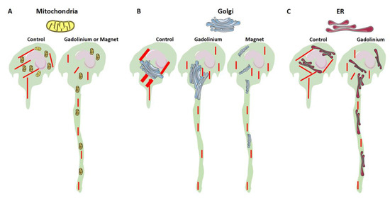
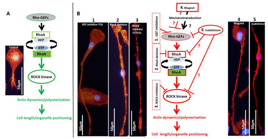
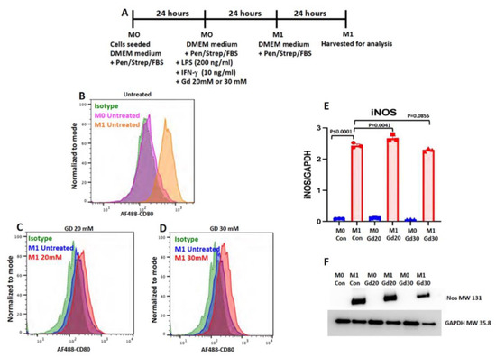
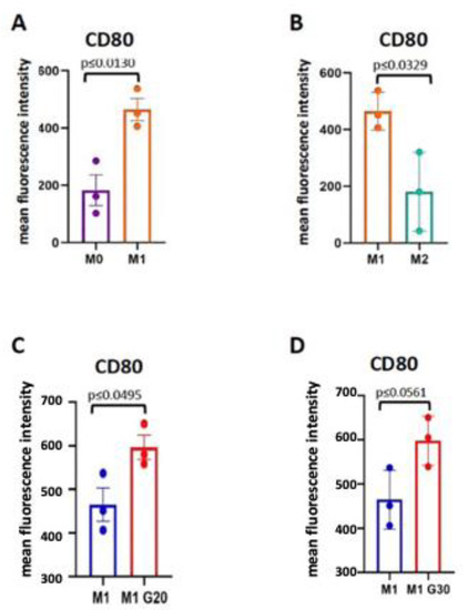
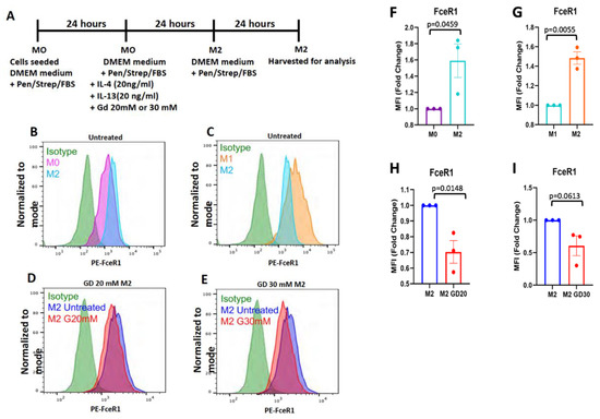
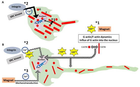
The Effect of Magnetic Field Gradient and Gadolinium-Based MRI Contrast Agent Dotarem on Mouse Macrophages
by
Priyanka Chanana, Ahmed Uosef, Nicole Vaughn, Martha Suarez-Villagran, Rafik M. Ghobrial, Malgorzata Kloc and Jarek Wosik
Cited by 6 | Viewed by 2753
Abstract
Magnetic resonance imaging (MRI) is widely used in diagnostic medicine. MRI uses the static magnetic field to polarize nuclei spins, fast-switching magnetic field gradients to generate temporal and spatial resolution, and radiofrequency (RF) electromagnetic waves to control the spin orientation. All these forms
[...] Read more.
Magnetic resonance imaging (MRI) is widely used in diagnostic medicine. MRI uses the static magnetic field to polarize nuclei spins, fast-switching magnetic field gradients to generate temporal and spatial resolution, and radiofrequency (RF) electromagnetic waves to control the spin orientation. All these forms of magnetic static and electromagnetic RF fields interact with human tissue and cells. However, reports on the MRI technique’s effects on the cells and human body are often inconsistent or contradictory. In both research and clinical MRI, recent progress in improving sensitivity and resolution is associated with the increased magnetic field strength of MRI magnets. Additionally, to improve the contrast of the images, the MRI technique often employs contrast agents, such as gadolinium-based Dotarem, with effects on cells and organs that are still disputable and not fully understood. Application of higher magnetic fields requires revisiting previously observed or potentially possible bio-effects. This article focuses on the influence of a static magnetic field gradient with and without a gadolinium-based MRI contrast agent (Dotarem) and the cellular and molecular effects of Dotarem on macrophages.
Full article
►▼
Show Figures
Open AccessArticle
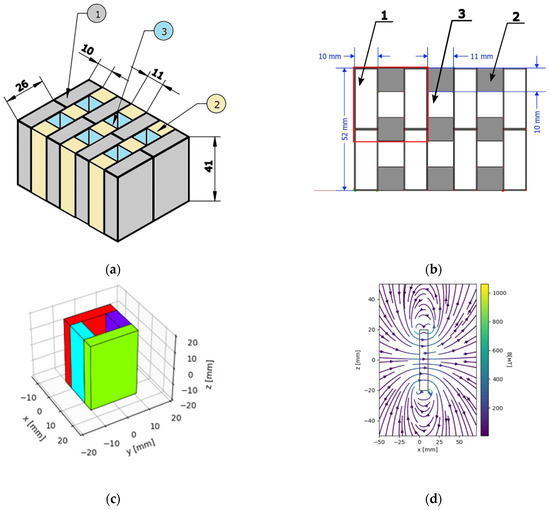
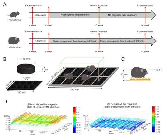
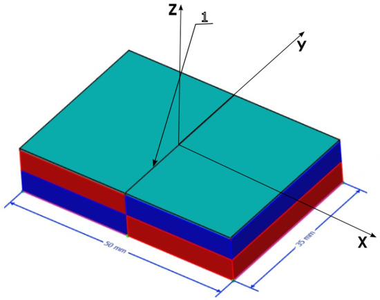
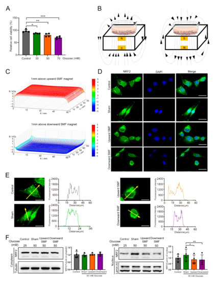
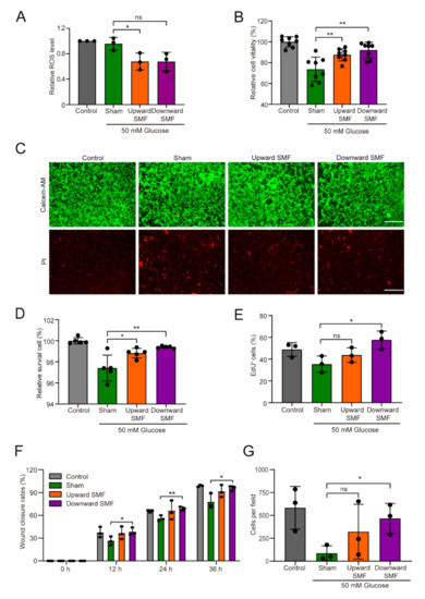
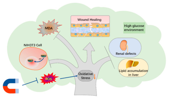
Static Magnetic Fields Reduce Oxidative Stress to Improve Wound Healing and Alleviate Diabetic Complications
by
Chuanlin Feng, Biao Yu, Chao Song, Junjun Wang, Lei Zhang, Xinmiao Ji, Ying Wang, Yanwen Fang, Zhongcai Liao, Min Wei and Xin Zhang
Cited by 14 | Viewed by 4317
Abstract
Although some studies have shown that some static magnetic fields (SMFs) can promote wound healing in diabetic mice, it is not clear whether the other diabetes complications, such as liver disease and diabetic nephropathy, can also be alleviated. Here, we constructed two simple
[...] Read more.
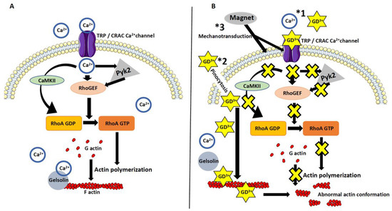
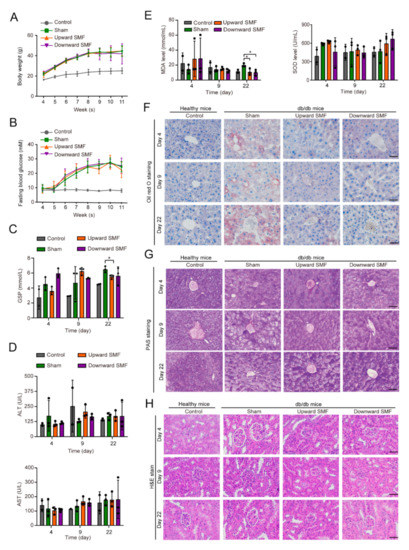
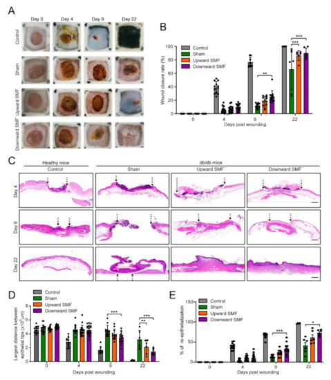
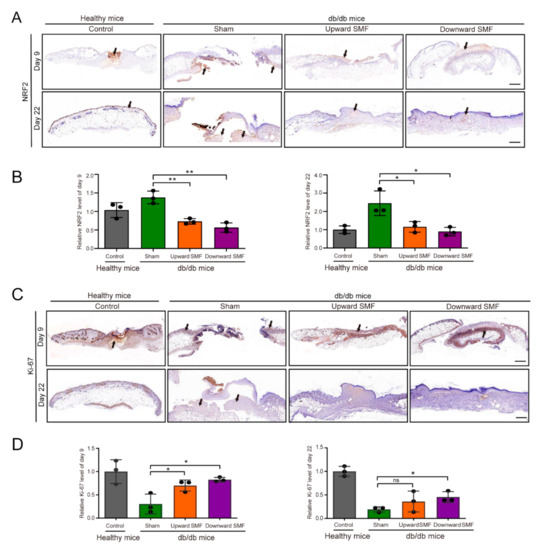
Although some studies have shown that some static magnetic fields (SMFs) can promote wound healing in diabetic mice, it is not clear whether the other diabetes complications, such as liver disease and diabetic nephropathy, can also be alleviated. Here, we constructed two simple magnetic plates using neodymium permanent magnets to examine the comprehensive effects of moderate SMFs on genetically obese leptin receptor-deficient db/db diabetic mice. We found that although the blood glucose was not obviously reduced by these two SMF settings, both of the glycated serum protein (GSP) and malondialdehyde (MDA) levels were significantly decreased (Cohen’s d = 2.57–3.04). Moreover, the wound healing, liver lipid accumulation, and renal defects were all significantly improved by SMF treatment (Cohen’s d = 0.91–2.05). Wound tissue examination showed obvious nuclear factor erythroid 2-related factor 2 (NRF2) level decrease (Cohen’s d = 2.49–5.40) and Ki-67 level increase (Cohen’s d = 2.30–3.40), indicating decreased oxidative stress and increased cell proliferation. In vitro cellular studies with fibroblast NIH3T3 cells showed that SMFs could reduce high glucose-induced NRF2 nucleus translocation (Cohen’s d = 0.87–1.15) and cellular reactive oxygen species (ROS) elevation (Cohen’s d = 0.92), indicating decreased oxidative stress. Consequently, high glucose-induced impairments in cell vitality, proliferation, and migration were all improved by SMF treatment. Therefore, our results demonstrate that these simple SMF devices could effectively reduce oxidative stress in diabetic mice and may provide a cost-effective physical therapy strategy to alleviate multiple diabetic complications in the future.
Full article
►▼
Show Figures
Open AccessReview
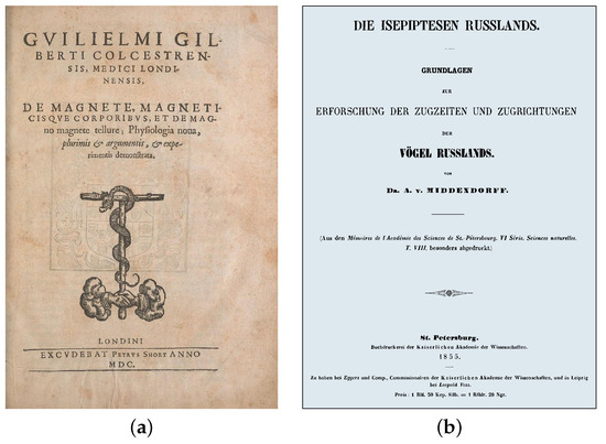
Theoretical Concepts in Magnetobiology after 40 Years of Research
by
Vladimir N. Binhi and Andrei B. Rubin
Cited by 26 | Viewed by 2751
Abstract
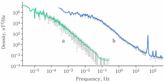
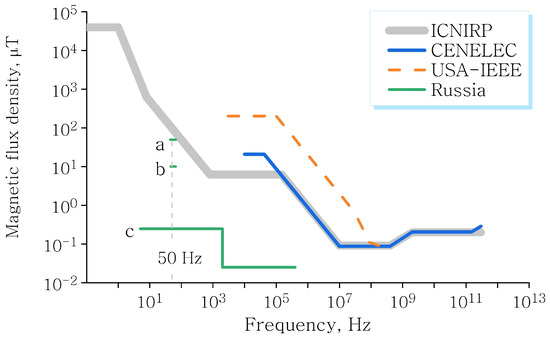
This review contains information on the development of magnetic biology, one of the multidisciplinary areas of biophysics. The main historical facts are presented and the general observed properties of magnetobiological phenomena are listed. The unavoidable presence of nonspecific magnetobiological effects in the everyday
[...] Read more.
This review contains information on the development of magnetic biology, one of the multidisciplinary areas of biophysics. The main historical facts are presented and the general observed properties of magnetobiological phenomena are listed. The unavoidable presence of nonspecific magnetobiological effects in the everyday life of a person and society is shown. Particular attention is paid to the formation of theoretical concepts in magnetobiology and the state of the art in this area of research. Some details are provided on the molecular mechanisms of the nonspecific action of a magnetic field on organisms. The prospects of magnetobiology for the near and distant future are discussed.
Full article
►▼
Show Figures
Open AccessArticle
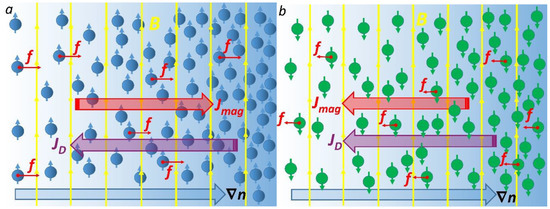
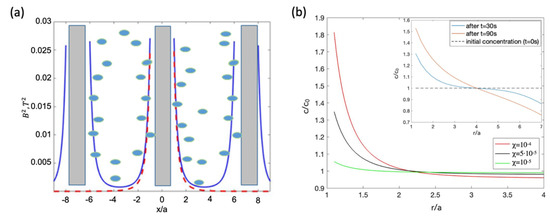
Effects of High Magnetic Fields on the Diffusion of Biologically Active Molecules
by
Vitalii Zablotskii, Tatyana Polyakova and Alexandr Dejneka
Cited by 9 | Viewed by 3004
Abstract
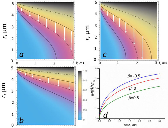
The diffusion of biologically active molecules is a ubiquitous process, controlling many mechanisms and the characteristic time scales for pivotal processes in living cells. Here, we show how a high static magnetic field (MF) affects the diffusion of paramagnetic and diamagnetic species including
[...] Read more.
The diffusion of biologically active molecules is a ubiquitous process, controlling many mechanisms and the characteristic time scales for pivotal processes in living cells. Here, we show how a high static magnetic field (MF) affects the diffusion of paramagnetic and diamagnetic species including oxygen, hemoglobin, and drugs. We derive and solve the equation describing diffusion of such biologically active molecules in the presence of an MF as well as reveal the underlying mechanism of the MF’s effect on diffusion. We found that a high MF accelerates diffusion of diamagnetic species while slowing the diffusion of paramagnetic molecules in cell cytoplasm. When applied to oxygen and hemoglobin diffusion in red blood cells, our results suggest that an MF may significantly alter the gas exchange in an erythrocyte and cause swelling. Our prediction that the diffusion rate and characteristic time can be controlled by an MF opens new avenues for experimental studies foreseeing numerous biomedical applications.
Full article
►▼
Show Figures
Open AccessArticle
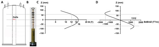
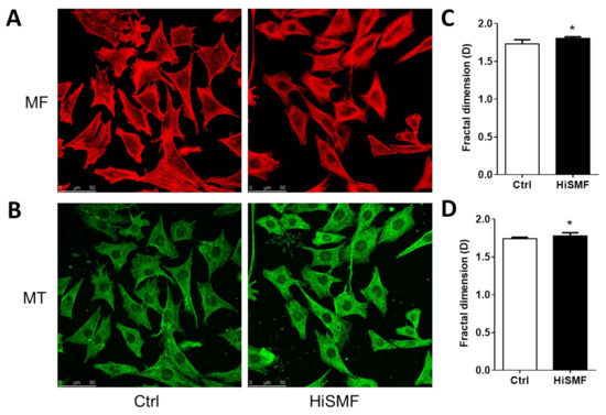
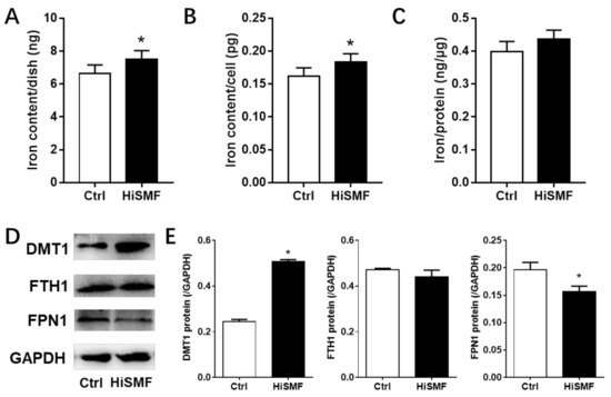
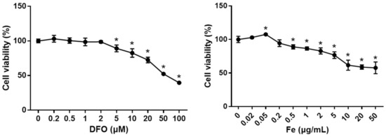
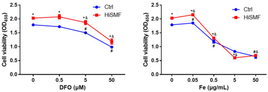
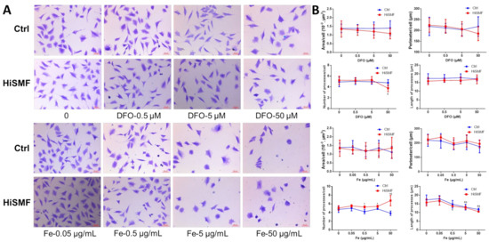
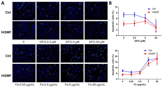
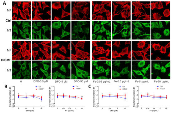
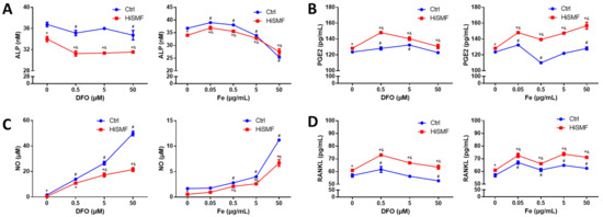
Effect of High Static Magnetic Fields on Biological Activities and Iron Metabolism in MLO-Y4 Osteocyte-like Cells
by
Jiancheng Yang, Gejing Zhang, Qingmei Li, Qinghua Tang, Yan Feng, Peng Shang and Yuhong Zeng
Cited by 6 | Viewed by 2690
Abstract
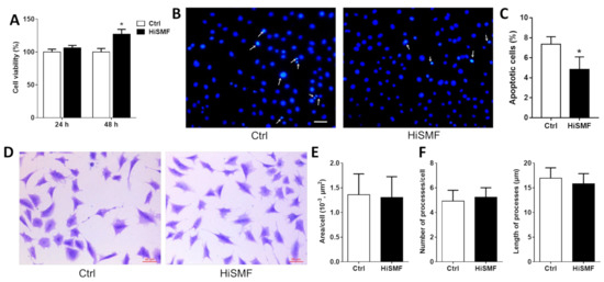
There are numerous studies that investigate the effects of static magnetic fields (SMFs) on osteoblasts and osteoclasts. However, although osteocytes are the most abundant cell type in bone tissue, there are few studies on the biological effects of osteocytes under magnetic fields. Iron
[...] Read more.
There are numerous studies that investigate the effects of static magnetic fields (SMFs) on osteoblasts and osteoclasts. However, although osteocytes are the most abundant cell type in bone tissue, there are few studies on the biological effects of osteocytes under magnetic fields. Iron is a necessary microelement that is involved in numerous life activities in cells. Studies have shown that high static magnetic fields (HiSMF) can regulate cellular iron metabolism. To illustrate the effect of HiSMF on activities of osteocytes, and whether iron is involved in this process, HiSMF of 16 tesla (T) was used, and the changes in cellular morphology, cytoskeleton, function-related protein expression, secretion of various cytokines, and iron metabolism in osteocytes under HiSMF were studied. In addition, the biological effects of HiSMF combined with iron preparation and iron chelator on osteocytes were also investigated. The results showed that HiSMF promoted cellular viability, decreased apoptosis, increased the fractal dimension of the cytoskeleton, altered the secretion of cytokines, and increased iron levels in osteocytes. Moreover, it was found that the biological effects of osteocytes under HiSMF are attenuated or enhanced by treatment with a certain concentration of iron. These data suggest that HiSMF-regulated cellular iron metabolism may be involved in altering the biological effects of osteocytes under HiSMF exposure.
Full article
►▼
Show Figures
Open AccessFeature PaperReview
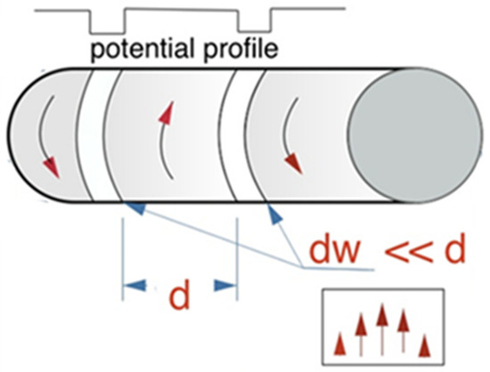
Magnetic Guiding with Permanent Magnets: Concept, Realization and Applications to Nanoparticles and Cells
by
Peter Blümler
Cited by 21 | Viewed by 5125
Abstract
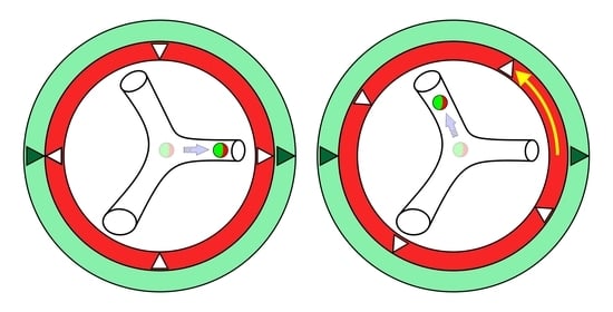
The idea of remote magnetic guiding is developed from the underlying physics of a concept that allows for bijective force generation over the inner volume of magnet systems. This concept can equally be implemented by electro- or permanent magnets. Here, permanent magnets are
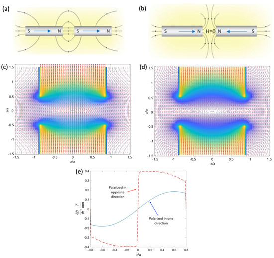
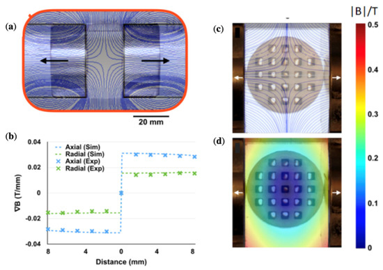
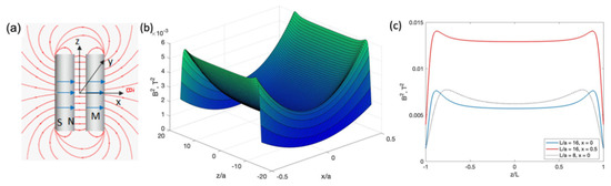
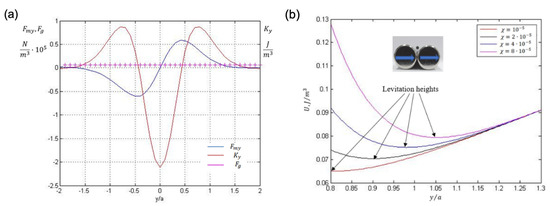
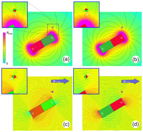
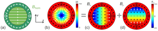
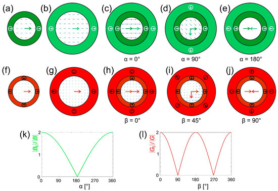
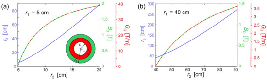
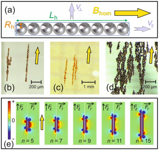
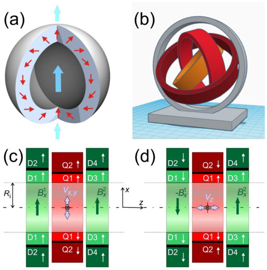
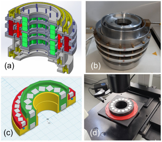
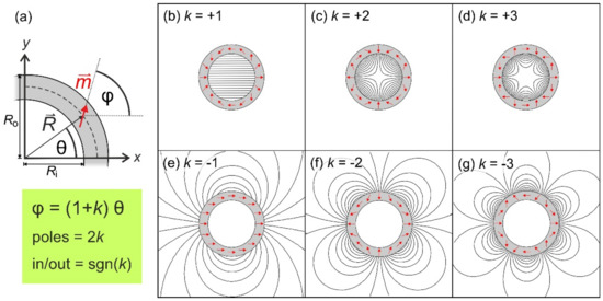
[...] Read more.
The idea of remote magnetic guiding is developed from the underlying physics of a concept that allows for bijective force generation over the inner volume of magnet systems. This concept can equally be implemented by electro- or permanent magnets. Here, permanent magnets are in the focus because they offer many advantages. The equations of magnetic fields and forces as well as velocities are derived in detail and physical limits are discussed. The special hydrodynamics of nanoparticle dispersions under these circumstances is reviewed and related to technical constraints. The possibility of 3D guiding and magnetic imaging techniques are discussed. Finally, the first results in guiding macroscopic objects, superparamagnetic nanoparticles, and cells with incorporated nanoparticles are presented. The constructed magnet systems allow for orientation, movement, and acceleration of magnetic objects and, in principle, can be scaled up to human size.
Full article
►▼
Show Figures
Open AccessReview
The Review of Bioeffects of Static Magnetic Fields on the Oral Tissue-Derived Cells and Its Application in Regenerative Medicine
by
Wei-Zhen Lew, Sheng-Wei Feng, Sheng-Yang Lee and Haw-Ming Huang
Cited by 19 | Viewed by 3156
Abstract
Magnets have been widely used in dentistry for orthodontic tooth movement and denture retention. Nevertheless, criticisms have arisen regarding the biosafety of static magnetic field (SMF) effects on surrounding tissues. Various controversial pieces of evidence have been discussed regarding SMFs on cellular biophysics,
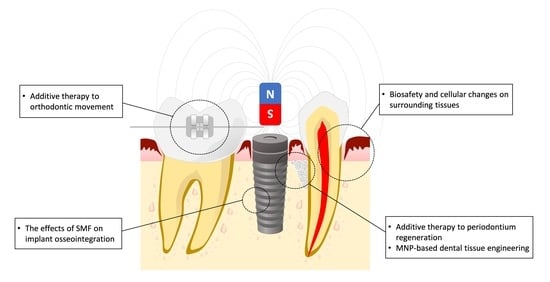
[...] Read more.
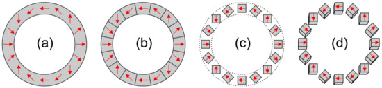
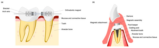
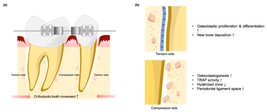
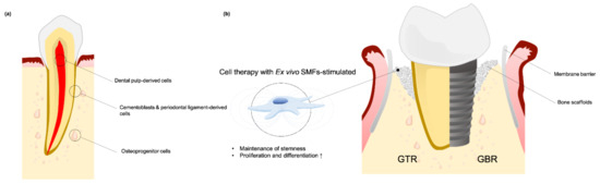
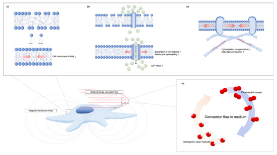
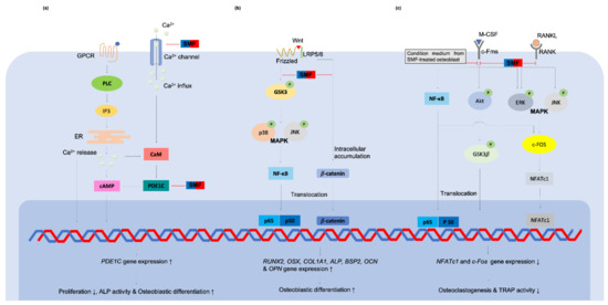
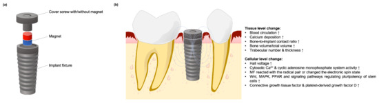
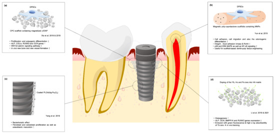
Magnets have been widely used in dentistry for orthodontic tooth movement and denture retention. Nevertheless, criticisms have arisen regarding the biosafety of static magnetic field (SMF) effects on surrounding tissues. Various controversial pieces of evidence have been discussed regarding SMFs on cellular biophysics, but little consensus has been reached, especially in the field of dentistry. Thus, the present paper will first review the safe use of SMFs in the oral cavity and as an additive therapy to orthodontic tooth movement and periodontium regeneration. Then, studies regarding SMF-incorporated implants are reviewed to investigate the advantageous effects of SMFs on osseointegration and the underlying mechanisms. Finally, a review of current developments in dentistry surrounding the combination of magnetic nanoparticles (MNPs) and SMFs is made to clarify potential future clinical applications.
Full article
►▼
Show Figures
Open AccessArticle
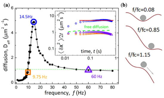
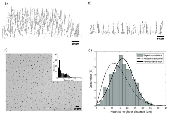
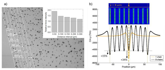
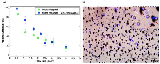
Self-Assembled Permanent Micro-Magnets in a Polymer-Based Microfluidic Device for Magnetic Cell Sorting
by
Lucie Descamps, Marie-Charlotte Audry, Jordyn Howard, Samir Mekkaoui, Clément Albin, David Barthelemy, Léa Payen, Jessica Garcia, Emmanuelle Laurenceau, Damien Le Roy and Anne-Laure Deman
Cited by 16 | Viewed by 3673
Abstract
Magnetophoresis-based microfluidic devices offer simple and reliable manipulation of micro-scale objects and provide a large panel of applications, from selective trapping to high-throughput sorting. However, the fabrication and integration of micro-scale magnets in microsystems involve complex and expensive processes. Here we report on
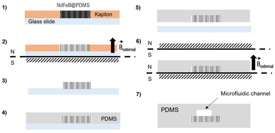
[...] Read more.
Magnetophoresis-based microfluidic devices offer simple and reliable manipulation of micro-scale objects and provide a large panel of applications, from selective trapping to high-throughput sorting. However, the fabrication and integration of micro-scale magnets in microsystems involve complex and expensive processes. Here we report on an inexpensive and easy-to-handle fabrication process of micrometer-scale permanent magnets, based on the self-organization of NdFeB particles in a polymer matrix (polydimethylsiloxane, PDMS). A study of the inner structure by X-ray tomography revealed a chain-like organization of the particles leading to an array of hard magnetic microstructures with a mean diameter of 4 µm. The magnetic performance of the self-assembled micro-magnets was first estimated by COMSOL simulations. The micro-magnets were then integrated into a microfluidic device where they act as micro-traps. The magnetic forces exerted by the micro-magnets on superparamagnetic beads were measured by colloidal probe atomic force microscopy (AFM) and in operando in the microfluidic system. Forces as high as several nanonewtons were reached. Adding an external millimeter-sized magnet allowed target magnetization and the interaction range to be increased. Then, the integrated micro-magnets were used to study the magnetophoretic trapping efficiency of magnetic beads, providing efficiencies of 100% at 0.5 mL/h and 75% at 1 mL/h. Finally, the micro-magnets were implemented for cell sorting by performing white blood cell depletion.
Full article
►▼
Show Figures
Planned Papers
The below list represents only planned manuscripts. Some of these
manuscripts have not been received by the Editorial Office yet. Papers
submitted to MDPI journals are subject to peer-review.
Title: /
Authors: Boelens
Affiliation: /
Abstract: Selectively-binding superparamagnetic carriers such as functionalized iron oxide nanoparticles and composite beads are widely used for magnetic separation processes in medicinal biotechnology. The broad range of their applications includes the diagnosis of pathogens, magnetic labelling of cancer cells and the purification of biomolecules such as proteins, antibodies, nucleic acids and peptides (Leong et al., 2016; Yildiz, 2016). Furthermore, there is interest in the scientific community for similar applications aimed at the removal and recovery of inorganic particles and metal ions by selective magnetic separation processes facilitated by peptide-functionalized suparparamagnetic carriers (Xu et al., 2020; Cetinel et al., 2018; Vreuls et al., 2011). However, the studies available on this topic so far have only focussed on the selective interactions at the peptide-functionalized particle surfaces. The potential for an industrially applicable process has been shown by the use of low-gradient magnetic-separation on a millileter-scale, but the role of the magnetic field and the fluid dynamics in an upscalable process has been neglected.
Recently, our group has succesfully identified selectively-binding peptides by Phage Surface Display and characterized their interaction with economically interesting materials such as rare earth element containing waste phosphor powders and gallium ions in industrial wastewater (Schönberger et al., 2021; Lederer et al., 2019; Lederer et al., 2016). Currently, we are investigating the functionalization of superparamagnetic nanoparticles and composite beads for selective interactions with rare earth element containing particles and their applicability in magnetic separation processes. By the use of a high-gradient magnetic-separator especially designed for biotechnological purposes (Schultz et al., 2007; Hoffman et al., 2002), we hope to give a first proof-of-concept for the upscaling of selective magnetic separation processes for resource recovery facilitated by peptide-functionalized carriers. In this interdisciplinary work, we hope to present important findings on the interplay of magnetic carrier design, particle surface interactions, magnetic field configuration and fluid dynamics.


